Understanding Young's Modulus of Human Soft Tissues: From kPa to MPa Ranges for Research & Drug Development
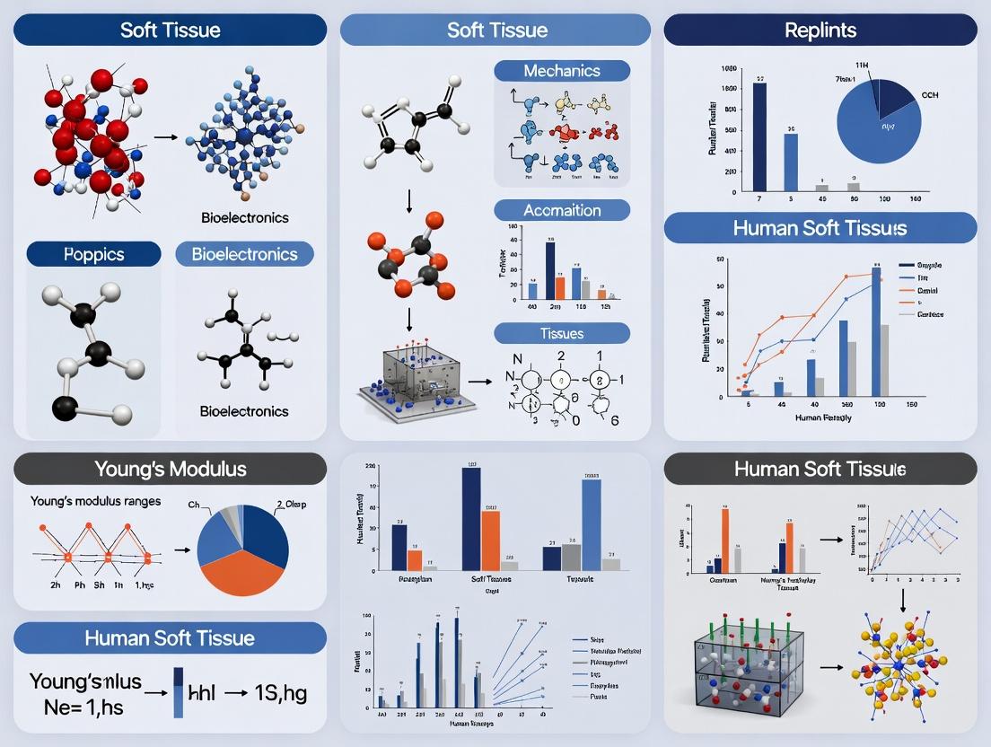
This comprehensive article examines the Young's modulus range of human soft tissues, a critical biomechanical property for biomedical research.
Understanding Young's Modulus of Human Soft Tissues: From kPa to MPa Ranges for Research & Drug Development
Abstract
This comprehensive article examines the Young's modulus range of human soft tissues, a critical biomechanical property for biomedical research. Tailored for researchers, scientists, and drug development professionals, it covers foundational definitions and tissue-specific values (kPa to MPa), explores measurement techniques (AFM, rheology, tensile testing) and their applications in disease modeling and drug screening. The guide addresses common measurement challenges, data variability, and optimization strategies, and provides a framework for validating results through comparative analysis with established literature and computational models. The synthesis aims to equip professionals with the knowledge to accurately characterize tissue mechanics for advancing therapeutic development and regenerative medicine.
What is Young's Modulus? Defining the Stiffness Spectrum of Human Soft Tissues
This technical guide provides a foundational overview of core biomechanical parameters—elastic modulus, stiffness, and compliance—within the context of human soft tissue research. Framed by the broader thesis of mapping the Young's modulus range of human soft tissues, this whitepaper details precise definitions, measurement methodologies, and the biological significance of these properties for researchers and drug development professionals. The content underscores the critical role of tissue mechanical properties in physiological function, disease progression, and therapeutic intervention.
Human soft tissues exhibit a vast and physiologically critical range of mechanical properties. Their elastic modulus (Young's modulus) can span from approximately 0.1 kPa for brain tissue to several GPa for tendon, reflecting specialized functional adaptation. Accurately defining and measuring stiffness (resistance to deformation) and its inverse, compliance (ease of deformation), is paramount for understanding tissue development, homeostasis, and pathology. This guide establishes the core concepts, measurement techniques, and current data within the framework of ongoing research to quantify the soft tissue modulus landscape.
Core Definitions in a Biological Context
Elastic Modulus (Young's Modulus, E): A fundamental intensive property of a material, defined as the ratio of stress (force per unit area) to strain (relative deformation) in the linear elastic regime. In biology, it describes the intrinsic local tissue firmness, independent of sample geometry.
Stiffness (k): An extensive property of a structure or object, defined as the force required to produce a unit displacement (k = F / Δx). It depends on both the material's elastic modulus and the object's geometry (e.g., length, cross-sectional area). For a simple prismatic specimen in uniaxial tension, k = (E * A) / L.
Compliance (C): The inverse of stiffness (C = 1/k = Δx / F), representing the displacement per unit applied force. It quantifies a structure's deformability. High compliance indicates easy deformation.
Key Experimental Protocols for Measurement
The following are standardized methodologies for determining the elastic modulus of soft tissues.
Atomic Force Microscopy (AFM) Indentation
Purpose: To map spatial variations in elastic modulus at micro- to nanoscale resolution. Protocol:
- Sample Preparation: Fresh or properly preserved tissue is cryosectioned (100-500 μm thick) or prepared as intact, excised specimens. Mounted in a physiological buffer to maintain hydration.
- Cantilever Calibration: The spring constant of the AFM cantilever is determined via thermal tuning or Sader method. Tip geometry (e.g., spherical bead radius) is characterized via SEM.
- Indentation: The tip is approached to the sample surface at a constant velocity (typically 1-10 μm/s). Force-displacement curves are recorded at multiple (>100) spatially referenced points.
- Data Analysis: Force-indentation curves are fit with an appropriate contact mechanics model (e.g., Hertz, Sneddon) to extract the apparent Young's Modulus. Assumptions include elasticity, homogeneity, and infinite sample thickness relative to indentation depth.
Shear Rheometry
Purpose: To characterize bulk viscoelastic properties (shear storage modulus G' and loss modulus G'') of soft, homogeneous tissues or hydrogels. Protocol:
- Geometry Selection: Parallel plate or cone-and-plate geometry is chosen based on sample consistency.
- Loading & Trimming: Tissue is placed on the lower plate, and the upper geometry is lowered to a defined gap (e.g., 500 μm). Excess material is trimmed.
- Strain Sweep: Performed at a constant frequency (e.g., 1 Hz) to identify the linear viscoelastic region (LVER).
- Frequency Sweep: Conducted within the LVER to measure G' and G'' as a function of frequency (e.g., 0.01 to 100 Hz).
- Modulus Conversion: For isotropic, incompressible materials, Young's Modulus is approximated as E ≈ 3G' within the LVER.
Uniaxial/Biaxial Tensile Testing
Purpose: To determine the tensile elastic modulus of tissue specimens with defined geometry under controlled strain. Protocol:
- Specimen Fabrication: Tissue is dissected into standardized dog-bone or rectangular shapes to minimize stress concentrations. Cross-sectional area is precisely measured.
- Mounting: Specimen ends are securely clamped or glued in a bath containing physiological saline at 37°C.
- Preconditioning: Sample is subjected to 10-20 cycles of low-load cyclic loading to achieve a repeatable mechanical response.
- Testing: A constant strain rate is applied until failure or a defined limit. Force and displacement are recorded.
- Analysis: Engineering stress vs. strain is plotted. The tensile elastic modulus is calculated as the slope of the linear (or linearized) portion of the curve.
Quantitative Data: Young's Modulus of Human Soft Tissues
Table 1: Representative Elastic Modulus (E) Ranges for Key Human Soft Tissues. Values are approximate and depend on measurement technique, location, and physiological state.
| Tissue Type | Approximate Elastic Modulus Range (kPa, unless noted) | Key Measurement Technique(s) | Physiological/Disease Relevance |
|---|---|---|---|
| Brain (Grey Matter) | 0.1 - 2.5 | AFM, MR Elastography | Altered in glioma, Alzheimer's, trauma. |
| Adipose | 0.5 - 5 | AFM, Compression Testing | Fibrosis in obesity, capsule mechanics. |
| Liver (parenchyma) | 0.5 - 8 | Shear Rheometry, Indentation | Stiffens in cirrhosis (can exceed 25 kPa). |
| Mammary Gland | 0.15 - 2.5 | AFM | Stroma stiffening promotes tumor invasion. |
| Skeletal Muscle (resting) | 8 - 18 | Tensile Testing, Shear Wave Elastography | Changes with fibrosis, dystrophy, contraction. |
| Articular Cartilage | 0.3 - 1 MPa | Unconfined/Confined Compression | Degrades in osteoarthritis. |
| Tendon | 0.2 - 1.5 GPa | Tensile Testing | High tensile strength for load transmission. |
The Scientist's Toolkit: Essential Research Reagents & Materials
Table 2: Key Research Reagent Solutions for Soft Tissue Biomechanics Studies.
| Item | Function & Explanation |
|---|---|
| Phosphate-Buffered Saline (PBS), pH 7.4 | Standard isotonic buffer for hydrating and rinsing tissue specimens during testing to prevent artifactual drying. |
| Protease/Phosphatase Inhibitor Cocktail | Added to lysis buffers during mechanobiology assays to preserve phosphorylation states and prevent protein degradation post-harvest. |
| Type I/II Collagenase | Enzyme used for tissue digestion to isolate cells for subsequent 2D/3D culture studies on substrates of controlled stiffness. |
| Polyacrylamide or PDMS Hydrogel Kits | Tunable-stiffness substrates for 2D cell culture. Stiffness is controlled by crosslinker concentration (acrylamide) or base-to-curing agent ratio (PDMS). |
| Fluorescent Microspheres (e.g., for Traction Force Microscopy) | Beads embedded in flexible substrates to quantify cellular traction forces, a downstream readout of mechanosensing. |
| Formalin or Paraformaldehyde (PFA) | Fixative for preserving tissue architecture and protein localization after mechanical testing or in situ analysis. |
| Triangular AFM Cantilevers with Colloidal Tips | Probes with defined spherical tip geometry (e.g., 5-20 μm diameter) for reliable nanoindentation on soft, heterogeneous tissues. |
Conceptual & Experimental Workflow Diagrams
Title: Workflow for Measuring Tissue Mechanical Properties
Title: Mechanotransduction Pathway from Stiffness to Cell Response
A rigorous and context-aware understanding of elastic modulus, stiffness, and compliance is non-negotiable for advancing human soft tissue research. The experimental data, standardized protocols, and conceptual frameworks presented here provide a foundation for consistent measurement and interpretation. Integrating precise biomechanical quantification with molecular biology is essential for the broader thesis of mapping tissue modulus, ultimately illuminating disease mechanisms (e.g., fibrosis, cancer) and informing the development of biomimetic materials and mechano-targeted therapeutics. Future directions necessitate in vivo validation and the establishment of standardized reporting guidelines for tissue mechanics data.
This whitepaper provides a hierarchical overview of the elastic modulus (Young's modulus) range of major human soft tissue groups, framed within the broader context of biomechanics and constitutive modeling research. Accurate quantification of tissue stiffness, spanning orders of magnitude from kilopascals (kPa) to megapascals (MPa), is critical for advancing tissue engineering, drug delivery system design, and understanding pathophysiology in fibrosis and cancer.
Hierarchical Stiffness Ranges of Major Soft Tissue Groups
The following table summarizes the reported Young's modulus ranges for major soft tissue groups, as established by contemporary research using techniques including atomic force microscopy (AFM), shear wave elastography, and tensile testing.
Table 1: Young's Modulus Ranges of Major Human Soft Tissue Groups
| Tissue Group | Typical Young's Modulus Range | Representative Tissues | Primary Structural Determinants |
|---|---|---|---|
| Parenchymal & Highly Cellular | 0.1 – 5 kPa | Brain, Bone Marrow, Adipose | Cell cortex, sparse reticulin network. |
| Mucosal & Glandular | 0.5 – 15 kPa | Liver, Kidney, Thyroid Parenchyma | Basement membrane, thin collagen stroma. |
| Dense Connective (Loose) | 2 – 50 kPa | Dermis (papillary), Submucosa, Lymph Node | Collagen I/III, elastin, proteoglycans. |
| Muscular | 10 – 150 kPa* | Myocardium, Skeletal Muscle (resting) | Sarcomeric apparatus, endomysium. |
| Fibrous & Dense Regular | 50 – 500 kPa | Tendon, Ligament, Cornea, Dura Mater | Highly aligned, dense collagen I bundles. |
| Cartilaginous | 0.5 – 1 MPa | Articular Cartilage, Meniscus | Collagen II fibrils, aggrecan, high osmotic pressure. |
*Note: Active muscle contraction can transiently increase effective modulus by an order of magnitude.
Core Experimental Methodologies for Modulus Characterization
Atomic Force Microscopy (AFM) Indentation
Protocol: Fresh or appropriately preserved tissue samples are sectioned (200-500 µm thick) and maintained in physiological buffer. A calibrated AFM tip (typically spherical, 5-20 µm diameter) is approached at a constant velocity (1-10 µm/s). Force-distance curves are recorded. The elastic modulus (E) is derived by fitting the retraction curve with an appropriate contact mechanics model (e.g., Hertz, Sneddon), assuming a Poisson's ratio (ν) of ~0.5. Key Considerations: Sample hydration, indentation depth (<10% sample thickness), tip geometry, and model selection are critical.
Shear Wave Elastography (SWE)
Protocol: An ultrasonic transducer array applies acoustic radiation force to generate shear waves in vivo. A high-frame-rate imaging sequence tracks shear wave propagation. The shear wave speed (cs) is calculated by time-to-peak or phase gradient methods. The shear modulus (G) is computed as G = ρcs², where ρ is tissue density (~1000 kg/m³). Young's modulus is then approximated as E ≈ 3G for incompressible tissues. Key Considerations: Assumes isotropy, homogeneity, and pure elasticity; sensitive to tissue boundaries and viscosity.
Uniaxial/Biaxial Tensile Testing
Protocol: Precisely machined tissue dog-bone or rectangular coupons are gripped in a mechanical testing system submerged in physiological saline at 37°C. A preload is applied. The sample is stretched at a constant strain rate until failure or to a defined limit. Engineering stress (load/original cross-sectional area) vs. strain (change in length/original length) is plotted. The elastic modulus is calculated as the slope of the linear (or linearized) region of the stress-strain curve. Key Considerations: Sample preconditioning (5-10 cycles), grip-induced stress concentrations, and true strain measurement are vital.
Signaling Pathways in Mechanotransduction
Cellular perception of extracellular matrix (ECM) stiffness triggers intracellular signaling that regulates phenotype.
Diagram Title: Core Stiffness-Sensing YAP/TAZ and MRTF-A Pathways
Research Reagent Solutions Toolkit
Table 2: Essential Reagents for Mechanobiology Research
| Reagent/Material | Function | Example Application |
|---|---|---|
| Polyacrylamide (PA) Hydrogels | Tunable substrate (0.1-100 kPa) for 2D cell culture. | Studying stiffness-dependent cell spreading, migration, and differentiation. |
| PDMS (Polydimethylsiloxane) | Elastomeric polymer for microfabricated devices and stretchable substrates. | Fabrication of microfluidic organs-on-chip or devices for cyclic strain studies. |
| Rho/ROCK Pathway Inhibitors (Y-27632) | Specific inhibitor of ROCK kinase activity. | Probing the role of cytoskeletal tension in mechanotransduction. |
| TRITC-Phalloidin | Fluorescent dye that binds filamentous actin (F-actin). | Visualizing and quantifying cytoskeletal organization in response to stiffness. |
| Collagen I, Matrigel | Natural ECM proteins for 3D cell encapsulation. | Creating biologically relevant 3D microenvironments of defined stiffness. |
| Blebbistatin | Myosin II ATPase inhibitor. | Reducing cellular contractility to decouple force from stiffness sensing. |
| Anti-YAP/TAZ Antibodies | For immunofluorescence and Western blotting. | Quantifying nuclear/cytoplasmic shuttling in response to mechanical cues. |
Experimental Workflow for Correlative Stiffness-Phenotype Analysis
Diagram Title: Workflow for Correlative Tissue Stiffness and Phenotype Analysis
The hierarchical landscape of soft tissue stiffness, from kPa to MPa, is a fundamental physical property that governs cellular behavior and tissue function. Integrating precise mechanical measurement protocols with molecular mechanobiology tools is essential for translating this knowledge into novel therapeutic strategies in fibrosis, cancer, and regenerative medicine.
Within the comprehensive study of human soft tissues' Young's modulus, neural tissues represent the most compliant extreme. Spanning approximately 0.1 to 2 kPa, this modulus range is critical for maintaining proper cellular function, influencing mechanotransduction, neurite outgrowth, and synaptic formation. This guide details the biophysical principles, experimental characterization, and research methodologies central to studying this delicate mechanical niche, which is foundational for neurodevelopmental studies, neurodegenerative disease modeling, and neural tissue engineering.
Quantitative Data on Neural Tissue Stiffness
Table 1: Reported Young's Modulus Values of Neural Tissues and Relevant Substrates
| Tissue or Material | Young's Modulus (kPa) | Measurement Technique | Notes / Developmental Stage |
|---|---|---|---|
| Adult Brain (Gray Matter) | 0.5 - 1.5 | Atomic Force Microscopy (AFM) | Varies by region (cortex, hippocampus). |
| Adult Brain (White Matter) | 1.0 - 2.0 | Magnetic Resonance Elastography (MRE) | Anisotropic due to axon tracts. |
| Developing Brain (Embryonic) | 0.1 - 0.5 | Micro-indentation | Highly compliant during neurogenesis. |
| Spinal Cord (Parenchyma) | 0.3 - 0.8 | AFM | |
| Peripheral Nerve | 0.4 - 0.9 | Tensile Testing | Epineurium contributes to higher range. |
| Matrigel | ~0.5 | Rheology | Common in vitro soft substrate. |
| Polyacrylamide Gel (8%) | ~2.0 | Shear Rheometry | Tunable for stiffness studies. |
| Alginate Hydrogel (0.5%) | ~0.7 - 1.2 | Compression Testing | Frequently used for 3D neural cultures. |
Table 2: Cellular Responses to Defined Substrate Stiffness (In Vitro Studies)
| Cell Type | Optimal Stiffness for Neurite Outgrowth | Pathogenic/Reactive Stiffness Cue | Key Measured Output |
|---|---|---|---|
| Primary Neurons (Cortical) | 0.5 - 1.0 kPa | > 2 kPa | Reduced branching, shorter neurites. |
| Neural Stem/Progenitor Cells (NSPCs) | 0.1 - 0.5 kPa | > 1 kPa | Promotes astroglial differentiation over neuronal. |
| Schwann Cells | ~0.4 kPa | > 2 kPa | Impaired process elongation and myelination. |
| Glioblastoma Cells | N/A (Migrate towards stiffness) | 0.6 kPa to > 2 kPa gradient | Directed migration (durotaxis). |
Experimental Protocols for Characterization
Protocol 1: Atomic Force Microscopy (AFM) for Ex Vivo Brain Tissue Modulus Mapping
Objective: To spatially map the elastic modulus of fresh or fixed brain tissue sections at micron resolution.
Materials: Fresh/frozen brain tissue, vibratome, AFM with liquid cell, spherical or pyramidal probes (2-10 μm diameter), PBS or appropriate culture medium, calibration cantilever.
Methodology:
- Sample Preparation: Section tissue to 200-400 μm thickness using a vibratome. Mount in AFM liquid chamber submerged in buffer.
- Probe Calibration: Determine the spring constant (k) of the cantilever using thermal tune or Sader method.
- Force Curve Acquisition: Program the AFM to acquire force-distance curves on a predefined grid (e.g., 50x50 points over 100x100 μm area). Use a trigger force of 0.5 - 2 nN to avoid damage.
- Data Analysis: Fit the retract portion of each force curve with the Hertz contact model (for spherical tips) to calculate the Young's Modulus (E). Spherical tip model: F = (4/3) * (E/(1-ν²)) * √R * δ^(3/2), where F is force, R is tip radius, δ is indentation depth, and ν is Poisson's ratio (assumed ~0.5 for soft tissue).
- Spatial Mapping: Generate a 2D stiffness map from the calculated modulus at each grid point.
Protocol 2: Fabrication of Stiffness-Tuned Polyacrylamide Hydrogels for 2D Neural Culture
Objective: To create ECM-coated substrates with precise elastic moduli in the 0.1-2 kPa range for mechanobiology studies.
Materials: 40% Acrylamide solution, 2% Bis-acrylamide solution, PBS, Ammonium persulfate (APS), Tetramethylethylenediamine (TEMED), Glass coverslips, Bind-silane (3-aminopropyltrimethoxysilane), Glutaraldehyde, Sulfo-SANPAH or Type I Collagen/Poly-D-Lysine for coating, UV ozone cleaner.
Methodology:
- Coverslip Activation: Clean coverslips with UV ozone for 15 min. Treat with bind-silane and glutaraldehyde to create a reactive aminoaldehyde surface for gel adhesion.
- Gel Solution Preparation: For a target stiffness, mix acrylamide and bis-acrylamide stocks in dH₂O to final concentrations (e.g., 5% acrylamide, 0.1% bis for ~0.5 kPa; 10% acrylamide, 0.15% bis for ~2 kPa). Degas for 10 minutes.
- Polymerization: Add APS (final 0.1%) and TEMED (final 0.01%), quickly pipette onto activated coverslip, and immediately top with a functionalized hydrophobic coverslip. Allow to polymerize for 30-45 min.
- Functionalization: Carefully separate coverslips. For Sulfo-SANPAH: Expose gel surface to UV light for photoactivation, incubate with 0.2 mg/ml Sulfo-SANPAH in PBS, then UV crosslink. Rinse and incubate with desired ECM protein (e.g., laminin, 10 μg/ml) overnight at 4°C.
- Validation: Confirm stiffness via AFM as per Protocol 1 on uncoated gel spots.
Protocol 3: Magnetic Resonance Elastography (MRE) ofIn VivoBrain Stiffness
Objective: To non-invasively measure global and regional brain stiffness in living organisms.
Materials: MRE-capable MRI scanner, pneumatic or electromechanical driver system, synchronized trigger hardware, custom head cradle for subject/driver.
Methodology:
- Wave Generation: A soft, passive driver is placed against the subject's head and connected to an active actuator generating continuous low-frequency shear waves (e.g., 50-100 Hz).
- Imaging Sequence: A modified phase-contrast MRI sequence (motion-encoding gradients synchronized to wave frequency) captures snapshots of wave propagation through the brain.
- Data Acquisition: Acquire wave images in multiple directions and phases.
- Inversion Processing: Using specialized algorithms (e.g., direct inversion or nonlinear fitting), the local wavelength is calculated from the acquired images. The shear modulus (G) is derived from G = ρf²λ², where ρ is density (~1,000 kg/m³), f is frequency, and λ is wavelength. Young's Modulus is approximated as E ≈ 3G for incompressible materials.
- Output: A quantitative elastogram map (in kPa) co-registered with anatomical MRI.
Visualizations of Key Concepts and Pathways
Diagram 1: Mechanosignaling on Compliant Neural Substrates
Diagram 2: Ex Vivo Tissue Stiffness Mapping Workflow
The Scientist's Toolkit: Research Reagent Solutions
Table 3: Essential Materials for Neural Tissue Mechanobiology Research
| Item / Reagent | Supplier Examples | Function in Research |
|---|---|---|
| Polyacrylamide Hydrogel Kits | Merck, Bio-Rad, Thermo Fisher | Provide pre-optimized components for creating 2D substrates with tunable stiffness (0.1-50 kPa range). |
| Recombinant Laminin-111 or Laminin-521 | Corning, Biolamina, Thermo Fisher | Gold-standard ECM coating for neural cultures; promotes adhesion and neurite outgrowth on soft gels. |
| YAP/TAZ Immunocytochemistry Kits | Cell Signaling, Santa Cruz | Detect localization (nuclear vs. cytoplasmic) of key mechanotransduction transcription factors. |
| Cantilevers for Soft Tissue AFM | Bruker, Asylum Research (Oxford Instruments) | Specialized spherical-tip probes (2-20 μm diameter) for accurate, non-destructive indentation of soft samples. |
| Traction Force Microscopy Beads | Micromod, Thermo Fisher | Fluorescent or plain microbeads embedded in gels to measure contractile forces exerted by neural cells. |
| Stiffness-Tunable 3D Hydrogels (Alginate, PEG, Hyaluronic Acid) | Cellink, Allevi, Advanced BioMatrix | Enable 3D neural culture in a defined, physiologically relevant mechanical microenvironment. |
| Phospho-FAK (Tyr397) Antibody | Cell Signaling, Abcam | Marker for early integrin-mediated mechanosignaling activation at focal adhesions. |
1. Introduction within the Thesis Context This whitepaper details a specific biomechanical niche within a broader thesis investigating the Young's modulus range of human soft tissues. The thesis posits that mechanical properties are not merely passive physical descriptors but are dynamic, biologically regulated parameters that influence cellular function, disease progression, and therapeutic targeting. Adipose tissue, particularly in the mammary gland, exemplifies this principle. Operating within a characteristic stiffness range of 0.5 to 5 kPa, it is not merely a passive energy reservoir but an active viscoelastic organ capable of mechanical energy storage and dissipation. This document provides a technical guide to its properties, measurement, and biological significance.
2. Quantitative Data Summary
Table 1: Reported Young's Modulus of Adipose and Mammary Tissues
| Tissue Type / State | Young's Modulus (kPa) | Measurement Technique | Key Condition / Note |
|---|---|---|---|
| Normal Mammary Adipose | 0.5 - 2 | Atomic Force Microscopy (AFM) | In vivo and ex vivo measurements |
| Mammary Adipose, Obese | 3 - 5 | AFM, Shear Rheology | Increased fibrosis and inflammation |
| Breast Cancer Adjacent Fat | 4 - 8 | AFM | Tumor-associated stromal remodeling |
| Subcutaneous Adipose | 1 - 3 | Magnetic Resonance Elastography | In vivo, frequency-dependent |
| Visceral Adipose | 2 - 4 | Rheology | Generally stiffer than subcutaneous |
Table 2: Key Viscoelastic Parameters of Mammary Adipose Tissue
| Parameter | Typical Range | Definition & Implication |
|---|---|---|
| Storage Modulus (G') | 0.3 - 4 kPa | Elastic (energy storage) component. Dominates at low frequencies. |
| Loss Modulus (G'') | 0.1 - 1.5 kPa | Viscous (energy dissipation) component. |
| Loss Tangent (tan δ = G''/G') | 0.3 - 0.5 | Ratio of viscous to elastic. <1 indicates solid-like behavior. |
| Stress Relaxation Time Constant | 10 - 100 seconds | Time for stress to decay after a step strain. Indicates fluidity of matrix. |
3. Core Experimental Protocols
Protocol 1: Atomic Force Microscopy (AFM) for Local Stiffness Mapping
- Sample Preparation: Fresh mammary adipose tissue is sectioned (300-500 µm thick) using a vibratome in PBS or DMEM at 4°C. Sections are adhered to a Petri dish using a biocompatible adhesive (e.g., Cell-Tak).
- Cantilever Selection: Use a spherical tip cantilever (diameter 5-20 µm) to prevent indentation damage. Pre-calibrate the spring constant (typically 0.01-0.1 N/m) using thermal tuning.
- Indentation: Perform force-volume mapping over a grid (e.g., 50x50 points) on adipocyte and stromal areas. Apply a minimum trigger force (~1 nN). Indentation depth should be limited to ≤10% of sample thickness.
- Data Analysis: Fit the retraction curve's contact region to the Hertzian contact model for a spherical indenter to extract the effective Young's modulus (E). Data is visualized as a stiffness heat map.
Protocol 2: Bulk Oscillatory Shear Rheometry
- Sample Preparation: Minced adipose tissue is loaded between parallel plates (e.g., 8 mm diameter) of a stress-controlled rheometer. A solvent trap with humidified gauze prevents dehydration.
- Strain Sweep: At a fixed frequency (e.g., 1 Hz), perform a strain sweep (0.1% - 10%) to identify the linear viscoelastic region (LVER).
- Frequency Sweep: Within the LVER (e.g., 1% strain), perform a frequency sweep from 0.01 Hz to 10 Hz to measure the frequency-dependent storage (G') and loss (G'') moduli.
- Stress Relaxation Test: Apply an instantaneous step strain (within LVER) and record the decay of shear stress over time (typically 300 s). Fit to a multi-exponential model to extract relaxation time constants.
4. Signaling Pathways in Mechanotransduction
Diagram 1: Stiffness-Driven Signaling in Adipose Tissue
5. Experimental Workflow for Mechano-Metabolic Studies
Diagram 2: Integrated Mechanobiology Workflow
6. The Scientist's Toolkit: Research Reagent Solutions
Table 3: Essential Materials for Adipose Tissue Mechanobiology
| Reagent / Material | Function / Application | Key Considerations |
|---|---|---|
| Polyacrylamide Hydrogels | Tunable substrate (0.5-50 kPa) for 2D/3D cell culture. Functionalized with collagen I. | Gold standard for in vitro stiffness studies. Use collagen-coated acrylamide. |
| Bis-Acrylamide Crosslinker | Varies crosslink density to adjust hydrogel stiffness. | Higher bis-acrylamide:acrylamide ratio increases stiffness. |
| Sulfo-SANPAH | Heterobifunctional crosslinker for covalent protein attachment to hydrogel surfaces. | UV activation required. Critical for cell adhesion. |
| Type I Collagen, Rat Tail | The primary ECM protein for coating hydrogels or creating 3D matrices. | Concentration and polymerization temperature affect final matrix stiffness. |
| YAP/TAZ Immunofluorescence Antibodies | Visualize nuclear vs. cytoplasmic localization as readout of mechano-activation. | Quantify nuclear/cytoplasmic intensity ratio. |
| Phospho-FAK (Tyr397) Antibody | Detect activated FAK, indicating integrin-mediated mechanosensing. | Key for Western blot or immunofluorescence. |
| FAK Inhibitor (PF-562271) | Small molecule inhibitor to disrupt FAK signaling in functional studies. | Validates the role of FAK in observed stiffness responses. |
| Verapamil or Blebbistatin | Small molecules to inhibit actomyosin contractility (via calcium channels or myosin II). | Tests the role of cellular tension in mechanotransduction. |
| Seahorse XF Analyzer Cartridge | Measure real-time extracellular acidification (glycolysis) and oxygen consumption (mitochondrial respiration). | Links substrate stiffness to adipocyte/stromal metabolic phenotype. |
This whitepaper serves as a technical guide on the biomechanical properties of key parenchymal organs, focusing on their Young's modulus range of 1 to 15 kilopascals (kPa). This range, signifying "functional softness," is a critical parameter in the broader thesis of human soft tissue biomechanics research. Understanding this mechanical microenvironment is not merely descriptive; it is foundational for elucidating organ physiology, the progression of fibrosis and cirrhosis, and the development of accurate disease models and therapeutic interventions. For researchers and drug development professionals, quantifying and replicating this softness in vitro is essential for creating physiologically relevant platforms for toxicity screening, disease modeling, and regenerative medicine.
Quantitative Data: Young's Modulus of Parenchymal Organs
The following tables compile quantitative data on the elastic modulus of healthy and diseased liver and kidney tissues, as measured by prominent techniques including Atomic Force Microscopy (AFM), Shear Wave Elastography (SWE), and Magnetic Resonance Elastography (MRE).
Table 1: Healthy Parenchymal Tissue Stiffness (Ex Vivo & In Vivo)
| Organ | Young's Modulus Range (kPa) | Measurement Technique | Key Notes |
|---|---|---|---|
| Liver (Healthy) | 1.0 - 5.0 kPa | AFM (ex vivo) | Dependent on region (periportal vs. pericentral); species-dependent. |
| Liver (Healthy) | 2.0 - 6.0 kPa | MRE / SWE (in vivo) | Gold-standard clinical non-invasive method. Affected by hydration, perfusion. |
| Kidney Cortex (Healthy) | 2.5 - 8.0 kPa | AFM (ex vivo) | Glomerular stiffness higher; tubulointerstitial matrix softer. |
| Kidney Medulla (Healthy) | 1.5 - 4.5 kPa | AFM (ex vivo) | Softer than cortex due to structural composition. |
| Spleen (Healthy) | 4.0 - 12.0 kPa | MRE (in vivo) | Highly vascular, stiffness varies with blood volume. |
| Pancreas (Healthy) | 3.0 - 10.0 kPa | MRE (in vivo) | Technically challenging to assess due to organ depth. |
Table 2: Diseased Tissue Stiffness Progression
| Organ / Condition | Young's Modulus Range (kPa) | Measurement Technique | Pathophysiological Correlation |
|---|---|---|---|
| Liver (Early Fibrosis) | 6.0 - 9.0 kPa | Transient Elastography (FibroScan) | F1-F2 METAVIR stage. Collagen deposition begins. |
| Liver (Advanced Fibrosis/Cirrhosis) | 9.0 - 75+ kPa | Transient Elastography / MRE | F3-F4 METAVIR stage. Architectural distortion, nodule formation. |
| Kidney (Fibrosis) | 8.0 - 20+ kPa | AFM / Ultrasound Elastography | Correlates with tubulointerstitial fibrosis grade, eGFR decline. |
| Renal Cell Carcinoma | 15 - 50 kPa | AFM (ex vivo) | Tumor tissue typically stiffer than surrounding parenchyma. |
| Non-Alcoholic Fatty Liver Disease (NAFLD) | 4.0 - 8.0 kPa | MRE | Inflammation (steatohepatitis) increases stiffness beyond simple steatosis. |
Experimental Protocols for Stiffness Measurement
Atomic Force Microscopy (AFM) on Tissue Sections
- Objective: To map micro-scale spatial variations in tissue stiffness with high resolution.
- Protocol:
- Sample Preparation: Fresh or frozen tissue is cryosectioned (5-20 µm thickness) and mounted on glass slides. Sections may be kept hydrated in PBS during measurement.
- Cantilever Selection: A borosilicate or silicon nitride spherical tip (diameter 2-10 µm) is preferred for parenchymal tissue to avoid indentation damage.
- Calibration: The cantilever's spring constant (k, typically 0.01-0.1 N/m) is determined via thermal tune or Sader method.
- Measurement: In force spectroscopy mode, the tip is brought into contact with the sample at multiple grid points (e.g., 32x32 over 50x50 µm area). A force-distance curve is recorded at each point.
- Data Analysis: The retraction curve is fit with a Hertzian contact mechanics model (spherical indenter) to extract the reduced Young's modulus (E). Poisson's ratio is typically assumed to be 0.4-0.5 for soft tissues.
Magnetic Resonance Elastography (MRE)
- Objective: To non-invasively measure global and regional liver stiffness in vivo.
- Protocol:
- Shear Wave Generation: A passive pneumatic driver is placed on the body wall adjacent to the liver. It transmits vibrations at a set frequency (e.g., 60 Hz) into the tissue.
- MRI Acquisition: A modified phase-contrast MRI sequence is used to acquire images of the propagating shear waves. Motion-encoding gradients (MEGs) are synchronized with the shear waves.
- Inversion Processing: The recorded wave images are processed using an inversion algorithm (e.g., direct inversion or wave equation solution) to generate a quantitative map of shear stiffness (µ) or shear modulus (G).
- Conversion: Shear modulus is often converted to Young's Modulus using the formula E = 3G, assuming tissue isotropy and incompressibility (ν ≈ 0.5). The mean stiffness is reported from a region of interest (ROI) placed in the right liver lobe, avoiding major vessels.
Signaling Pathways in Mechanotransduction
The functional softness of the parenchyma is actively sensed by cells via mechanotransduction. This pathway converts mechanical cues into biochemical signals.
Diagram 1: Mechanosensing via YAP/TAZ in Soft Parenchyma
Research Reagent Solutions Toolkit
Table 3: Essential Materials for Mechanobiology Research in Parenchymal Tissues
| Research Reagent / Material | Function / Application | Example Vendor(s) |
|---|---|---|
| Polyacrylamide Hydrogels | Tunable 2D substrates to culture cells at physiologically relevant stiffnesses (1-15 kPa). | BioVision, Merck Millipore |
| Collagen I, Matrigel | Natural ECM components for 3D cell culture, providing biochemical and mechanical cues. | Corning, R&D Systems |
| YAP/TAZ Antibodies | Immunofluorescence and Western Blot analysis of mechanotransduction effector localization/expression. | Cell Signaling Technology, Santa Cruz |
| Phalloidin (Actin Stain) | Visualize cytoskeletal architecture (F-actin) which reorganizes in response to substrate stiffness. | Thermo Fisher, Abcam |
| TGF-β1 Recombinant Protein | Key fibrogenic cytokine; used to induce myofibroblast differentiation in stiffness assays. | PeproTech |
| Atomic Force Microscope | Gold-standard instrument for nanomechanical mapping of tissues and engineered substrates. | Bruker, Asylum Research |
| Silicon Cantilevers (Spherical Tips) | AFM probes designed for soft biological samples to prevent piercing. | Novascan, Bruker |
Experimental Workflow forIn VitroStiffness Studies
A standard workflow to investigate the impact of parenchymal-like softness on cell phenotype.
Diagram 2: Workflow for Cell-Stiffness Response Assays
Thesis Context: This whitepaper details the micromechanical and functional properties of skeletal muscle within the 10-100 kPa range of Young's modulus, a critical segment in the comprehensive mapping of human soft tissue biomechanics. Understanding this specific niche is fundamental for advancing research in myopathies, regenerative medicine, and mechanobiology-driven drug discovery.
Skeletal muscle is a paradigmatic anisotropic soft tissue. Its stiffness is not a single value but a range (10-100 kPa) that varies with fiber orientation, activation state, and disease condition. This stiffness range, derived from techniques like Atomic Force Microscopy (AFM) and shear wave elastography, positions muscle as a stiffer tissue compared to adipose tissue (<5 kPa) but more compliant than tendon (>100 MPa). Its anisotropic nature—stiffer along the fiber axis than transversely—is intrinsic to its contractile function and is governed by a highly organized extracellular matrix (ECM) and cytoskeletal architecture.
Quantitative Data on Skeletal Muscle Biomechanics
Table 1: Reported Young's Modulus of Skeletal Muscle Under Various Conditions
| Condition / Measurement Technique | Young's Modulus Range (kPa) | Direction | Notes |
|---|---|---|---|
| Healthy, Passive (AFM, indentation) | 10 - 25 | Transverse | Varies with muscle type and location. |
| Healthy, Passive (Tension Testing) | 50 - 100 | Longitudinal (along fiber) | Reflects contribution of aligned myofibers and perimysium. |
| Healthy, Active (Maximal contraction) | 100 - 300+ | Longitudinal | Stiffness increases dramatically with cross-bridge formation. |
| Murine Duchenne Muscular Dystrophy (mdx) model (AFM) | 2 - 5 | Transverse | Severe softening due to ECM degradation and necrosis. |
| Early-Stage Fibrosis (e.g., post-injury) | 30 - 50 | Transverse | Initial increase from collagen deposition. |
| Advanced Fibrosis | 50 - 150+ | Isotropic | Loss of anisotropy, widespread ECM scarring. |
Table 2: Key Structural Contributors to Muscle Stiffness
| Component | Primary Contributor to Modulus (kPa range) | Role in Anisotropy |
|---|---|---|
| Myofibrils (Actin/Myosin) | 100+ (when active) | Primary source of longitudinal stiffness and force. |
| Intermediate Filaments (Desmin) | 1 - 10 | Maintains lateral alignment of sarcomeres; contributes to transverse stiffness. |
| Basal Lamina (Collagen IV, Laminin) | 10 - 20 | Provides local transverse structural support to fibers. |
| Perimysial Collagen (Collagen I, III) | 20 - 60 (longitudinal) | Key determinant of passive longitudinal stiffness and tissue integrity. |
| Endomysial ECM | 5 - 15 | Governs local transverse micromechanical environment. |
Core Mechanotransduction Pathways
Mechanical cues within the 10-100 kPa range are transduced into biochemical signals via specific pathways.
Diagram Title: Key Mechanotransduction Pathways in Skeletal Muscle (97 chars)
Detailed Experimental Protocols
Protocol 1: Atomic Force Microscopy (AFM) for Transverse Muscle Stiffness Measurement
- Objective: To measure the local, transverse Young's modulus of skeletal muscle tissue sections or engineered muscle in a physiologically relevant hydrated state.
- Materials: Fresh/frozen muscle tissue section (< 200 µm thick) or engineered muscle construct on a glass slide, AFM with liquid cell, spherical or pyramidal probes (5-20 µm sphere recommended for tissue), calibrated cantilever (spring constant: 0.01-0.1 N/m), PBS (pH 7.4, with protease inhibitors).
- Procedure:
- Sample Preparation: Mount tissue section or construct in AFM liquid cell. Immerse in PBS to prevent dehydration. Locate region of interest (e.g., perimysium, endomysium) using optical microscope.
- Probe Calibration: Perform thermal tune to determine cantilever's exact spring constant and sensitivity.
- Force Mapping: Program a grid of indentation points (e.g., 32x32 over 50x50 µm area). At each point, approach the surface at 1-2 µm/s, trigger a force setpoint (0.5-5 nN), and retract. Record force-distance curves.
- Data Analysis: Fit the retraction curve's contact region with the Hertzian contact model for a spherical indenter to extract the Young's Modulus (E). Use Poisson's ratio (ν) assumed at 0.5 (incompressible). Generate spatial stiffness maps.
- Key Output: 2D map and histogram distribution of Young's Modulus (kPa) across the measured area.
Protocol 2: Uniaxial Tensile Testing of Passive Muscle Tissue
- Objective: To determine the bulk, longitudinal passive stress-strain relationship and Young's modulus of a muscle fascicle or whole muscle.
- Materials: Fresh muscle biopsy, dissection tools, physiological saline (Krebs solution), tensile testing machine with a 5-50N load cell, cryo-glue or custom clamps with sandpaper to prevent slippage, calipers.
- Procedure:
- Sample Preparation: Dissect a muscle fascicle bundle (~1-2 mm diameter, ~10-15 mm length) along the fiber direction under saline. Measure cross-sectional area via diameter or histological section.
- Mounting: Secure each end of the sample in the machine's clamps using cryo-glue or gentle pressure with sandpaper-lined clamps. Ensure the sample is taut but unstressed.
- Testing: Immerse sample bath in Krebs solution at room temp or 37°C. Pre-condition with 5 cycles of 1-2% strain. Perform a final ramp to failure at a constant strain rate (e.g., 0.1 %/s). Record force and displacement.
- Data Analysis: Convert force to engineering stress (Force/Initial Area). Convert displacement to engineering strain (ΔL/L0). Calculate the Young's Modulus (E) as the slope of the linear (toe) region of the stress-strain curve (typically between 2-5% strain).
- Key Output: Stress-strain curve, ultimate tensile strength, and Young's Modulus in the linear region (kPa or MPa).
The Scientist's Toolkit: Research Reagent Solutions
Table 3: Essential Reagents and Materials for Skeletal Muscle Mechanobiology Research
| Item | Function/Application | Example/Catalog Consideration |
|---|---|---|
| Tunable Hydrogels (PA, PEG, Fibrin) | To create 2D or 3D substrates with stiffness precisely controlled within the 1-100 kPa range for cell culture. | Matrigen/Corning Life Sciences PA gels; BioRod PEG-based kits; Fibrinogen from Sigma. |
| α7β1 Integrin Inhibitors/Agonists | To specifically probe the role of the primary muscle integrin in mechanosensing. | Agonist: Laminin-211 (α2-chain). Inhibitor: small molecule or blocking antibody. |
| Piezo1 Modulators | To activate (Yoda1) or inhibit (GsMTx4) the major mechanosensitive ion channel in myocytes. | Yoda1 (Tocris); Grammostola spider venom (GsMTx4, Peptide Institute). |
| FAK/Src Pathway Inhibitors | To dissect the role of focal adhesion signaling in mechanotransduction. | PF-573228 (FAK inhibitor); PP2 (Src inhibitor). |
| Myogenic Induction Media | To differentiate human primary myoblasts or pluripotent stem cells (iPSCs) into aligned, contractile myotubes in vitro. | SkGM-2/SkDM-2 BulletKit (Lonza); Gibco Human Skeletal Muscle Cell Differentiation Kit. |
| Collagen Probes | To quantify and visualize ECM deposition and fibrosis. | Sirius Red/Fast Green stain; Antibodies against Collagen I, III, VI. |
| Live-Cell Tension Sensors | To visualize and measure intracellular contractile forces. | FRET-based tension sensor modules (e.g., Vinculus, TSmod). |
| Decellularized Muscle ECM | Provides a biologically relevant, tissue-specific 3D scaffold with native stiffness and biochemical cues. | Commercial rat/muscle-derived powder (e.g., from Matricel) or lab-prepared. |
This whitepaper details the mechanical and biological properties of tendons and ligaments, which occupy the highest range of Young's modulus (100 to over 1000 MPa) within the human soft tissue hierarchy. This positioning frames them as critical, high-stiffness connectors within the musculoskeletal system. Understanding their unique biomechanics is central to a broader thesis on the Young's modulus range of human soft tissues, informing research on tissue engineering, injury repair, and pharmacotherapeutic interventions targeting matrix homeostasis.
Structural Composition & Hierarchical Organization
The extreme stiffness of tendons and ligaments derives from a highly organized, hierarchical extracellular matrix (ECM) dominated by type I collagen. While both are dense, regular connective tissues, key compositional differences exist.
Table 1: Comparative Composition of Tendon and Ligament
| Component | Tendon (Function: Muscle-to-Bone Force Transmission) | Ligament (Function: Bone-to-Bone Stabilization) |
|---|---|---|
| Collagen Type I | ~85-90% of dry weight, highly aligned, large diameter fibrils | ~70-80% of dry weight, less uniform alignment, smaller fibrils |
| Collagen Type III | <5% | Higher proportion (~15%) than tendon |
| Elastin | Very low (~2%) | Higher content (5-15%), especially in elastic ligaments |
| Proteoglycans | Predominantly decorin, aggrecan in compression zones | More versican, biglycan; modulate viscoelasticity |
| Water Content | ~55-70% of wet weight | ~60-70% of wet weight |
| Cellularity | Low; fibroblasts (tenocytes) in aligned rows | Slightly higher; fibroblasts (ligamentocytes) more scattered |
Quantitative Biomechanical Properties
Mechanical testing via uniaxial tensile testing is standard. Properties vary significantly with anatomical site, age, and loading history.
Table 2: Representative Young's Modulus (Stiffness) Ranges
| Tissue Type / Specific Example | Young's Modulus (MPa) | Ultimate Tensile Strength (MPa) | Strain at Failure (%) | Key Notes |
|---|---|---|---|---|
| Human Patellar Tendon | 660 - 1200 | 60 - 100 | 12 - 18 | High-stiffness, central graft source |
| Human Achilles Tendon | 800 - 1500+ | 70 - 110 | 10 - 15 | Highest modulus in human body |
| Human Flexor Tendon | 300 - 800 | 50 - 90 | 15 - 25 | Lower modulus, greater range of motion |
| Human ACL (Ligament) | 100 - 300 | 35 - 50 | 20 - 40 | Viscoelastic, anisotropic behavior |
| Human MCL (Ligament) | 150 - 350 | 40 - 60 | 15 - 20 | Higher healing potential vs. ACL |
| Other Soft Tissues (Context) | ||||
| Articular Cartilage | 0.5 - 20 (compressive) | - | - | Hyaluronic acid & aggrecan dependent |
| Skin (Dermis) | 0.1 - 20 | 5 - 30 | 50 - 100 | Highly elastic & nonlinear |
Detailed Experimental Protocol: Uniaxial Tensile Testing of Tendon/Ligament Explants
Objective: To determine the quasi-static tensile mechanical properties (Young's modulus, ultimate tensile strength, failure strain) of a tendon or ligament sample.
Materials & Equipment:
- Fresh or properly thawed (from -80°C, saline-moistened) tendon/ligament explant.
- Phosphate-buffered saline (PBS) or physiological saline solution.
- Material testing system (e.g., Instron, Bose) with a calibrated load cell (e.g., 500 N).
- Environmental chamber or bath for PBS at 37°C (optional but preferred).
- Custom or pneumatic serrated grips to prevent slippage.
- Calipers or non-contact video extensometer (highly recommended).
- Data acquisition software.
Methodology:
- Sample Preparation: Dissect tissue to a uniform gauge region (e.g., dog-bone shape or rectangular strip). Avoid nicks or crush damage. Measure cross-sectional area accurately using calipers or laser scanning; assume elliptical or rectangular geometry.
- Mounting: Secure sample ends firmly in grips, ensuring the long axis is perfectly aligned with the direction of loading. The gauge length (distance between grips) is recorded. Keep sample moist with PBS throughout.
- Preconditioning: Apply 10-20 cycles of low-load (sub-failure) cyclic loading (e.g., 0.5-2% strain) to achieve a repeatable mechanical response, minimizing hysteresis.
- Quasi-Static Test: Perform a tensile test to failure at a constant strain rate (typical for soft collagenous tissues: 0.1% to 1% per second of the gauge length).
- Data Analysis: From the resulting engineering stress (Load/Initial Area) vs. strain (ΔLength/Initial Length) curve:
- Young's Modulus: Calculate the slope of the linear region (the "toe" region is nonlinear and excluded).
- Ultimate Tensile Strength: The maximum stress sustained.
- Failure Strain: The strain at the point of failure.
Key Signaling Pathways in Tendon/Ligament Homeostasis & Healing
Healing and adaptation in tendons and ligaments involve a complex interplay of growth factors and signaling pathways. Dysregulation leads to pathology (tendinopathy) or failed repair.
Diagram 1: Key Signaling Pathways in Tendon/Ligament Homeostasis
The Scientist's Toolkit: Essential Research Reagent Solutions
Table 3: Key Reagents for Tendon/Ligament Research
| Reagent Category | Specific Item/Example | Primary Function in Research |
|---|---|---|
| Cell Isolation & Culture | Collagenase Type I/II Blend | Enzymatic digestion of tissue to isolate primary tenocytes/ligamentocytes. |
| Dulbecco's Modified Eagle Medium (DMEM), High Glucose | Basal culture medium for fibroblastic cells, supports matrix production. | |
| L-Ascorbic Acid 2-Phosphate | Stable vitamin C derivative; essential cofactor for collagen synthesis and secretion. | |
| Growth Factors & Cytokines | Recombinant Human TGF-β1 | To stimulate collagen production, myofibroblast differentiation, and model fibrotic pathways. |
| Recombinant Human BMP-12 (GDF-7) | To induce tenogenic differentiation from mesenchymal stem cells (MSCs). | |
| Recombinant Human FGF-2 (bFGF) | To promote cell proliferation in vitro, often used in expansion phases. | |
| Histology & Imaging | Picrosirius Red Stain | Specific for collagen; under polarized light, distinguishes collagen fiber organization and maturity. |
| Antibody to Collagen Type I (e.g., COL1A1) | Immunohistochemistry/immunofluorescence to visualize and quantify primary collagen matrix. | |
| Antibody to Scleraxis (SCX) | Key transcription factor marker for tenogenic and ligamentogenic lineage commitment. | |
| Biochemical Assays | Hydroxyproline Assay Kit | Quantifies collagen content in tissue samples or cell culture lysates. |
| DMMB (Dimethylmethylene Blue) Assay | Quantifies sulfated glycosaminoglycan (sGAG) content in tissue or medium. | |
| Gene Expression Analysis | TaqMan Assays for COL1A1, COL3A1, TNMD (Tenomodulin), SCX, DCN (Decorin) | Quantitative PCR for profiling expression of key matrix and lineage-specific genes. |
Advanced Experimental Workflow: From Isolation to 3D Engineered Construct
A common workflow for in vitro modeling involves creating 3D engineered tissue constructs to study mechanobiology or test therapeutic compounds.
Diagram 2: Workflow for Engineering 3D Tendon/Ligament Constructs
Tendons and ligaments, as the high-stiffness extremum of the soft tissue modulus spectrum, present unique challenges and targets for intervention. Their limited vascularity and dense ECM complicate drug delivery, while their high mechanosensitivity dictates that therapeutic strategies must consider the physical microenvironment. Future research directions critical for drug development include targeted biologics (e.g., anti-TGF-β, pro-GDF), advanced delivery systems (nanoparticles, hydrogels) for sustained release at the injury site, and combinatorial approaches that pair pharmacotherapies with controlled rehabilitation protocols. A precise understanding of their structure-function relationship is indispensable for advancing regenerative therapies for tendinopathy and ligament rupture.
Abstract This whitepaper examines the biomechanical properties of the human dermis, focusing on its Young's modulus range of 10-200 kPa. Framed within broader research on human soft tissue elasticity, this document details the significant variations stemming from anatomical region and age. We present consolidated quantitative data, standardize key experimental methodologies for comparability, and elucidate the underlying biological mechanisms driving these mechanical changes. This guide serves as a technical resource for researchers and development professionals in biomechanics, dermatology, and transdermal drug delivery.
1. Introduction: Context within Soft Tissue Biomechanics The Young's modulus (E) of human soft tissues spans orders of magnitude, from brain parenchyma (~0.1-1 kPa) to pre-tensed fascia (>1 MPa). The dermis occupies a critical middle range (10-200 kPa), serving as the primary mechanical barrier and load-bearing layer of the integumentary system. Precise characterization of its modulus is not only fundamental to understanding skin physiology and pathology but is also paramount for designing medical devices, biomimetic materials, and optimizing drug delivery systems where mechanical interaction with tissue is key.
2. Quantitative Data Synthesis: Regional and Age-Dependent Variation
Table 1: Regional Variation of Dermal Young's Modulus (Adult)
| Anatomical Region | Typical Young's Modulus Range (kPa) | Primary Measurement Technique | Key Influencing Factors |
|---|---|---|---|
| Forehead | 15 - 40 kPa | Suction Cutometry, AFM | High sebaceous content, constant muscular activity |
| Forearm (volar) | 20 - 60 kPa | Tensiometry, Rheometry | Moderate sun exposure, common site for testing |
| Cheek | 10 - 30 kPa | Suction Cutometry, Indentation | High elasticity, thinner dermal layer |
| Back | 25 - 80 kPa | Torsional Shear, Indentation | Thicker reticular dermis, lower elasticity |
| Palm/Sole | 100 - 200+ kPa | Uniaxial Tension, Indentation | Hyperkeratinization, thick stratum corneum & dense collagen |
Table 2: Age-Dependent Variation of Dermal Young's Modulus
| Age Cohort | Typical Modulus Trend & Range (kPa) | Key Structural Changes |
|---|---|---|
| Neonatal/Child | Low (≈10-30 kPa), High Elasticity | High HA content, less cross-linked, organized collagen |
| Young Adult | Medium-High (≈20-80 kPa), Peak Function | Optimal collagen/elastin network, balanced synthesis/degradation |
| Aged (>70 yrs) | Highly Variable (≈15-200 kPa), Often Increased | Collagen fragmentation, elastosis, reduced HA, increased cross-linking |
3. Underlying Biological Mechanisms and Signaling Pathways
The mechanical properties of the dermis are governed by the extracellular matrix (ECM) composition, primarily Collagen I/III fibrils (providing tensile strength) and elastin fibers (providing elasticity), embedded in a glycosaminoglycan (e.g., Hyaluronan) ground substance. Aging and regional differences are driven by shifts in the synthesis/degradation equilibrium of these components.
TGF-β Signaling in Dermal ECM Homeostasis
Age-Related Shift in ECM Regulation
4. Key Experimental Protocols for Modulus Measurement
4.1. Atomic Force Microscopy (AFM) Nanoindentation
- Principle: A microfabricated tip on a cantilever indents the sample. Force-displacement curves are analyzed via Hertzian or other contact models to calculate E.
- Protocol: 1) Sample Prep: Fresh/frozen human skin sections (100-500 µm thick) mounted in PBS. 2) Calibration: Cantilever spring constant (k) determined via thermal tune. 3) Indentation: Multiple force curves (e.g., 256x256 grid) acquired over region of interest (≈100x100 µm²). 4) Analysis: Fit approach curve with Hertz model: F = (4/3) * (E/(1-ν²)) * √R * δ^(3/2), where F=force, R=tip radius, δ=indentation, ν=Poisson's ratio (~0.5).
4.2. Suction Cutometry (Commercial: Cutometer)
- Principle: Negative pressure deforms skin; an optical system measures deformation. E is derived from pressure-deformation relationship.
- Protocol: 1) Calibration: Use manufacturer's standard. 2) Measurement: Apply probe (e.g., 2mm aperture) to skin. Run cyclic (e.g., 500 mbar for 5s on/off) or constant suction. 3) Analysis: Use parameters like Uf (final deformation) and Ur (immediate retraction). Apparent Elastic Modulus can be estimated as E ≈ (P * R) / (2 * Uf), where P=pressure, R=aperture radius.
4.3. Uniaxial Tensile Testing
- Principle: A standardized dermal strip is stretched until failure. Stress (σ)-Strain (ε) curve provides E from the linear elastic region.
- Protocol: 1) Sample Prep: Harvest full-thickness skin, separate dermis, cut dumbbell strips (e.g., 20x4mm). 2) Mounting: Secure ends in clamps, submerge in isotonic saline at 37°C. 3) Testing: Apply constant strain rate (e.g., 10 mm/min). 4) Analysis: E = Δσ / Δε from the linear region (typically 0-15% strain).
5. The Scientist's Toolkit: Key Research Reagent Solutions
Table 3: Essential Reagents for Dermal Biomechanics Research
| Item / Reagent | Function / Application |
|---|---|
| Type I Collagen Antibody | Immunohistochemistry to visualize collagen density and architecture in tissue sections. |
| MMP-1 (Collagenase) Activity Assay Kit | Quantify collagen degradation activity in tissue lysates, crucial for aging studies. |
| Recombinant Human TGF-β1 | Stimulate ECM production in dermal fibroblast cultures to model pro-fibrotic conditions. |
| BAPN (β-Aminopropionitrile) | Lysyl Oxidase (LOX) inhibitor; used in vitro to prevent collagen cross-linking and study its direct mechanical impact. |
| Elastase (from porcine pancreas) | Enzyme used ex vivo to selectively digest elastin fibers, isolating their contribution to elastic recoil. |
| Phalloidin (F-Actin Stain) | Fluorescent label for cytoskeleton; assesses fibroblast contractility and morphology in response to substrate stiffness. |
| Polyacrylamide Hydrogels with tuned stiffness (1-200 kPa) | 2D cell culture substrates to study dermal fibroblast mechanotransduction (phenotype, gene expression). |
| Dispase II | Enzyme for clean epidermal-dermal separation to isolate native dermal tissue for tensile testing. |
6. Conclusion and Research Implications The dermis is a dynamic mechanical compartment. Its 10-200 kPa modulus range is not a fixed value but a variable outcome of locale and lifespan, driven by definable molecular pathways. Standardized measurement protocols and targeted reagents are essential for advancing this field. This knowledge directly informs the development of age- and site-specific dermatological treatments, realistic skin models, and transdermal delivery systems that account for mechanical tissue heterogeneity.
Thesis Context: This whitepaper is situated within a broader research thesis aimed at mapping the Young's modulus range of human soft tissues. Arteries represent a critical case study due to their dynamic mechanical properties, which are essential for understanding vascular physiology, pathophysiology, and the mechanobiology underpinning drug delivery and therapeutic development.
Arteries are not passive conduits but complex, multilayer composites exhibiting significant mechanical nonlinearity. Their stress-strain relationship is J-shaped, with low stiffness at physiological pressures (low strain) and rapidly increasing stiffness at higher pressures (high strain). This nonlinear elastic behavior, characterized by a tangent Young's modulus ranging from approximately 100 kPa to 1 MPa across the physiological pressure range, is crucial for pulse wave damping and efficient cardiac workload.
Structural Basis of Nonlinearity
The nonlinear response originates from the sequential engagement of distinct structural components within the arterial wall:
- At low pressures/intraluminal pressures: Elastin fibers, arranged in concentric lamellae, are primarily engaged, providing compliant, rubber-like elasticity.
- At higher pressures: Wavy collagen fibers progressively uncrimp, bear load, and dramatically increase wall stiffness. Smooth muscle cells within the media also contribute actively and passively to the mechanical response.
Quantitative Data on Arterial Mechanical Properties
The following table summarizes key mechanical parameters for major human arteries, highlighting the pressure-dependent range of the tangent modulus.
Table 1: Biomechanical Properties of Major Human Arteries
| Artery | Typical Diameter (mm) | Wall Thickness (mm) | Physiological Pressure Range (mmHg) | Low-Pressure Tangent Modulus (kPa) | High-Pressure Tangent Modulus (MPa) | Primary Structural Contributor at High Strain |
|---|---|---|---|---|---|---|
| Thoracic Aorta | 25-30 | 1.5-2.0 | 70-120 | 80-150 | 0.8-1.2 | Collagen (Type I/III) |
| Abdominal Aorta | 15-20 | 1.2-1.8 | 70-120 | 100-200 | 1.0-1.5 | Collagen (Type I/III) |
| Common Carotid | 6-8 | 0.7-1.0 | 70-100 | 150-300 | 1.2-1.8 | Collagen & Smooth Muscle |
| Femoral Artery | 6-8 | 0.8-1.2 | 70-100 | 200-400 | 1.5-2.5 | Collagen |
Note: Data is compiled from recent ex vivo biaxial testing and ultrasonic studies. Values are population averages and subject to inter-individual variation based on age, health, and sex.
Key Experimental Protocols for Characterization
Protocol: Biaxial Tensile Testing of Arterial Tissue
Objective: To characterize the anisotropic, nonlinear stress-strain relationship of arterial wall samples. Sample Preparation: Arterial segments are dissected and cleaned of perivascular tissue. Rectangular specimens (e.g., 10x10mm) or ring segments are prepared. Thickness is measured via optical or digital calipers. Methodology:
- The sample is mounted in a biaxial testing system with sutures or hooks connected to load cells in two orthogonal directions (circumferential and axial).
- The sample is submerged in a physiological saline bath (37°C, pH 7.4).
- A preconditioning protocol (10-15 cycles of loading/unloading) is applied to achieve a repeatable mechanical state.
- The specimen is subjected to controlled displacement or force protocols in both axes, often using a ratio mimicking in vivo stretch.
- Simultaneous force and displacement data are recorded.
- Cauchy stress (force/current cross-sectional area) and Green-Lagrange strain are calculated.
- The tangent modulus is derived as the slope of the stress-strain curve at specified stress or pressure points.
Protocol: Pressure-Diameter Testing with Simultaneous Ultrasonography
Objective: To measure the in vivo or ex vivo pressure-diameter relationship and calculate the incremental elastic modulus. Methodology:
- An arterial segment (ex vivo) or an accessible artery in a human/animal model (in vivo) is identified.
- Ex vivo: The vessel is cannulated, placed in an organ bath, and connected to a pressure servo-controller.
- In vivo: A blood pressure cuff and high-resolution ultrasound probe are positioned.
- Pressure is varied in a controlled ramp or step-wise manner (e.g., 40-180 mmHg).
- Concurrently, outer diameter is tracked via ultrasonic echo wall tracking or external video dimension analysis.
- Data is used to plot pressure-diameter curves.
- The Incremental Elastic Modulus (Einc) is calculated using the formula derived from Laplace's law for a thin-walled cylinder:
E_inc = (ΔP * D_i^2 * D_o) / (ΔD * (D_o^2 - D_i^2))where ΔP/ΔD is the slope of the pressure-diameter curve, and Di and D_o are inner and outer diameters.
Signaling Pathways in Mechanotransduction
Arterial cells translate mechanical stretch into biochemical signals (mechanotransduction). Key pathways involve Integrin-mediated signaling and calcium influx.
Diagram 1: Arterial Mechanotransduction Pathways
Research Reagent Solutions Toolkit
Table 2: Essential Reagents & Materials for Arterial Biomechanics Research
| Item | Function/Application |
|---|---|
| Physiological Salt Solution (PSS) | Maintains ionic balance and tissue viability during ex vivo testing (e.g., Krebs-Henseleit buffer). |
| Pressure Servo System | Precisely controls and measures intraluminal pressure in ex vivo vessel perfusion setups. |
| Biaxial/Tensile Testing System | Applies controlled independent loads in two axes to characterize anisotropic material properties. |
| High-Frequency Ultrasound System | Enables non-invasive, real-time measurement of arterial wall diameter and thickness in vivo/ex vivo. |
| Collagen & Elastin Assay Kits | Quantifies changes in extracellular matrix composition in response to mechanical or pharmacological stimuli. |
| Phospho-Specific Antibodies | Western blot detection of activated mechanotransduction proteins (e.g., phospho-FAK, phospho-ERK). |
| Calcium-Sensitive Fluorescent Dyes | Visualizes and quantifies intracellular Ca²⁺ flux in vascular smooth muscle cells under stretch. |
| Piezo1 Channel Agonists/Antagonists | Pharmacological tools to probe the role of specific mechanosensitive ion channels. |
| Elastase/Collagenase | Enzymes for selective digestion to study the contribution of specific ECM components to mechanics. |
Diagram 2: Experimental Workflow for Arterial Studies
Understanding the pressure-dependent nonlinear elasticity of arteries (100 kPa - 1 MPa) is foundational for the broader thesis on soft tissue mechanics. This knowledge directly informs computational modeling of cardiovascular dynamics, the design of drug delivery systems that respond to vascular mechanics, and the development of treatments for pathologies like hypertension and atherosclerosis, where arterial stiffening is a central feature. Future research integrating advanced biomaterials, gene expression profiling, and patient-specific modeling will continue to refine this critical parameter range.
This document is framed within a broader research thesis aiming to map and understand the Young's modulus range of human soft tissues, which spans approximately 0.1 kPa (brain parenchyma) to over 100 MPa (dense tendon). This extreme mechanical diversity is not arbitrary but is exquisitely governed by the relative composition, spatial organization, and cross-linking of three primary extracellular matrix (ECM) macromolecules: collagen, elastin, and proteoglycans. Understanding the quantitative and qualitative rules that link this molecular composition to tissue-scale modulus is fundamental for fields ranging from regenerative medicine and biomaterials design to drug development targeting fibrotic or degenerative diseases.
Core Macromolecular Determinants of Modulus
Collagen: The Load-Bearing Scaffold
Collagen, primarily Type I, provides tensile strength and stiffness. Its modulus contribution is non-linear, characterized by a toe region (straightening of crimped fibers), a linear region (elastic deformation of aligned fibers), and a failure region. The modulus in the linear region is dictated by fibril density, diameter, alignment, and crucially, cross-link density (enzymatic vs. non-enzymatic).
Elastin: The Elastic Recoil Element
Elastin provides long-range elasticity and resilience, enabling tissues to withstand repeated deformation. It governs the low-strain modulus (toe region) and recovery. Its contribution is more linear at low strains compared to collagen. A reduction in functional elastin increases stiffness disproportionately at low strains.
Proteoglycans/Glycosaminoglycans (PGs/GAGs): The Hydrated Matrix
PGs (e.g., aggrecan, decorin) with their GAG side chains (e.g., chondroitin sulfate, hyaluronic acid) create a hydrated, viscous gel. They resist compressive loads via Donnan osmotic pressure and fluid-flow-dependent viscoelasticity. The fixed charge density (FCD) of GAGs is a key parameter governing compressive modulus (aggregate modulus, H~A~).
Table 1: Representative Composition and Modulus of Human Soft Tissues
| Tissue | Approx. Collagen (dry wt%) | Approx. Elastin (dry wt%) | Proteoglycan/GAG Content | Typical Young's Modulus (Tension, Small Strain) | Key Determinant for Modulus |
|---|---|---|---|---|---|
| Articular Cartilage | 60-70% (Type II) | <1% | High (Aggrecan, CS, HA) | 0.2 - 0.8 MPa (perpendicular to surface) | Compression: PGs/GAGs. Tension: Collagen fibril alignment. |
| Skin (Dermis) | 70-80% (Type I) | 2-4% | Low (Decorin, CS/DS) | 2 - 20 MPa | Low-strain: Elastin network. High-strain: Collagen density/alignment. |
| Tendon (Achilles) | 85-90% (Type I) | <2% | Very Low (Decorin) | 200 - 800 MPa | Collagen fibril density, alignment, and cross-linking. |
| Lung Parenchyma | 10-20% (Type I/III) | 20-30% | Moderate | 1 - 10 kPa | Elastin network integrity; Collagen prevents over-distension. |
| Arterial Wall (Media) | 20-30% (Type I/III) | 40-50% | Moderate | 0.5 - 1.5 MPa (circumferential) | Elastin: low-strain stiffness. Collagen: high-strain stiffness. |
| Brain (Gray Matter) | Very Low (Type IV in BM) | Very Low | Moderate (Hyaluronan) | 0.1 - 1 kPa | Cell-cell adhesion, PGs, and fluid content. |
Table 2: Effect of Specific Compositional Changes on Tissue Modulus
| Compositional Change | Experimental/Pathologic Context | Observed Effect on Modulus | Primary Mechanism |
|---|---|---|---|
| Increased Pyridinoline Cross-links | Aging, Diabetes | Modulus Increases (Tendon, Skin) | Reduced collagen fibril slippage, increased fibrillar stiffness. |
| Elastin Degradation | Elastase treatment, AAA, Emphysema | Low-Strain Modulus Decreases; High-Strain Modulus Increases | Loss of elastic recoil, leading to collagen engagement at lower strains. |
| GAG Depletion | Chondroitinase treatment, Osteoarthritis | Compressive Modulus Decreases Dramatically | Loss of osmotic pressure and hydration. |
| Collagen Alignment | Tendon vs. Skin, Scarring | Modulus Increases with Alignment | More efficient load transfer along fiber direction. |
| Increased HA | Fibrosis, Tumor Stroma | Modulus Often Increases | Increased hydration and swelling pressure, cell signaling effects. |
Experimental Protocols for Key Investigations
Protocol: Biaxial Mechanical Testing to Decouple Collagen/Elastin Contributions
Objective: To characterize the non-linear, anisotropic stress-strain relationship of planar tissues (e.g., skin, arterial wall) and model contributions.
- Tissue Preparation: Harvest fresh tissue, maintain hydration. Cut into square specimens (~10x10 mm).
- Marking: Apply a fiducial grid of dots on the surface for digital image correlation (DIC).
- Mounting: Secure edges in a biaxial testing system with rakes or clamps.
- Preconditioning: Apply 10-15 cycles of equibiaxial strain (e.g., 5-10%) to achieve repeatable response.
- Testing Protocol:
- Perform multiple testing ratios (e.g., 1:1, 1:0.75, 0.75:1 of axial1:axial2 stretch).
- Record force from each load cell and full-field strain via DIC.
- Model Fitting: Fit data to a constitutive model (e.g., Holzapfel-Gasser-Ogden) to extract parameters representing isotropic matrix (elastin/PGs) and anisotropic fiber family (collagen) contributions.
Protocol: Enzymatic Degradation to Isolate Component Function
Objective: To directly assess the mechanical role of a specific ECM component.
- Sample Groups: Assign matched tissue specimens (e.g., cartilage explants, tendon fascicles) to Control (buffer only), Collagenase, Elastase, or Chondroitinase groups.
- Enzyme Treatment: Incubate samples in specific enzymatic solution at physiologic temperature and pH.
- Collagenase (Type I): 100-200 U/mL in PBS + 5mM CaCl~2~, 24-48h.
- Elastase (Pancreatic): 10-50 U/mL in Tris buffer, 4-24h.
- Chondroitinase ABC: 2-5 U/mL in Tris-acetate buffer, 4-24h.
- Mechanical Testing: Post-incubation, rinse samples and subject to unconfined compression (for PGs) or tension (for collagen/elastin).
- Biochemical Assay: Post-testing, quantify residual component (e.g., hydroxyproline for collagen, desmosine for elastin, DMMB for GAGs) to confirm digestion.
Protocol: Atomic Force Microscopy (AFM) Nanomechanical Mapping
Objective: To measure local modulus at the micro/nano scale, correlating with microstructural features.
- Sample Preparation: Cryosection fresh/fixed tissue (10-50 µm thick) or use intact surface. Mount on glass slide.
- Cantilever Selection: Use a tipless cantilever with a spherical silica bead (5-10 µm diameter) to avoid sharp indentation artifacts. Calibrate spring constant (~0.1 N/m).
- Mapping: Perform force-volume or peak-force tapping mode over a defined region (e.g., 50x50 µm).
- Data Analysis: Fit each force-indentation curve to the Hertzian contact model (for spherical tip) to calculate local Young's modulus (E). Generate spatial stiffness maps.
Visualization Diagrams
(Diagram 1: ECM Components to Modulus Relationship)
(Diagram 2: Enzymatic Decoupling Experimental Workflow)
The Scientist's Toolkit: Research Reagent Solutions
Table 3: Essential Reagents and Materials for ECM Modulus Research
| Item | Function/Application | Key Consideration |
|---|---|---|
| Type I Collagenase (Clostridium histolyticum) | Selective digestion of native collagen fibrils for functional assessment. | Activity varies by lot; require calcium; control time/temp to prevent complete dissolution. |
| Chondroitinase ABC (Proteus vulgaris) | Specific cleavage of chondroitin/dermatan sulfate GAG chains from PGs. | Essential for isolating the compressive role of PGs; verify buffer (Tris-acetate). |
| Pancreatic Elastase | Hydrolysis of elastin fibers to study elastic network function. | Concentration critical to avoid non-specific proteolysis; use specific inhibitors in controls. |
| Hydroxyproline Assay Kit | Quantitative biochemical measure of total collagen content. | Gold standard; requires acid hydrolysis of tissue first. |
| Dimethylmethylene Blue (DMMB) Dye | Colorimetric quantification of sulfated GAG content. | Simple, sensitive; can be affected by polyanions and pH. |
| Pentosan Polysulfate | A GAG-mimetic used as a positive control for charge-based mechanical effects. | Modulates osmotic pressure and viscoelasticity. |
| β-Aminopropionitrile (BAPN) | Lysyl oxidase inhibitor used in vivo/in vitro to reduce enzymatic collagen cross-linking. | Creates a model of reduced tissue stiffness. |
| Atomic Force Microscope with Spherical Tip Probes | Nanomechanical mapping of tissue sections or cell-ECM interfaces. | Spherical tips prevent sample damage; choice of radius and spring constant is key. |
| Biaxial/Tensile Testing System with Environmental Chamber | Macroscopic mechanical characterization under physiologic conditions. | Must maintain tissue hydration (PBS bath/humidity) at 37°C for relevant data. |
| Digital Image Correlation (DIC) System | Non-contact, full-field strain measurement during mechanical testing. | Requires surface patterning; critical for anisotropic tissues and validating homogeneity. |
How to Measure and Apply Tissue Stiffness: Techniques for Research & Pharma
This whitepaper provides a technical analysis of measurement techniques for determining the Young's modulus of human soft tissues. Accurate quantification of this elastic modulus range (typically 0.1 kPa to 1 GPa) is critical for advancing biomechanical models, diagnostic tool development, and regenerative medicine strategies within pharmaceutical research.
Core Principles of Measurement
Young's modulus (E) is defined as the ratio of tensile stress to tensile strain in the linear elastic region of a material. For anisotropic, viscoelastic, and often heterogeneous soft tissues, measurement resolution is contingent on technique-specific interactions at the micro- and nano-scale.
Gold-Standard Techniques
Uniaxial Tensile Testing
Principle: A standardized tissue sample is stretched at a constant rate while force and displacement are recorded. Resolution: Macroscopic, providing bulk tissue properties (mm to cm scale). Protocol: Fresh or preserved tissue is cut into a dog-bone shape to minimize stress concentrations. Mounted in a mechanical tester in a physiologically relevant buffer (e.g., PBS at 37°C). Pre-conditioned with 10-20 loading cycles to achieve a repeatable stress-strain response. Loaded to failure at a strain rate of 1-10% per second. E is calculated from the slope of the linear region of the engineering stress-strain curve.
Atomic Force Microscopy (AFM) Nanoindentation
Principle: A calibrated cantilever with a spherical or pyramidal tip indents the tissue surface. Force-displacement curves are analyzed via Hertzian or other contact models. Resolution: Nanoscale spatial resolution (µm to nm), mapping local stiffness. Protocol: Fresh or lightly fixed tissue sections are immobilized on a Petri dish. AFM is performed in liquid. A minimum of 50 force curves are obtained per region of interest at a 1-10 µm/s approach rate. The contact point is identified, and the slope of the force-indentation curve is fit with the appropriate model (e.g., Hertz model for a spherical tip) to derive the reduced modulus, which is related to Young's modulus.
Shear Rheometry
Principle: Oscillatory shear stress is applied to a tissue sample, and the resultant strain is measured to determine the complex shear modulus G. Resolution: Bulk viscoelastic properties. Young's modulus is derived as E ≈ 3G for incompressible materials. Protocol: Tissue is placed between parallel plates or a cone-and-plate geometry. A frequency sweep (0.1-100 Hz) at a fixed, low strain (within the linear viscoelastic region) is performed. Storage (G') and loss (G'') moduli are recorded. Temperature control (e.g., 37°C) and hydration are maintained.
Emerging Techniques
Brillouin Microscopy
Principle: Measures frequency shifts in scattered light caused by interaction with inherent acoustic phonons in tissue, related to the longitudinal modulus. Resolution: ~µm spatial, non-contact, label-free. Requires conversion to Young's modulus. Protocol: A confocal microscope is integrated with a high-contrast tandem Fabry–Pérot interferometer. Tissue is scanned point-by-point. The Brillouin shift (GHz) is recorded at each voxel. Data is calibrated using materials of known modulus and converted using empirical or theoretical relationships between longitudinal and elastic moduli.
Optical Coherence Elastography (OCE)
Principle: Combines OCT imaging with a mechanical loading source (air-puff, acoustic radiation force) to track micron-scale displacements and calculate strain maps. Resolution: Tens of µm in 3D, depth-resolved. Protocol: Tissue is imaged with OCT. A controlled mechanical stimulus is applied. Cross-correlation of pre- and post-loading OCT scans yields displacement fields. Spatial differentiation generates strain maps, which are inversely related to stiffness (qualitative elastogram) or fed into an inverse model for quantitative E.
Acoustic Radiation Force Impulse (ARFI) Imaging
Principle: Uses focused ultrasound pulses to generate localized, micron-scale displacements in tissue, tracked via ultrasonic correlation methods. Resolution: Sub-mm, capable of deep tissue (>cm) interrogation. Protocol: A diagnostic ultrasound transducer delivers a "pushing" beam followed by tracking beams to monitor displacement over time. The time-to-peak displacement and recovery time constants are measured, which correlate with tissue stiffness. Requires calibration with phantom materials of known modulus.
Table 1: Quantitative Comparison of Measurement Techniques
| Technique | Typical Young's Modulus Range Measured | Spatial Resolution | Depth Penetration | Throughput | Key Limitation |
|---|---|---|---|---|---|
| Uniaxial Tensile | 1 kPa - 1 GPa | Bulk (mm) | Full sample | Low | Destructive; requires excision |
| AFM Nanoindentation | 100 Pa - 100 kPa | ~50 nm - 10 µm | Surface (<10 µm) | Very Low | Surface-sensitive; slow mapping |
| Shear Rheometry | 10 Pa - 100 kPa | Bulk (mm) | Full sample | Medium | Requires geometric uniformity |
| Brillouin Microscopy | 100 kPa - 10 GPa* | ~0.5 - 2 µm | ~200 µm (confocal) | Medium | Measures longitudinal modulus; conversion needed |
| OCE | 1 kPa - 100 MPa | 10 - 50 µm | 1 - 3 mm | High | Requires mechanical loading model |
| ARFI | 1 kPa - 100 kPa | 0.5 - 1 mm | Several cm | High | Qualitative or semi-quantitative |
*Interpreted value after conversion; native Brillouin shift corresponds to longitudinal modulus.
Signaling Pathways in Mechanotransduction Research
Studying modulus requires understanding cellular response. A key pathway is Integrin-Mediated Mechanotransduction.
Title: Integrin-Mediated Mechanotransduction Pathway
Experimental Workflow for Comparative Modulus Analysis
Title: Multi-Scale Modulus Measurement Workflow
The Scientist's Toolkit: Research Reagent Solutions
Table 2: Essential Materials for Soft Tissue Biomechanics Research
| Item | Function & Rationale |
|---|---|
| Phosphate-Buffered Saline (PBS) | Isotonic buffer for tissue hydration and maintenance of ionic balance during ex-vivo testing. Prevents desiccation artifacts. |
| Protease/Phosphatase Inhibitors | Added to preservation buffers to halt post-excision degradation of extracellular matrix and signaling proteins, preserving native mechanical state. |
| Polyacrylamide Hydrogels | Tunable-stiffness substrates for 2D cell culture calibration. Coated with collagen/fibronectin to allow integrin binding. Standard for in vitro mechanobiology studies. |
| Fluorescent Beads (µm & nm scale) | Used as tracers for Digital Image Correlation (DIC) in tensile testing or embedded in gels for displacement tracking in OCE and rheology. |
| Collagenase Type I/II | Enzymes for tissue dissociation to isolate cells for subsequent culture on engineered substrates, linking tissue modulus to cellular response. |
| Calibration AFM Cantilevers | Pre-calibrated with defined spring constants (e.g., 0.01 - 1 N/m) and tip geometries (spherical, pyramidal) for quantitative nanoindentation. |
| Reference Elasticity Phantoms | Silicone or polyvinyl alcohol gels with certified, stable Young's moduli (e.g., 1-50 kPa range). Essential for calibrating AFM, OCE, ARFI, and Brillouin systems. |
| Fibrin/Collagen Type I Gels | Biologically relevant 3D matrices for encapsulating cells to study mechanosensing in a more physiologically representative environment. |
This whitepaper details the application of Atomic Force Microscopy (AFM) for quantifying the mechanical properties of human soft tissues, a core methodology within the broader thesis research on establishing a definitive Young's modulus range for physiological and pathological states. Accurate mapping of micro- and nano-scale mechanics is critical for understanding disease progression (e.g., fibrosis, cancer metastasis) and evaluating the efficacy of drug candidates targeting tissue stiffness.
Core AFM Principles for Biomechanics
AFM operates by scanning a sharp tip (probe) attached to a flexible cantilever across a sample surface. Tip-sample interactions cause cantilever deflection, measured via a laser-photodiode system. For force spectroscopy—the primary mode for mechanical measurement—the tip is indented into the sample at a single location while force (F) vs. indentation depth (δ) is recorded. The Young's modulus (E) is derived by fitting the retract curve with an appropriate contact mechanics model, most commonly the Hertzian model for elastic, isotropic materials.
Experimental Protocols for Tissue & Cell Mechanics
Protocol: AFM Nanoindentation on Fresh Human Tissue Sections
- Sample Preparation: Fresh soft tissue (e.g., liver, breast, kidney) is embedded in Optimal Cutting Temperature (OCT) compound and cryo-sectioned to 10-20 µm thickness onto glass slides or Petri dishes. Sections are kept hydrated in appropriate physiological buffer (e.g., PBS) at all times.
- AFM Setup: The sample is mounted on the AFM stage equipped with a fluid cell. Silicon nitride cantilevers with spherical tips (diameter: 2.5-10 µm) are used to reduce local strain and prevent tissue damage. The cantilever spring constant (k, typically 0.01-0.1 N/m) is calibrated via thermal tune method.
- Measurement: In force spectroscopy mode, a grid of 32x32 or 64x64 points is defined over the region of interest. At each point, a force-distance curve is acquired with set parameters: maximum trigger force (0.5-5 nN), approach/retract velocity (1-10 µm/s), and dwell time (0-1 s). Minimum 3 biological replicates are required.
- Data Analysis: Force curves are processed (baseline subtraction, contact point identification). The Hertz model for a spherical indenter is applied:
F = (4/3) * (E/(1-ν²)) * √R * δ^(3/2)where F is force, E is Young's modulus, ν is Poisson's ratio (assumed 0.5 for incompressible tissue), R is tip radius, and δ is indentation depth. Fitting yields a Young's modulus value for each pixel, generating a stiffness map.
Protocol: Live Cell Mechanics in Culture
- Cell Preparation: Cells are cultured directly on 35 mm plastic or glass dishes to ~70% confluence. Prior to measurement, growth medium is replaced with imaging medium (e.g., CO₂-independent medium).
- AFM Setup & Measurement: Sharp pyramidal tips (spring constant ~0.1 N/m) or colloidal probes are used. The AFM is coupled with an optical microscope for tip positioning on specific cell regions (e.g., nucleus, periphery). A lower force trigger (0.1-1 nN) is used. The Sneddon modification of the Hertz model is used for pyramidal tips.
- Data Analysis: Similar to tissue analysis, but often includes temporal studies to monitor dynamic changes in response to drug treatment.
Quantitative Data: Young's Modulus of Human Soft Tissues
The following table synthesizes Young's modulus values reported in recent literature for healthy and diseased human soft tissues, as measured by AFM.
Table 1: AFM-Derived Young's Modulus of Selected Human Soft Tissues
| Tissue Type | Physiological State | Approx. Young's Modulus (kPa) | Measurement Conditions (Tip Radius, Indentation) | Key Pathological Change |
|---|---|---|---|---|
| Liver | Healthy parenchyma | 0.2 - 2 kPa | Spherical, 5 µm, 500 nm | Fibrosis: 5 - 25 kPa |
| Breast Tissue | Normal adipose/stroma | 0.2 - 1 kPa | Spherical, 2.5-5 µm, 300 nm | Invasive Carcinoma: 2 - 10 kPa |
| Artery (Media) | Healthy | 3 - 10 kPa | Spherical, 10 µm, 1000 nm | Atherosclerosis: 20 - 100 kPa |
| Brain (Cortex) | Healthy | 0.1 - 0.5 kPa | Spherical, 5 µm, 300 nm | Glioblastoma: 1 - 5 kPa |
| Skin (Dermis) | Healthy | 2 - 8 kPa | Spherical, 5 µm, 500 nm | Scleroderma: 15 - 50 kPa |
| Cardiac Muscle | Healthy | 10 - 50 kPa | Spherical, 5 µm, 500 nm | Post-MI Scar: 80 - 200 kPa |
Note: Values are highly dependent on measurement parameters, hydration, and sample preparation. Data compiled from current literature.
Signaling Pathways in Mechanotransduction
Mechanical cues sensed via AFM can trigger intracellular signaling. The following diagram outlines a core mechanotransduction pathway relevant to tissue stiffness research.
Title: Core Mechanotransduction Pathway from ECM Stiffness to Gene Expression
AFM Experimental Workflow for Tissue Mechanics
Title: AFM Nanoindentation Workflow for Tissue Stiffness Mapping
The Scientist's Toolkit: Key Research Reagent Solutions
Table 2: Essential Materials for AFM-Based Tissue & Cell Mechanics
| Item | Function/Brief Explanation | Example Product/Category |
|---|---|---|
| AFM with Fluid Cell | Core instrument. Must allow force spectroscopy under liquid. | Bruker BioResolve, JPK NanoWizard, Asylum MFP-3D-BIO |
| Cantilevers (Spherical Tips) | Probes for indentation. Spherical tips reduce sample damage and enable Hertz model use. | Novascan Pyrex-Nitride (PNP-TR), Bruker SL-BI-10 (5µm sphere) |
| Cantilever Calibration Kit | For accurate determination of spring constant (k) and deflection sensitivity. | Bruker Probe Calibration Sample, thermal tune software |
| OCT Compound | For embedding and cryo-preserving fresh tissue prior to sectioning. | Tissue-Tek O.C.T. Compound |
| Physiological Buffers | To maintain tissue/cell hydration and viability during measurement. | Phosphate-Buffered Saline (PBS), CO₂-Independent Medium |
| Hertz Model Fitting Software | Specialized software to batch-process force curves and extract modulus. | AtomicJ, NanoScope Analysis (Bruker), JPK DP, custom Igor/Matlab scripts |
| Live-Cell Dyes (Optional) | For correlating mechanics with cellular structures (e.g., actin, nucleus). | Phalloidin (F-actin), Hoechst (DNA) |
| Anti-Vibration Table | Critical to isolate the AFM from ambient mechanical noise. | Active or passive air isolation systems |
This technical guide is framed within the context of a comprehensive thesis dedicated to establishing and interpreting the Young's modulus (E) range of human soft tissues. Rheology, the study of material deformation and flow, provides the theoretical framework for understanding viscoelastic properties, while Shear Wave Elastography (SWE) serves as the critical non-invasive measurement tool. Together, they enable the characterization of bulk tissue mechanical properties, which are increasingly recognized as potent biomarkers for physiological state, pathological progression, and therapeutic response. This synergy is pivotal for research in oncology, fibrosis, musculoskeletal disorders, and drug development, where mechanical changes often precede or accompany molecular ones.
Rheological Fundamentals for Soft Tissue
Human soft tissues are viscoelastic, exhibiting both solid-like (elastic) and fluid-like (viscous) behaviors. Key rheological models applied include:
- Kelvin-Voigt Model: A spring (elastic element, E) and dashpot (viscous element, η) in parallel. Good for stress relaxation.
- Maxwell Model: A spring and dashpot in series. Good for creep.
- Standard Linear Solid (Zener) Model: Combines features of both, offering a more accurate representation for many tissues.
The complex shear modulus, G, is the primary rheological property measured by SWE:
G* = G' + iG''
where G' is the storage modulus (elastic component) and G'' is the loss modulus (viscous component). Under certain assumptions (incompressibility, isotropic, homogeneous material), Young's modulus is derived as E ≈ 3G, where G is the shear modulus (often taken as |G| or G').
Shear Wave Elastography: Principles and Protocols
SWE generates and tracks transient shear waves within tissue to quantify its stiffness.
Core Experimental Protocol for Ultrasound-based SWE:
Shear Wave Generation:
- Method A (Acoustic Radiation Force Impulse - ARFI): A focused ultrasonic "push" beam is applied transiently, inducing localized tissue displacement that propagates as a shear wave.
- Method B (External Mechanical Actuator): A surface vibrator or light tap induces low-frequency shear waves.
Shear Wave Imaging:
- A high-frame-rate, ultrafast ultrasound imaging sequence (e.g., plane wave imaging) is used to track the propagation of the shear wave in real-time across a region of interest (ROI).
Wave Speed Estimation & Elasticity Map Generation:
- Algorithms (e.g., time-to-peak, cross-correlation, Radon transform) calculate the shear wave velocity (Vs) at multiple points.
- Vs is related to shear modulus:
Vs = √(G/ρ), where ρ is tissue density (assumed ~1000 kg/m³). - A 2D quantitative color-coded elastogram (E-map) is generated, superimposed on a B-mode image.
Critical Experimental Controls:
- Pre-compression must be minimized.
- Probe must be held steady, with light coupling.
- ROI selection must avoid large vessels, cysts, or artifacts.
- Measurements should be repeated multiple times for reliability.
Young's Modulus Data for Human Soft Tissues
The following table synthesizes Young's modulus ranges reported in recent literature for healthy and pathological states, as measured by SWE.
Table 1: Young's Modulus of Human Soft Tissues via SWE
| Tissue / Organ | Condition | Young's Modulus (E) Range (kPa) | Key Notes |
|---|---|---|---|
| Liver | Normal | 3 - 7 | Highly dependent on measurement depth and method. |
| Fibrosis (F2-F4) | 8 - 40 | Staging correlates with E; confounded by congestion. | |
| Breast | Normal Fat | 10 - 30 | Heterogeneous parenchyma. |
| Fibroglandular | 20 - 65 | ||
| Invasive Carcinoma | 45 - 300+ | Stiffer than benign lesions (e.g., fibroadenomas). | |
| Thyroid | Normal Parenchyma | 10 - 30 | |
| Malignant Nodule | 30 - 150 | Overlap exists with benign calcified nodules. | |
| Skeletal Muscle | Resting | 8 - 20 | Highly anisotropic; varies with fiber orientation and contraction. |
| Contracted | 50 - 200 | ||
| Prostate | Normal Peripheral Zone | 20 - 40 | |
| Carcinoma | 60 - 200 | ||
| Kidney Cortex | Normal | 5 - 15 | Affected by hydration and pressure. |
| Myocardium | Normal (ex vivo) | 20 - 100 | Anisotropic and challenging in vivo. |
Advanced Rheological Characterization via SWE
Modern SWE systems can extract viscoelastic parameters by analyzing shear wave dispersion (frequency-dependence of Vs).
Experimental Protocol for Viscoelastic SWE:
- Acquire shear wave propagation data at an ultra-high frame rate.
- Perform spatio-temporal Fourier analysis to decompose the wave field into its frequency components.
- Calculate Vs as a function of frequency (dispersion curve).
- Fit the dispersion data to a rheological model (e.g., Kelvin-Voigt, Springpot).
- Solve for
G'andG''or the model parameters (e.g., E, η, α).
Visualization of Concepts
Title: SWE Workflow: From Push to Modulus
Title: Model-Based Viscoelastic Parameter Extraction
The Scientist's Toolkit: Research Reagent Solutions
Table 2: Essential Materials for SWE Research & Validation
| Item / Reagent | Function / Purpose |
|---|---|
| Phantom Materials | |
| Polyvinyl Alcohol (PVA) Cryogel | Tissue-mimicking material with tunable, stable stiffness and scattering properties. Can undergo freeze-thaw cycles to adjust E. |
| Agarose-Gelatin Phantoms | Common, easily formulated phantoms for basic stiffness calibration and system testing. |
| Silicone-based Phantoms | Commercial, durable phantoms with known, stable mechanical properties for long-term system QA. |
| In Vivo Coupling Medium | |
| Ultrasound Gel (Water-Based) | Provides acoustic coupling between transducer and tissue with minimal pre-compression artifact. |
| Biomarkers (Correlative Studies) | |
| Histology Stains (H&E, Trichrome) | Gold standard for correlating tissue stiffness with structural pathology (e.g., collagen deposition in fibrosis). |
| α-SMA Antibodies (IHC/IF) | Marker for activated myofibroblasts, key cellular mediators of stiffness in fibrosis and cancer. |
| Software & Analysis | |
| DICOM Viewing & ROI Analysis Software | For offline, standardized measurement of E within elastograms (e.g., mean, max, SD of kPa values). |
| Custom MATLAB/Python Scripts | For advanced analysis of raw shear wave data, dispersion fitting, and custom viscoelastic modeling. |
| Finite Element Modeling Software | To model complex wave propagation, understand boundary effects, and design experiments. |
The macroscopic mechanical characterization of soft biological tissues via tensile and compression testing is a cornerstone of biomechanics and constitutive modeling. This guide is framed within a broader research thesis aiming to define and understand the extensive range of Young's modulus values reported for human soft tissues—spanning from approximately 0.1 kPa for very soft tissues like brain parenchyma to several GPa for stiff, load-bearing tissues like tendon. This variability arises from intrinsic tissue heterogeneity, anisotropic fiber architectures, testing methodologies, and environmental conditions. For researchers in drug development, accurate mechanical data is critical for modeling tissue response to pharmaceutical interventions, designing drug delivery systems, and understanding disease progression that alters tissue mechanics.
Foundational Principles & Data Synthesis
Young's Modulus Ranges in Human Soft Tissues
A synthesis of current literature reveals the following ranges for the elastic modulus (often the initial or low-strain modulus in tensile testing).
Table 1: Representative Elastic Modulus Ranges of Human Soft Tissues
| Tissue Type | Approximate Elastic Modulus Range | Key Structural Determinants | Typical Test Mode |
|---|---|---|---|
| Brain (Grey Matter) | 0.1 - 2 kPa | Cell density, extracellular matrix (ECM) viscosity | Unconfined Compression, Indentation |
| Adipose Tissue | 1 - 10 kPa | Lipid vacuole content, collagen septa density | Compression, Tensile (small strain) |
| Liver Parenchyma | 0.5 - 8 kPa | Vascular and lobular architecture | Compression, Shear |
| Skeletal Muscle (Resting, along fiber) | 10 - 50 kPa | Myofibril density, perimysial collagen | Uniaxial/Biaxial Tensile |
| Articular Cartilage (Superficial zone) | 5 - 25 MPa (tension) | Collagen II fibril density & orientation | Uniaxial Tensile, Confined Compression |
| Skin (Dermis) | 1 - 80 MPa | Collagen I/III ratio, elastin network | Uniaxial/Biaxial Tensile |
| Tendon (Achilles, along fiber) | 0.5 - 2.0 GPa | Highly aligned, cross-linked collagen I fibers | Uniaxial Tensile |
Note: Ranges are indicative and depend heavily on strain rate, hydration, specimen orientation, and preconditioning.
Detailed Experimental Protocols
Standard Uniaxial Tensile Test for Tendon (as a model anisotropic tissue)
Objective: To determine the stress-strain relationship, ultimate tensile strength, failure strain, and elastic modulus.
Protocol:
- Specimen Preparation: Dissect tendon (e.g., human cadaveric or animal model) into uniform dog-bone shaped coupons to prevent grip failure. Hydrate in phosphate-buffered saline (PBS) at 37°C.
- Dimensional Measurement: Use digital calipers to measure width and thickness at multiple points. Mark a gauge length (typically 10-20mm) on the specimen.
- Mounting: Secure specimen in mechanical testing grips (e.g., pneumatic or screw-action), ensuring alignment to avoid shear. Pre-load to 0.01N.
- Preconditioning: Apply 10-20 cycles of loading-unloading to a low strain (e.g., 2-3%) to achieve a repeatable mechanical response.
- Testing: Conduct a tensile test to failure at a constant strain rate (e.g., 0.1-1.0 %/s for quasi-static properties). Simultaneously record force (N) and grip-to-grip displacement (mm) or use a non-contact video extensometer for accurate strain measurement.
- Data Analysis: Convert force to engineering stress (Force/Initial Cross-sectional Area). Convert displacement to engineering strain (ΔL/Gauge Length). The elastic modulus is calculated as the slope of the linear region of the stress-strain curve (typically the low-strain, toe region for soft tissues).
- Environmental Control: Perform test in a bath or chamber with PBS at 37°C to maintain physiological conditions.
Unconfined Compression Test for Liver Parenchyma
Objective: To characterize the compressive modulus and time-dependent viscoelastic properties.
Protocol:
- Specimen Preparation: Core cylindrical samples (e.g., 5mm diameter x 3mm height) using a biopsy punch. Keep submerged in culture medium or PBS.
- Mounting: Place specimen on the base plate of the testing system. Lower a polished, impermeable platen to just contact the specimen surface (defined by a minimal contact force, e.g., 0.001N).
- Stress Relaxation Test: Apply a rapid compressive strain step (e.g., 5-15% strain) at a high strain rate. Hold the strain constant and record the decaying force over time (typically 300-600 seconds until equilibrium is approximated).
- Analysis: Calculate the equilibrium compressive modulus from the equilibrium stress divided by the applied strain. Fit the relaxation data to a viscoelastic model (e.g., quasi-linear viscoelastic (QLV) theory, Prony series for a generalized Maxwell model).
- Confined Compression: For proteoglycan-rich tissues like cartilage, a confining chamber is used to measure the aggregate modulus and hydraulic permeability.
Mandatory Visualizations
Workflow for Soft Tissue Mechanical Testing
Mechanobiological Signaling Cascade from Load
The Scientist's Toolkit
Table 2: Essential Research Reagent Solutions for Ex Vivo Soft Tissue Testing
| Item | Function & Rationale |
|---|---|
| Phosphate-Buffered Saline (PBS), 1X | Maintains physiological pH and ionic strength, prevents tissue desiccation during testing. Often used as a hydration bath. |
| Protease Inhibitor Cocktail | Added to storage or testing buffers to minimize post-harvest enzymatic degradation (e.g., from matrix metalloproteinases) that alters mechanical properties. |
| Hank's Balanced Salt Solution (HBSS) | A more complex physiological saline, often used for longer-duration tests to better maintain cell viability within the tissue. |
| Bovine Serum Albumin (BSA), 0.1-1% | Added to bathing solutions to reduce tissue adhesion to testing platens or grips. |
| Sodium Azide (0.02%) | A preservative for long-term storage of cadaveric tissues, though it may cross-link proteins and affect mechanics if used pre-testing. |
| Collagenase Type I/II | Used for controlled tissue digestion in protocols aiming to isolate specific mechanical contributions of the ECM or for creating tissue-derived constructs. |
This whitepaper situates the comparative analysis of Optical Coherence Elastography (OCE) and Magnetic Resonance Elastography (MRE) within a broader thesis on quantifying the Young's modulus range of human soft tissues. Accurate biomechanical profiling is critical for advancing fundamental pathophysiology research, developing mechano-biological disease models, and screening drug efficacy. Both OCE and MRE have emerged as leading non-invasive imaging techniques for elasticity mapping, yet they operate on fundamentally different physical principles, offering complementary clinical and research insights.
Fundamental Principles and Technical Comparison
Optical Coherence Elastography (OCE) leverages the interferometric detection of backscattered light from tissue microstructure. It provides micron-scale spatial resolution by measuring the tissue's displacement in response to a mechanical excitation (e.g., air-puff, acoustic radiation force, or contact probe). The displacement is encoded into phase changes within the Optical Coherence Tomography (OCT) signal, allowing for strain and elasticity calculation.
Magnetic Resonance Elastography (MRE) utilizes a phase-contrast MRI sequence to visualize the propagation of externally induced shear waves (typically 20-1000 Hz) within tissue. The resulting wavelength patterns are inversely proportional to the shear modulus, which can be converted to Young's modulus under the assumption of tissue incompressibility.
Table 1: Core Technical Specifications of OCE vs. MRE
| Parameter | Optical Coherence Elastography (OCE) | Magnetic Resonance Elastography (MRE) |
|---|---|---|
| Excitation Source | Acoustic radiation force, air-puff, contact actuator | External pneumatic or electromechanical driver |
| Excitation Frequency | 10 Hz - 4 kHz | 20 Hz - 1 kHz |
| Imaging Depth | 1 - 2 mm | Unlimited depth, whole organ |
| Spatial Resolution | 1 - 15 µm (axial), 5 - 50 µm (lateral) | 1 - 3 mm (isotropic) |
| Field of View | Typically < 10 x 10 mm | Unlimited, typically full organ |
| Primary Output | Micro-strain, Elasticity (Young's modulus) map | Shear wave speed, Shear modulus, Elasticity map |
| Key Assumption | Local stress estimation or known loading | Tissue isotropy and incompressibility (ν ≈ 0.5) |
| Exam Time | Seconds to minutes | 5 - 20 minutes |
Table 2: Representative Young's Modulus Ranges in Human Tissues from OCE & MRE
| Tissue Type | Pathological State | Typical Young's Modulus Range (kPa) | Primary Correlative Modality |
|---|---|---|---|
| Brain Tissue | Healthy (in vivo) | 1 - 5 kPa | MRE |
| Glioblastoma | 10 - 20 kPa | MRE | |
| Liver | Healthy | 2 - 5 kPa | MRE |
| Fibrosis (F2-F4) | 6 - 15+ kPa | MRE | |
| Cornea | Healthy | 50 - 200 kPa | OCE |
| Keratoconus | 20 - 80 kPa | OCE | |
| Skin (Epidermis/Dermis) | Healthy | 5 - 100 kPa (layer-dependent) | OCE |
| Basal Cell Carcinoma | 20 - 500 kPa (stiffer nodule) | OCE | |
| Breast | Healthy Adipose | 1 - 10 kPa | MRE |
| Invasive Carcinoma | 15 - 100+ kPa | MRE/OCE (intraoperative) | |
| Arterial Wall | Non-atherosclerotic | 50 - 200 kPa | OCE (ex vivo) |
| Atherosclerotic Plaque | 0.5 - 1000 kPa (highly heterogeneous) | OCE |
Detailed Experimental Protocols
Protocol 2.1: Air-Puff OCE for Corneal Biomechanics
Objective: To map the spatially resolved elastic modulus of the cornea in vivo for diagnosing ectatic disorders.
- System Setup: A spectral-domain OCT system is integrated with a calibrated, computer-controlled air-puff nozzle. The system trigger synchronizes the air-puff delivery with OCT B-scan acquisition.
- Data Acquisition: The cornea is subjected to a low-pressure, short-duration (~1 ms) air impulse. A series of M-B scans (repeated B-scans at the same location) are acquired at a high line rate (~50-100 kHz) during the induced tissue deformation and recovery.
- Displacement Calculation: Phase-sensitive OCT analysis is performed. The phase difference between sequential A-lines, Δφ(z,t), is computed and converted to axial displacement: d(z,t) = (λ₀ * Δφ(z,t)) / (4πn), where λ₀ is the central wavelength and n is the refractive index.
- Elasticity Estimation: A biomechanical model (often a linear elastic, semi-infinite half-space model) is applied to the spatio-temporal displacement field. Young's modulus E is derived from the peak deformation or by inverse fitting of the deformation profile.
Protocol 2.2: Hepatic MRE for Liver Fibrosis Staging
Objective: To quantitatively assess liver stiffness as a biomarker for fibrosis stage (F0-F4).
- Patient Preparation & Hardware: The patient lies supine in a 1.5T or 3.0T MRI scanner. A passive pneumatic driver is placed against the anterior body wall over the liver.
- Wave Generation & Imaging: The active driver generates continuous harmonic shear waves at 60 Hz. A modified 2D or 3D gradient-echo phase-contrast sequence with motion-encoding gradients (MEGs) synchronized to the wave frequency is used. Typical parameters: TR/TE = 50/20 ms, MEG frequency = 60 Hz, 4 phase offsets, 4-8 slices.
- Wave Field Processing: The acquired phase images are converted to displacement fields. A directional filter separates the propagating wave front.
- Inversion Algorithm: The filtered wave data is processed using a local frequency estimation (LFE) or direct inversion algorithm to generate a pixel-by-pixel map of the shear modulus (μ). Young's modulus is calculated as E = 3μ (assuming incompressibility).
- Clinical Analysis: A region of interest (ROI) is drawn in the liver parenchyma, avoiding large vessels and artifacts. The mean stiffness value (in kPa) is correlated with the METAVIR fibrosis score via established cut-off values.
Visualizing Workflows and Correlations
OCE Workflow: From Excitation to Elasticity Map
MRE Workflow: From Wave Generation to Stiffness Map
Clinical Correlation Framework for Tissue Elasticity
The Scientist's Toolkit: Key Research Reagent Solutions
Table 3: Essential Materials and Reagents for OCE and MRE Research
| Item | Function/Description | Typical Application |
|---|---|---|
| Phantom Materials (Agarose/Gelatin) | Tissue-mimicking materials with tunable elastic properties via concentration. Used for system calibration and validation. | Protocol development for both OCE & MRE. |
| Polyacrylamide (PA) Phantoms | Covalently cross-linked, stable hydrogels with precisely controlled stiffness. Serve as gold-standard calibration phantoms. | Quantitative validation of elasticity measurements. |
| Silicone-Based Elastomers | Stable, non-hydrating materials for long-term use as elasticity standards. | MRE driver coupling, reference phantoms. |
| Fiducial Markers (Microspheres) | High-reflectance or MRI-visible beads embedded in phantoms. Enable tracking of non-affine motion and validation. | OCE displacement validation, MRE landmarking. |
| Commercial MRE Drivers | FDA-cleared pneumatic or electromechanical actuators for clinical MRI systems. | Standardized hepatic MRE exams. |
| ARF-OCE Transducers | Focused ultrasound transducers integrated into OCT probes to generate localized tissue excitation. | High-resolution, depth-resolved OCE in deeper layers. |
| Motion Encoding Gradient (MEG) Kits | Specialized MRI pulse sequence packages optimized for MRE wave encoding. | Enabling MRE on research and clinical MRI systems. |
| Inverse Problem Solver Software | Custom or commercial algorithms (e.g., local frequency estimation, finite element model inversion) to convert displacement to elasticity. | Core data processing for both MRE and OCE. |
| Mathematical Phantoms (e.g., FEM Models) | Digital simulations of wave propagation in complex media. Used to test inversion algorithms and understand artifacts. | Method development and error analysis. |
Within the broader thesis on the Young's modulus range of human soft tissues (0.1 kPa to 1 GPa), the specific mechanical property known as modulus has emerged as a critical, yet historically underappreciated, parameter in drug development. This whitepaper delineates how substrate stiffness—a direct proxy for tissue modulus—influences cellular phenotype, drug response, and therapeutic efficacy, thereby establishing its non-negotiable role in creating predictive preclinical models.
The Mechanobiology-Drug Response Nexus
Cells sense their mechanical microenvironment via integrins and focal adhesion complexes, activating key signaling pathways that dictate fate and function. In diseased states such as fibrosis, cancer, or atherosclerosis, tissue modulus can change by orders of magnitude, creating a pathological mechanical niche.
Central Mechanotransduction Pathways
Diagram Title: Core Mechanosensing Pathways Influencing Drug Response
Quantitative Impact of Modulus on Drug Efficacy
Recent studies quantify how modulus dictates IC50, proliferation, and apoptosis rates.
Table 1: Impact of Substrate Modulus on Cancer Drug Efficacy (in vitro)
| Cell Type | Drug | Soft Substrate (0.5 kPa) IC50 | Stiff Substrate (25 kPa) IC50 | Fold Change | Key Outcome |
|---|---|---|---|---|---|
| MDA-MB-231 (Breast CA) | Paclitaxel | 45 nM | 12 nM | 3.75x | Increased efficacy on stiff matrix |
| HSCs (Hepatic Stellate) | Nintedanib | 18 nM | 52 nM | 2.9x | Decreased efficacy on stiff matrix |
| Lung Fibroblasts | Bleomycin | High Apoptosis | Low Apoptosis | N/A | Stiffness confers survival |
Table 2: Human Soft Tissue Modulus Reference
| Tissue Type | Healthy Modulus Range | Diseased/ Fibrotic Modulus Range | Common In Vitro Model Stiffness |
|---|---|---|---|
| Brain Parenchyma | 0.1 - 1 kPa | 2 - 5 kPa (Gliosis) | 0.5 kPa |
| Mammary Gland | 0.15 - 0.5 kPa | 4 - 15 kPa (Carcinoma) | 0.5 kPa, 5 kPa |
| Liver | 0.2 - 0.8 kPa | 8 - 25 kPa (Cirrhosis) | 1 kPa, 12 kPa |
| Lung | 0.5 - 2 kPa | 10 - 30 kPa (IPF) | 2 kPa, 20 kPa |
| Artery (Smooth Muscle) | 10 - 50 kPa | 50 - 100 kPa (Atherosclerosis) | 15 kPa, 75 kPa |
Experimental Protocols for Mechano-Pharmacology
Protocol 1: Fabricating Tunable Stiffness Hydrogels for HTS
Objective: Create polyacrylamide (PA) gels of defined modulus for high-throughput drug screening. Materials: 40% acrylamide, 2% bis-acrylamide, ammonium persulfate (APS), TEMED, glass-bottom plates. Procedure:
- Mix Precursors: Combine acrylamide/bis-acrylamide solutions to achieve desired stiffness (e.g., 0.5 kPa: 3% AA, 0.1% BA; 25 kPa: 10% AA, 0.3% BA).
- Polymerize: Add 1/100 volume of 10% APS and 1/1000 volume TEMED. Piper onto activated coverslips.
- Functionalize: Coat with 0.2 mg/mL sulfo-SANPAH under UV light (365 nm, 10 min).
- Protein Coat: Incubate with 10 µg/mL collagen I or fibronectin (1 hr, 37°C).
- Seed Cells: Plate cells at optimized density (e.g., 10,000 cells/cm²).
- Drug Treatment: After 24h, apply compound library. Assess viability at 48-72h via ATP-based assay.
Protocol 2: Measuring Cellular Contractile Forces on Fibrotic Matrices
Objective: Quantify traction forces exerted by fibroblasts on substrates mimicking healthy and fibrotic lung. Materials: PA gels with embedded 0.2 µm red fluorescent beads (580/605 nm), TGF-β1. Procedure:
- Prepare Bead-Embedded Gels: Mix fluorescent beads into PA solution prior to polymerization.
- Image Acquisition: Using a confocal microscope, take z-stacks of beads with and without cells at 24h post-seeding.
- Force Calculation: Use Fourier Transform Traction Cytometry (FTTC) in open-source software (e.g., ImageJ plugin "TFM"). Displacement fields are calculated from bead displacements.
- Stimulate: Treat cells with 5 ng/mL TGF-β1 for 48h to induce myofibroblast differentiation. Re-measure forces.
- Inhibit: Co-treat with anti-fibrotic drug (e.g., Pirfenidone, 1 mM) and compare force generation.
The Scientist's Toolkit: Essential Research Reagents
Table 3: Key Reagent Solutions for Mechano-Pharmacology Studies
| Reagent/Material | Function | Key Consideration |
|---|---|---|
| Polyacrylamide Hydrogel Kits (e.g., CytoSoft) | Provide physiologically tuned, tunable-stiffness substrates. | Ensure consistent collagen I functionalization across stiffnesses. |
| Traction Force Microscopy Beads (FluoSpheres) | Serve as fiduciary markers for calculating cellular displacement fields. | Choose diameter (0.1-0.2 µm) to avoid phagocytosis. |
| YAP/TAZ Immunofluorescence Kit | Visualize mechanosensitive transcriptional co-activator localization. | Nuclear/cytosolic ratio is the primary quantitative readout. |
| Rho/ROCK Pathway Inhibitors (Y-27632, Blebbistatin) | Chemically decouple mechanical signaling to establish causality. | Use at low doses (e.g., 10 µM Y-27632) to avoid off-target effects. |
| Tunable Stiffness 3D Matrices (e.g., PEGDA, Alginate) | Model modulus in a three-dimensional microenvironment. | Polymerization method must not generate cytotoxic byproducts. |
Advanced Workflow: Integrating Modulus into Drug Discovery
Diagram Title: Mechano-Aware Drug Discovery Pipeline
Integrating the physiological and pathological modulus ranges of human soft tissues into drug development pipelines is no longer optional. It is a critical factor that governs predictive validity. From screening to lead optimization, accounting for substrate stiffness elucidates mechanisms, identifies context-dependent efficacy, and ultimately derisks candidates destined for clinical trials. The future of precision medicine hinges on embracing this mechanical dimension.
1.0 Introduction: The Stiffness Imperative in a Broader Thesis Context
The central thesis of contemporary human soft tissue research posits that the Young's modulus (E), a measure of stiffness, is not merely a passive structural property but a dynamic bioactive signal. Cells perceive and respond to substrate stiffness through mechanotransduction, dictating lineage commitment, proliferation, migration, and extracellular matrix (ECM) production. Consequently, the design of biomimetic scaffolds must transcend architectural mimicry to precisely replicate the mechanical niche. This whitepaper provides a technical guide to scaffold design centered on matching the native stiffness range of human soft tissues, a parameter foundational to the broader thesis on mechanobiology.
2.0 Young's Modulus of Native Human Soft Tissues: A Quantitative Baseline
Accurate scaffold design requires benchmarking against the physiological stiffness range. The following table consolidates current data on the Young's modulus of key soft tissues, typically measured via techniques like atomic force microscopy (AFM), shear rheometry, or tensile testing.
Table 1: Young's Modulus Range of Select Human Soft Tissues
| Tissue Type | Young's Modulus (E) Range | Measurement Technique (Typical) | Physiological Significance |
|---|---|---|---|
| Brain | 0.1 - 1 kPa | AFM Indentation, Shear Rheometry | Critical for neuronal growth, astrocyte reactivity. |
| Adipose | 1 - 5 kPa | Uniaxial Compression, AFM | Regulates adipogenic stem cell differentiation. |
| Skeletal Muscle | 10 - 15 kPa (relaxed) | Tensile Testing, AFM | Myoblast alignment, fusion, and contractile function. |
| Liver (Parenchyma) | 0.5 - 8 kPa | Shear Rheometry, AFM | Hepatocyte function, fibrosis progression marker. |
| Articular Cartilage | 0.5 - 2 MPa (in compression) | Unconfined/Confined Compression | Chondrocyte phenotype maintenance, load-bearing. |
| Skin (Dermis) | 10 - 100 kPa | Tensile Testing, Suction | Fibroblast to myofibroblast transition, wound healing. |
| Blood Vessel (Artery) | 0.1 - 1 MPa (circumferential) | Biaxial Tensile Testing | Smooth muscle cell contractility, vascular remodeling. |
3.0 Material Systems and Tuning Strategies for Target Stiffness
Biomaterial scaffolds are engineered to fall within the ranges specified in Table 1. Stiffness is tuned by manipulating polymer concentration, crosslinking density, and composition.
Table 2: Common Biomaterial Systems and Stiffness Tuning Parameters
| Material Class | Example Materials | Key Tuning Parameter(s) | Achievable E Range | Primary Crosslinking Method |
|---|---|---|---|---|
| Natural Hydrogels | Collagen I, Fibrin, Alginate | Polymer concentration, Ionic crosslinker concentration (e.g., Ca²⁺ for alginate). | 0.1 Pa - 50 kPa | Physical / Ionic / Enzymatic. |
| Synthetic Hydrogels | Polyacrylamide (PA), Polyethylene Glycol (PEG) | Monomer:Crosslinker ratio, Polymer weight percent. | 0.1 kPa - 300 kPa | Covalent (e.g., radical polymerization, click chemistry). |
| Hybrid/Composite | Gelatin-Methacrylate (GelMA), PEG-RGD | Polymer concentration, Photoinitiator dose & UV exposure. | 1 kPa - 100 kPa | Covalent (Photo-crosslinking). |
| Electrospun Fibers | Polycaprolactone (PCL), Polylactic acid (PLA) | Polymer molecular weight, Fiber diameter & alignment, Porosity. | 1 MPa - 4 GPa (bulk), tissue-like when highly porous. | Solvent Evaporation / Thermal. |
4.0 Experimental Protocol: Fabricating and Characterizing a Tunable PEGDA Hydrogel System
This protocol details the creation of a poly(ethylene glycol) diacrylate (PEGDA) hydrogel library with stiffness spanning the soft tissue range.
4.1 Materials Synthesis:
- Prepare a 10 mM HEPES buffered saline (HBS) solution, pH 7.4.
- Dissolve PEGDA (Mn 6kDa or 10kDa) in HBS to final w/v percentages of 5%, 10%, and 15%.
- Add the photoinitiator Irgacure 2959 to each PEGDA solution at a concentration of 0.1% w/v. Protect from light.
- For cell-adhesive formulations, supplement with 1 mM of the peptide Acrylate-PEG-RGDS prior to polymerization.
4.2 Polymerization & Scaffold Formation:
- Pipet the precursor solution between two sterile glass plates separated by a 0.5 mm spacer.
- Expose to 365 nm UV light (intensity: 5 mW/cm²) for 3-5 minutes.
- Wash the resulting hydrogel slab in sterile PBS for 24 hours to remove unreacted monomers.
4.3 Mechanical Characterization via Atomic Force Microscopy (AFM):
- Use a colloidal probe AFM tip (e.g., 10 μm diameter silica sphere).
- Hydrate samples in PBS at 37°C. Perform indentation in force spectroscopy mode.
- Acquire force-distance curves at >10 random locations per sample.
- Fit the retraction curve using the Hertzian contact model for a spherical indenter to calculate the reduced Young's modulus (E). Convert to sample Young's modulus (E) using the Poisson's ratio (ν ~0.5 for hydrogels): *E = E x (1-ν²)*.
5.0 The Mechanotransduction Signaling Pathway
Scaffold stiffness is transduced into biochemical signals via integrin-mediated pathways.
Diagram 1: Core stiffness-sensitive mechanotransduction pathway.
6.0 Research Reagent Solutions Toolkit
Table 3: Essential Reagents for Stiffness-Matched Scaffold Research
| Reagent/Material | Supplier Examples | Primary Function in Experiment |
|---|---|---|
| PEG-Diacrylate (PEGDA) | Sigma-Aldrich, Laysan Bio | Synthetic hydrogel precursor; stiffness tuned by MW and concentration. |
| Irgacure 2959 | BASF, Sigma-Aldrich | UV photoinitiator for radical polymerization of PEGDA and other hydrogels. |
| Acrylate-PEG-RGDS | Peptides International, Nanosoft | Incorporates cell-adhesive ligand into synthetic hydrogels via copolymerization. |
| Type I Collagen, Rat Tail | Corning, Thermo Fisher | Natural polymer for reconstituted fibrillar hydrogels; stiffness tuned by pH and concentration. |
| Sulfo-SANPAH | ProteoChem, Thermo Fisher | Heterobifunctional crosslinker for covalent immobilization of peptides onto amine-free hydrogels (e.g., PA). |
| Polyacrylamide (PA) Kit | Advanced BioMatrix | Ready-to-use system for fabricating stiffness-tuned 2D substrates for mechanobiology studies. |
| Gelatin Methacryloyl (GelMA) | Cellink, Allevi | Photocrosslinkable natural polymer combining biocompatibility of gelatin with tunable stiffness. |
7.0 Workflow: From Tissue Target to Functional Scaffold
The design process integrates target identification, material selection, fabrication, and validation.
Diagram 2: Iterative workflow for designing stiffness-matched scaffolds.
8.0 Conclusion
The precise matching of scaffold stiffness to native tissue, as delineated in the broader thesis on human soft tissue mechanobiology, is a non-negotiable design criterion. By adhering to the quantitative benchmarks, material tuning strategies, and validation protocols outlined herein, researchers can engineer biomimetic scaffolds that provide the correct mechanical cues to direct cell behavior and ultimately achieve functional tissue regeneration.
This technical guide explores the pathophysiological remodeling of extracellular matrix (ECM) stiffness—quantified by Young's modulus—in three major disease classes. It is situated within a broader thesis mapping the Young's modulus range of human soft tissues, from ~0.1 kPa (brain) to >10 GPa (bone). Fibrosis, cancer, and atherosclerosis represent critical paradigms where stiffness is both a cause and consequence of disease, creating mechanobiological feedback loops that drive progression.
Biomechanical Foundations and Quantitative Ranges
Tissue stiffness is primarily governed by the composition and cross-linking of the ECM. The key structural proteins involved are collagen (types I, III), elastin, and glycosaminoglycans. Pathological remodeling alters the density, orientation, and cross-linking of this network.
Table 1: Young's Modulus Ranges in Health and Disease
| Tissue / Disease State | Typical Young's Modulus Range | Primary ECM Drivers of Change | Key Measurement Technique(s) |
|---|---|---|---|
| Normal Soft Tissues | |||
| Brain (Grey Matter) | 0.1 - 1 kPa | Proteoglycans, low collagen | AFM, MRE |
| Adipose Tissue | 0.5 - 2 kPa | Collagen IV (basement membrane) | AFM |
| Liver (Healthy Parenchyma) | 0.5 - 3 kPa | Collagen III (reticulin) | AFM, Shear Wave Elastography |
| Breast Tissue (Normal) | 0.5 - 4 kPa | Collagen I, III | Compression Testing, MRE |
| Arterial Media (Healthy) | 50 - 150 kPa | Elastin, Collagen I/III | Tensile Testing |
| Fibrosis (e.g., Liver, Lung) | 5 - 50 kPa | Collagen I deposition, LOX-mediated cross-linking | AFM, Transient Elastography (FibroScan) |
| Desmoplastic Tumors | |||
| Breast Carcinoma Stroma | 1 - 20 kPa | Collagen I alignment, HA, TNC | AFM, Ultrasound Elastography |
| Pancreatic Ductal Adenocarcinoma (PDAC) | 2 - 15 kPa | Dense collagen I fibrosis | AFM |
| Atherosclerotic Plaque | |||
| Fibrous Cap | 100 - 1000 kPa | Collagen I/III, smooth muscle cells | nanoindentation, Intravascular Ultrasound Palpography |
| Lipid-Rich Necrotic Core | 0.5 - 5 kPa | Apoptotic debris, low collagen | AFM |
Mechanotransduction Signaling Pathways
Disease progression is fueled by cellular responses to altered stiffness via mechanosensors (e.g., integrins, focal adhesion kinase (FAK), YAP/TAZ).
Fibrosis Mechanotransduction Feedback Loop
Cancer Cell Stiffness-Induced Invasion Pathway
Core Experimental Protocols
Atomic Force Microscopy (AFM) for Ex Vivo Tissue Stiffness Mapping
Objective: Quantify spatially resolved Young's modulus of healthy and diseased tissue sections. Protocol:
- Tissue Preparation: Flash-freeze fresh tissue in OCT. Cryosection at 5-10 µm thickness onto glass slides. Keep hydrated in PBS.
- Cantilever Selection: Use silicon nitride cantilevers with spherical silica tips (diameter 5-10 µm) for biological samples. Calibrate spring constant (k, typically 0.01-0.1 N/m) via thermal fluctuation method.
- Measurement: Mount sample on AFM stage in fluid. Use force spectroscopy mode. Approach surface at 1-2 µm/s, trigger force setpoint 1-5 nN. Acquire 10x10 to 100x100 force curves over selected area (e.g., 50x50 µm²).
- Data Analysis: Fit the retraction curve's linear contact region to the Hertzian model for a spherical indenter: E = (3(1-ν²)F) / (4√R δ^(3/2)), where E is Young's modulus, ν is Poisson's ratio (assume 0.5), F is force, R is tip radius, and δ is indentation depth. Generate stiffness heatmaps.
3D Cell Culture in Tunable Stiffness Hydrogels
Objective: Model disease-specific stiffness to study in vitro cellular responses (activation, proliferation, migration). Protocol (Polyacrylamide Gel Preparation):
- Gel Solution: Mix acrylamide (40% w/v) and bis-acrylamide (2% w/v) to achieve desired final stiffness (e.g., 1 kPa: 5% acrylamide, 0.1% bis; 10 kPa: 10% acrylamide, 0.3% bis). Add PBS, 10% APS (ammonium persulfate), and TEMED to initiate polymerization.
- Functionalization: Coat glass coverslips with 0.1 M NaOH, APTES, and glutaraldehyde. Coat gel surface with 0.2 mg/mL Sulfo-SANPAH under UV light, then incubate with ECM protein (e.g., 50 µg/mL collagen I, fibronectin).
- Cell Seeding: Plate primary cells (e.g., hepatic stellate cells for fibrosis, cancer-associated fibroblasts, vascular smooth muscle cells) at 5,000-20,000 cells/cm² in complete medium.
- Endpoint Analysis: After 24-72h, fix for immunostaining (α-SMA, YAP localization), extract RNA for qPCR (COL1A1, CTGF), or perform migration assays.
In Vivo Stiffness Modulation and Assessment
Objective: Test causality of stiffness in disease progression using animal models. Protocol (LOX Inhibition in Liver Fibrosis Model):
- Disease Induction: Administer carbon tetrachloride (CCl₄, 0.5 µL/g body weight in corn oil, i.p., twice weekly) to mice for 6-8 weeks to induce fibrosis.
- Therapeutic Intervention: Treat experimental group with LOX inhibitor β-aminopropionitrile (BAPN, 100 mg/kg/day in drinking water) from week 4 onwards.
- In Vivo Stiffness Measurement: At endpoint, use shear wave elastography (Vevo LAZR, Fujifilm) under anesthesia. Acquire multiple measurements in liver lobe, reporting velocity (m/s) converted to kPa.
- Validation: Sacrifice, harvest liver. Perform AFM on fresh/fixed tissue (as in 3.1), histology (Masson's Trichrome, Picrosirius Red), and hydroxyproline assay for collagen content.
The Scientist's Toolkit: Key Research Reagents & Materials
Table 2: Essential Reagents for Mechanobiology Research
| Item | Function & Application | Example Product/Catalog # |
|---|---|---|
| Tunable Hydrogels | Provide substrates with physiologically relevant and adjustable stiffness for 2D/3D culture. | Cytosoft plates (Advanced BioMatrix), Polyacrylamide kits (Cell Guidance Systems) |
| Lysyl Oxidase (LOX) Inhibitor | Blocks collagen/elastin cross-linking to dissect the role of matrix stabilization in stiffening. | β-aminopropionitrile (BAPN), Sigma A3134 |
| FAK/YAP Inhibitors | Small molecules to disrupt key mechanotransduction signaling nodes. | PF-573228 (FAK inhibitor), Verteporfin (YAP inhibitor) |
| Collagen Hybridizing Peptide (CHP) | Binds to unfolded collagen strands, labeling newly deposited or degraded collagen in tissue. | 3Helix F-CHP |
| Traction Force Microscopy Beads | Fluorescent beads embedded in hydrogel to quantify cellular contractile forces. | Fluorosphere beads (0.2 µm), Invitrogen F8807 |
| Atomic Force Microscope Probes | Cantilevers with defined tips for nanomechanical measurements. | MLCT-Bio-DC (Bruker), with 5 µm spherical tip |
| Phospho-Specific Antibodies | Detect activation of mechanosensing pathways via immunofluorescence/Western. | p-FAK (Tyr397), p-MLC2 (Ser19), anti-YAP/TAZ |
Integrated Experimental Workflow
Disease Stiffness Research Pipeline
Translational Implications and Drug Development
Targeting stiffness and mechanotransduction offers novel therapeutic avenues:
- Anti-Fibrotics: LOXL2 inhibitors (simtuzumab), integrin αvβ1/β6 blockers.
- Cancer Therapy: FAK inhibitors (defactinib) combined with chemotherapy or immunotherapy to disrupt the protective stromal niche.
- Plaque Stabilization: Collagen synthesis promoters (P4H inhibition) or MMP inhibitors to strengthen the fibrous cap.
Quantifying tissue Young's modulus transitions from a descriptive biomarker to a central, targetable driver in the pathophysiology of fibrosis, cancer, and atherosclerosis, enabling the development of mechano-based therapeutics.
This whitepaper provides a technical guide for investigating mechanotransduction—the process by which cells convert mechanical cues from their microenvironment into biochemical signals. The core thesis situates these studies within the essential context of the Young's modulus (elastic modulus) range of human soft tissues. This physical parameter, measured in Pascals (Pa) or kilopascals (kPa), defines the stiffness that cells naturally experience in vivo. Reconstituting this physiological stiffness range in vitro is critical for generating biologically relevant data on stem cell differentiation, proliferation, and function for drug development and regenerative medicine.
Physiological Stiffness Ranges of Human Soft Tissues
The following table summarizes the elastic modulus of key human soft tissues, providing the target range for biologically relevant substrate design.
Table 1: Young's Modulus of Human Soft Tissues
| Tissue Type | Approximate Young's Modulus Range | Physiological Context |
|---|---|---|
| Bone Marrow | 0.1 - 0.3 kPa | Hematopoietic stem cell niche |
| Brain | 0.1 - 1 kPa | Neural tissue |
| Adipose | 0.5 - 2 kPa | Fat tissue |
| Muscle | 8 - 17 kPa | Skeletal and cardiac muscle |
| Cartilage | 300 - 800 kPa | Articular surfaces |
| Pre-calcified Bone | 15 - 30 GPa | Mineralized tissue |
Note: Values are approximate and can vary based on measurement technique, age, and health status. Most soft tissue mechanobiology studies focus on the 0.1 kPa to 50 kPa range.
Core Signaling Pathways in Stiffness-Mediated Fate Decisions
Cells sense substrate stiffness primarily through integrin-mediated adhesion formations. Force-dependent changes in the cytoskeleton and focal adhesion (FA) assembly activate key mechanosensitive pathways.
Diagram 1: Core mechanotransduction pathways from stiffness to fate.
Experimental Protocols for 2D Substrate Fabrication & Characterization
Polyacrylamide Hydrogel (PA) Preparation
PA gels are the gold standard for 2D tunable-stiffness substrates.
Protocol:
- Surface Activation: Prepare glass coverslips (e.g., 18 mm) by cleaning and treating with bind-silane (3-aminopropyltrimethoxysilane) and glutaraldehyde to enable gel adhesion.
- Gel Solution Preparation: For a specific stiffness (e.g., ~1 kPa or ~10 kPa), mix acrylamide and bis-acrylamide in precise ratios in dH₂O. Refer to Table 2 for formulations.
- Functionalization: Add sulfo-SANPAH (N-sulfosuccinimidyl 6-(4'-azido-2'-nitrophenylamino)hexanoate) to the solution, or coat the polymerized gel with it. This photosensitive crosslinker enables covalent coupling of ECM proteins (e.g., collagen I, fibronectin) via UV exposure (352 nm, 5-10 min).
- Polymerization: Pipette the solution onto activated coverslips, cover with a hydrophobic-treated glass slide, and allow to polymerize (30-45 min).
- Protein Coating: After polymerization and UV-crosslinking, incubate gels with the desired ECM protein solution (e.g., 0.1 mg/ml collagen I, 1-2 hours, 37°C).
- Validation: Characterize stiffness via Atomic Force Microscopy (AFM) indentation.
Table 2: Example Polyacrylamide Formulations for Target Stiffness
| Target Elastic Modulus | % Acrylamide | % Bis-Acrylamide | Approx. Young's Modulus (kPa)* |
|---|---|---|---|
| Soft (Brain-like) | 5% | 0.06% | 0.2 - 0.5 kPa |
| Intermediate (Muscle-like) | 7.5% | 0.15% | 8 - 12 kPa |
| Stiff (Bone-like) | 10% | 0.3% | 30 - 50 kPa |
Stiffness is highly sensitive to bis-acrylamide concentration and polymerization conditions. AFM validation is mandatory.
Atomic Force Microscopy (AFM) for Stiffness Validation
Protocol:
- Probe Selection: Use a colloidal probe (sphere-tipped cantilever, diameter 5-20 µm) for indentation on soft hydrogels.
- Calibration: Calibrate the cantilever spring constant (k, typically 0.01-0.1 N/m for soft samples) using the thermal fluctuation method.
- Indentation: Perform force-displacement curves across multiple gel locations (n > 20) at a controlled speed (0.5-2 µm/s). Ensure indentation depth is ≤ 10% of gel thickness.
- Analysis: Fit the retract curve's linear region to the Hertzian contact model to calculate the elastic modulus (E). Report mean ± SD.
Standardized Cell Fate Assay Workflow
Diagram 2: Workflow for stiffness-mediated fate assays.
Protocol for Mesenchymal Stem Cell (MSC) Differentiation:
- Seed human MSCs (passage 3-5) onto characterized gels and TCP controls in growth medium (e.g., α-MEM + 10% FBS).
- After 24h, switch to differentiation media (osteogenic: β-glycerophosphate, ascorbic acid, dexamethasone; adipogenic: IBMX, indomethacin, insulin, dexamethasone) or maintenance media.
- Culture for 7-21 days, changing media every 2-3 days.
- Assess Fate:
- Proliferation: At defined time points (e.g., day 3), perform a 4-6 hour EdU (5-ethynyl-2’-deoxyuridine) pulse, then fix and detect using click chemistry.
- Osteogenesis: Fix at day 14. Stain for alkaline phosphatase (ALP) activity or perform immunofluorescence for Osteocalcin. Quantify mineral deposition with Alizarin Red S (day 21).
- Adipogenesis: Fix at day 14. Stain lipid droplets with Oil Red O or Bodipy. Perform qPCR for markers (PPARγ, FABP4).
- Mechanosensing: Fix at 24h. Perform immunofluorescence for YAP/TAZ (nuclear/cytoplasmic ratio), vinculin (focal adhesion size), and F-actin (stress fiber formation).
The Scientist's Toolkit: Research Reagent Solutions
Table 3: Essential Materials for Mechanotransduction Studies
| Item | Function & Rationale |
|---|---|
| Acrylamide/Bis-Acrylamide | Monomer and crosslinker for PA hydrogel fabrication. Ratio dictates final stiffness. |
| Sulfo-SANPAH | Heterobifunctional crosslinker. Couples acrylamide gel to ECM proteins via UV activation for stable cell adhesion. |
| Type I Collagen / Fibronectin | ECM proteins coating gels. Provide integrin-binding ligands (e.g., α2β1, α5β1). Concentration affects ligand density, a separate cue from stiffness. |
| Atomic Force Microscope (AFM) | Gold-standard instrument for nanomechanical characterization of hydrogel and tissue stiffness. |
| YAP/TAZ Antibody | Key readout for mechanosensing. Nuclear localization indicates active mechanotransduction on stiff substrates. |
| Cytoskeletal Dyes (Phalloidin) | High-affinity probe for F-actin. Visualizes stress fiber formation, which increases with substrate stiffness. |
| EdU Click-iT Kit | Superior alternative to BrdU for quantifying cell proliferation. Allows for co-staining with other IF markers. |
| Soft Lithography PDMS | Polydimethylsiloxane used for microcontact printing or creating micropatterned substrates to control cell shape independently of stiffness. |
| Rho/ROCK Inhibitor (Y-27632) | Pharmacological tool to inhibit actomyosin contractility. Used to dissect the role of cellular tension in mechanotransduction. |
| Traction Force Microscopy (TFM) Beads | Fluorescent microbeads embedded in gels to quantify the magnitude and direction of cellular traction forces. |
This technical guide explores the integration of tissue stiffness—quantified by Young's modulus—as a critical, tunable parameter in high-throughput organ-on-a-chip (OoC) platforms. It is framed within a broader thesis research on the Young's modulus range of human soft tissues, which spans from approximately 0.1 kPa (brain) to over 100 kPa (stiffer cartilages). The pathophysiological relevance of substrate stiffness is well-established, influencing cell differentiation, migration, proliferation, and organ-level function. Incorporating this mechanical biomarker into OoC platforms enables more physiologically relevant models for drug screening and disease modeling.
Young's Modulus of Human Soft Tissues: A Reference Framework
The following table summarizes the Young's modulus ranges for key human soft tissues, providing the target benchmarks for OoC platform design.
Table 1: Young's Modulus of Representative Human Soft Tissues
| Tissue/Organ | Approximate Young's Modulus Range | Pathophysiological Relevance |
|---|---|---|
| Brain (Grey Matter) | 0.1 - 1 kPa | Neurodegeneration, glioma progression |
| Adipose Tissue | 0.5 - 2 kPa | Obesity, tumor microenvironment |
| Lung Parenchyma | 1 - 10 kPa | Fibrosis, asthma, COPD |
| Liver | 0.5 - 8 kPa | Cirrhosis, fibrosis, metastatic niche |
| Skeletal Muscle (Resting) | 10 - 50 kPa | Dystrophies, regeneration, fibrosis |
| Cardiac Muscle | 10 - 100 kPa | Post-MI remodeling, hypertrophy |
| Cartilage (Articular) | 0.5 - 2 MPa | Osteoarthritis, degeneration |
Techniques for Stiffness-Tunable Substrates in OoC
Material Systems & Fabrication Protocols
Creating dynamically stiff microenvironments requires materials whose elastic modulus can be precisely controlled and potentially altered in real-time.
Protocol 3.1.1: Fabrication of PDMS Substrates with Tunable Stiffness
- Objective: Create polydimethylsiloxane (PDMS) membranes with stiffness varying from 10 kPa to 2 MPa for OoC applications.
- Materials: Sylgard 527 silicone gel (soft component), Sylgard 184 base & curing agent (stiff component), hexane, spin coater, plasma cleaner.
- Procedure:
- Prepare separate pre-polymers of Sylgard 527 (1:1 ratio of Parts A and B) and Sylgard 184 (base:curing agent ratios from 10:1 to 50:1).
- Mix the two pre-polymers at defined weight ratios (e.g., 0%, 20%, 40%, 60%, 80%, 100% Sylgard 184 pre-polymer) to create a stiffness gradient. Use hexane to adjust viscosity if needed.
- Spin-coat the mixture onto a silicon wafer or plasma-treated glass substrate to achieve a uniform thickness (50-200 µm).
- Cure at 80°C for 2 hours.
- Characterize the Young's modulus of each formulation via atomic force microscopy (AFM) nanoindentation (see Protocol 3.2.1).
Protocol 3.1.2: Synthesis of Phototunable Polyacrylamide (PAAm) Hydrogels
- Objective: Synthesize hydrogels whose stiffness can be dynamically increased via secondary photo-crosslinking.
- Materials: Acrylamide (AAm), Bis-acrylamide (Bis), methacrylated gelatin (GelMA), photoinitiator (LAP), PBS, 3-(Trimethoxysilyl)propyl methacrylate (bind-silane).
- Procedure:
- Treat glass coverslips or OoC substrate with bind-silane to enable hydrogel adhesion.
- Prepare a precursor solution of AAm (e.g., 5-10% w/v), Bis (0.05-0.3% w/v, primary crosslinker), and GelMA (1-3% w/v, bioactive component) in PBS. Add LAP (0.1% w/v).
- Pipette the solution into the OoC chamber or onto the functionalized substrate.
- Cover with a hydrophobic mask and expose to 405 nm light (5-20 mW/cm², 30-60 sec) for primary crosslinking to achieve an initial modulus (e.g., 2-5 kPa).
- For secondary stiffening, expose specific regions or the entire gel to a higher intensity 365 nm UV light (50-100 mW/cm², 1-5 min) to activate further crosslinking in the methacrylate groups of GelMA, increasing stiffness to 10-20 kPa.
Stiffness Characterization Protocols
Protocol 3.2.1: Atomic Force Microscopy (AFM) Nanoindentation for OoC Substrates
- Objective: Quantify the local Young's modulus of fabricated substrates within an OoC device.
- Materials: AFM with liquid cell, spherical or pyramidal colloidal probes (diameter 5-20 µm), PBS, temperature controller.
- Procedure:
- Mount the OoC device or substrate in the AFM liquid cell and immerse in PBS at 37°C.
- Calibrate the AFM cantilever spring constant and sensitivity.
- Program a force map over the area of interest (e.g., 10x10 grid over a 100x100 µm area).
- At each point, perform a force-distance curve with a controlled indentation depth (typically 1-2 µm, <10% of gel thickness).
- Fit the retraction curve to the Hertz contact model (for spherical tips) or Sneddon model (for pyramidal tips) using AFM software to extract the Young's modulus (E).
High-Throughput Screening (HTS) Workflow Integrating Stiffness
A robust HTS workflow requires the parallelization of stiffness conditions, automated readouts, and integrated data analysis.
Diagram Title: High-Throughput Screening Workflow with Stiffness Variation
Key Mechanosensitive Signaling Pathways in OoC Models
Substrate stiffness is transduced into biochemical signals via mechanotransduction pathways. Key pathways relevant to drug screening are illustrated below.
Diagram Title: Core Mechanotransduction Pathway: Stiffness to Proliferation
The Scientist's Toolkit: Essential Research Reagent Solutions
Table 2: Key Reagents and Materials for Stiffness-Modulated OoC Experiments
| Item Name | Supplier Examples | Function in Experiment |
|---|---|---|
| Sylgard 184 & 527 Kits | Dow, Ellsworth Adhesives | Tunable PDMS elastomers for fabricating device components and substrates with a range of stiffnesses. |
| Methacrylated Gelatin (GelMA) | Advanced BioMatrix, Cellink | A photopolymerizable bioactive hydrogel that allows UV-controlled stiffening and presents cell-adhesive motifs (RGD). |
| Lithium Phenyl-2,4,6-trimethylbenzoylphosphinate (LAP) | Sigma-Aldrich, TCI | A biocompatible, water-soluble photoinitiator for visible/UV light crosslinking of hydrogels like GelMA. |
| Collagen I, Rat Tail | Corning, Thermo Fisher | The most common natural extracellular matrix (ECM) protein; its polymerization density and crosslinking can be adjusted to vary gel stiffness. |
| YAP/TAZ Inhibitor (e.g., Verteporfin) | Selleckchem, Tocris | Pharmacological tool to inhibit the key mechanosensitive transcriptional co-activators YAP/TAZ, used to validate pathway involvement. |
| ROCK Inhibitor (Y-27632) | Sigma-Aldrich, Cayman Chemical | Inhibits Rho-associated kinase (ROCK), a key mediator of actomyosin contractility, to decouple cellular force from substrate stiffness. |
| CellRox Deep Red Reagent | Thermo Fisher | A live-cell probe for reactive oxygen species (ROS), a common downstream readout of mechanochemical signaling in fibrosis and cancer. |
| AFM Cantilevers (Spherical Tips) | Bruker, Novascan | Specialized probes with defined geometry (5-20 µm diameter) for accurate nanoindentation and local stiffness measurement of soft hydrogels. |
| Polyacrylamide Hydrogel Kits | MicroSurfaces, Matrigen | Commercial kits providing pre-formulated acrylamide/bis-acrylamide solutions for creating hydrogels with precise, validated stiffness. |
Data Integration & Application in Drug Development
Quantitative outputs from stiffness-encoded HTS campaigns must be structured for actionable insight.
Table 3: Example Multi-Parametric Output from a Stiffness-Varied Liver-on-Chip Tox Screen
| Drug Candidate | Substrate Stiffness | Albumin Secretion (% of Ctrl) | CYP3A4 Activity (% of Ctrl) | ROS Induction (Fold Change) | Nuclear YAP Localization (% Cells) | Phenotype Score |
|---|---|---|---|---|---|---|
| Control | 1 kPa (Healthy) | 100 ± 5 | 100 ± 7 | 1.0 ± 0.2 | 15 ± 3 | 0 |
| Control | 25 kPa (Fibrotic) | 82 ± 6 | 75 ± 8 | 1.8 ± 0.3 | 65 ± 7 | N/A |
| Drug A | 1 kPa | 98 ± 4 | 95 ± 6 | 1.1 ± 0.2 | 18 ± 4 | 0.1 (Safe) |
| Drug A | 25 kPa | 85 ± 5 | 80 ± 7 | 1.9 ± 0.3 | 68 ± 6 | 0.3 (Ineffective) |
| Drug B | 1 kPa | 45 ± 8 | 30 ± 10 | 3.5 ± 0.5 | 80 ± 10 | 0.9 (Toxic) |
| Drug B | 25 kPa | 20 ± 10 | 15 ± 12 | 5.0 ± 0.8 | 92 ± 5 | 1.0 (Highly Toxic) |
| Anti-fibrotic C | 25 kPa | 95 ± 6 | 88 ± 7 | 1.3 ± 0.3 | 25 ± 6 | 0.8 (Effective) |
Phenotype Score: A composite metric (0-1) integrating functional, toxicity, and mechanistic readouts, where 0 = healthy control profile and 1 = severe dysfunction.
Integrating physiologically relevant, tunable stiffness into HTS OoC platforms is no longer an aspiration but a necessary evolution for predictive pharmacology and disease modeling. By anchoring platform design in the established Young's modulus ranges of human tissues, employing robust fabrication and characterization protocols, and interrogating conserved mechanotransduction pathways, researchers can generate compound efficacy and toxicity data with unprecedented physiological context. This approach promises to de-risk drug development by identifying stiffness-dependent effects and compounds effective against disease-specific microenvironments early in the pipeline.
Navigating Challenges: Overcoming Variability in Soft Tissue Modulus Measurement
This guide examines the critical sources of error and data scatter in reported literature values for the Young's modulus of human soft tissues. This analysis is framed within the context of a broader thesis seeking to establish a reliable, standardized range for these values, a pursuit essential for advancements in biomechanical modeling, surgical simulation, medical device design, and drug delivery system development. The significant scatter in reported data—often spanning orders of magnitude for a single tissue type—hinders consensus and translational progress.
Biological and Sample-Related Variability
- Inter-Donor Variability: Age, sex, genetics, BMI, and health status.
- Tissue State: Post-mortem interval, preservation method (fresh, frozen, fixed), hydration state, and preconditioning.
- Anatomical Location: Precise site of tissue harvest (e.g., medial vs. lateral liver lobe).
- Anisotropy & Heterogeneity: Direction-dependent properties and layered/non-uniform composition.
Experimental Methodological Divergence
- Testing Technique: Different fundamental principles (e.g., tensile testing vs. atomic force microscopy (AFM) indentation vs. shear wave elastography) probe different length scales and tissue responses.
- Loading Conditions: Strain rate (viscoelasticity), applied strain magnitude (non-linearity), and loading history (preconditioning cycles).
- Environmental Control: Temperature, pH, and immersion solution (e.g., PBS, saline) during testing.
Data Processing and Reporting Inconsistencies
- Definition of "Modulus": The chosen linear region from a non-linear stress-strain curve is subjective. Reporting of tangent, secant, or incremental modulus varies.
- Contact Mechanics Model: For indentation, choice of model (Hertz, Oliver-Pharr) and assumptions (poisson's ratio, tissue infinity) affect results.
- Lack of Reporting: Omission of critical parameters like strain rate, sample dimensions, preconditioning protocol, or post-mortem time.
Quantitative Data Comparison of Reported Young's Modulus
Table 1: Young's Modulus Range of Select Human Soft Tissues from Key Methodologies
| Tissue Type | Tensile Testing (kPa) | AFM Micro-indentation (kPa) | Shear Wave Elastography (kPa) | Major Source of Discrepancy |
|---|---|---|---|---|
| Liver (Cortex) | 100 - 600 | 300 - 3,000 | 2 - 10 | Length scale: macroscopic vs. microscopic (collagen-rich capsule). |
| Myocardium (Cross-fiber) | 20 - 500 | 10 - 100 | 2 - 50 | Anisotropy, strain rate dependency, and ex-vivo degradation. |
| Arterial Wall (Aorta) | 100 - 2,000 | 1,000 - 10,000 | 30 - 200 | Non-linearity: reported modulus highly dependent on chosen strain level. |
| Skin (Dermis) | 1,000 - 20,000 | 1 - 100 (single fiber) | 20 - 200 | Hierarchy: AFM probes single fibrils, tensile tests whole tissue composite. |
| Brain (Grey Matter) | 0.5 - 2 | 0.1 - 5 | 0.5 - 3 | Minimal scatter when methodology is controlled; sensitive to handling. |
Note: Ranges are synthesized from recent literature (2020-2024) and are illustrative of core discrepancies.
Detailed Experimental Protocols
Protocol: Uniaxial Tensile Testing for Arterial Tissue
Objective: To determine the non-linear stress-strain relationship and tangent modulus at a physiological strain.
- Sample Preparation: Harvest fresh human arterial tissue (e.g., saphenous vein) within 48 hours post-mortem. Dissect into 10mm x 5mm rectangular strips. Maintain hydration in PBS at 4°C. Measure cross-section with digital calipers.
- Mounting: Secure ends in mechanical grips with sandpaper interfaces to prevent slippage. Ensure gauge length is 5mm.
- Preconditioning: Apply 20 cycles of 0-10% strain at a strain rate of 5% per minute to achieve a repeatable mechanical response.
- Testing: Perform a monotonic tensile test to failure at a quasi-static strain rate of 1% per minute.
- Data Analysis: Calculate engineering stress and strain. Plot stress-strain curve. Report the tangent modulus at the physiological strain range (e.g., 5-7% strain).
Protocol: Atomic Force Microscopy (AFM) Indentation on Liver Tissue
Objective: To map the micro-elasticity of liver tissue at the cellular level.
- Sample Preparation: Flash-freeze fresh human liver biopsy in OCT compound. Cryosection at 300μm thickness. Thaw in PBS and immobilize on a Petri dish coated with poly-L-lysine.
- Cantilever Selection: Use a colloidal tip cantilever (sphere diameter 5μm, spring constant ~0.01 N/m), calibrated via thermal tune method.
- Indentation: In fluid, approach the surface at 1μm/s. Acquire force-distance curves on a 10x10 grid over a 100μm x 100μm area.
- Model Fitting: Fit the retraction curve with the Hertz contact model for a spherical indenter, assuming a Poisson's ratio of 0.5.
- Reporting: Report the reduced Young's modulus (Er) distribution and median value. Clearly state the indentation depth (e.g., 500 nm).
Visualizations
The Scientist's Toolkit: Essential Research Reagents & Materials
Table 2: Key Reagent Solutions for Ex-Vivo Soft Tissue Mechanical Testing
| Item | Function & Rationale |
|---|---|
| Phosphate-Buffered Saline (PBS) | Isotonic solution to maintain tissue hydration and ionic balance during testing, preventing artificial stiffening from dehydration. |
| Protease/Phosphatase Inhibitor Cocktail | Added to immersion solution to minimize post-mortem degradation and alteration of ECM proteins during preparation and testing. |
| Optimal Cutting Temperature (OCT) Compound | For cryosectioning samples for micro-indentation. Provides structural support during freezing without excessive hardening. |
| Poly-L-Lysine or Cell-Tak | Coating agents to securely immobilize thin tissue sections or samples to substrate plates for AFM or tensile testing, preventing slippage. |
| Collagenase Type I/II | For tissue digestion in studies correlating bulk properties with specific ECM composition (e.g., before biochemical assay). |
| Digital Caliper & Microscope | For accurate measurement of sample dimensions (cross-sectional area), the single largest source of error in stress calculation. |
| Standard Reference Gel (e.g., PDMS) | Elastic material with known modulus for calibration and validation of indentation systems (AFM, micro-indenters). |
Accurate quantification of the Young's modulus range of human soft tissues is fundamental to biomechanics, surgical simulation, implant design, and understanding disease pathophysiology. A significant source of discrepancy in reported modulus values (spanning orders of magnitude for tissues like liver or brain) stems not from measurement technique alone, but from pre-analytical variables. This guide details how post-mortem changes, hydration control, and pre-strain state introduce critical pitfalls, corrupting data fidelity and confounding cross-study comparisons.
Post-Mortem Changes: Autolysis and Cold Storage
Tissue excision initiates irreversible biochemical and structural decay (autolysis), directly altering viscoelastic properties.
Key Processes:
- ATP Depletion & Rigor: Loss of ATP causes actomyosin cross-bridging in cells with contractile elements, increasing initial stiffness.
- Proteolytic Degradation: Calpain and matrix metalloproteinase (MMP) activity cleaves cytoskeletal and extracellular matrix proteins, leading to tissue softening over time.
- Ionic Imbalance & Swelling: Failure of membrane pumps causes cellular edema, changing local compressive properties.
Experimental Protocol for Time-Delay Analysis:
- Tissue Source: Obtain fresh porcine liver or tendon samples (similar to human) with IRB/ethics approval for model studies.
- Preparation: Immediately section into identical cubes (e.g., 10x10x10 mm) using a sharp vibratome in chilled phosphate-buffered saline (PBS).
- Groups: Randomly assign samples to different post-mortem delay groups (0, 30, 60, 120, 240 minutes) at room temperature (22°C).
- Storage Control Group: Parallel samples stored in isotonic saline at 4°C.
- Testing: Perform unconfined compression or tensile testing on all samples using a calibrated rheometer or materials testing system at a fixed strain rate.
- Analysis: Fit the linear region of the stress-strain curve to calculate apparent Young's Modulus (E).
Table 1: Impact of Post-Mortem Delay on Apparent Young's Modulus (Sample: Porcine Liver)
| Post-Mortem Delay (min) | Storage Condition | Apparent E (kPa) Mean ± SD | % Change from Baseline | Primary Mechanism |
|---|---|---|---|---|
| 0 (Baseline) | Immediate Test | 12.5 ± 1.8 | - | Native State |
| 60 | 22°C, Air | 18.3 ± 2.4 | +46% | Cellular Rigor, Edema |
| 120 | 22°C, Air | 10.1 ± 2.1 | -19% | Onset of Proteolysis |
| 240 | 22°C, Air | 6.7 ± 1.5 | -46% | Advanced Proteolysis |
| 240 | 4°C, Isotonic Saline | 11.2 ± 1.9 | -10% | Cold-Slowed Autolysis |
Hydration and Osmotic Environment
Soft tissues are ~70-80% water. The osmotic environment during preparation and testing critically modulates modulus by affecting cell volume and interstitial pressure.
Experimental Protocol for Hydration Control:
- Solution Preparation: Prepare bathing solutions of varying osmolarity: Hypotonic (200 mOsm), Isotonic (300 mOsm), and Hypertonic (400 mOsm) using NaCl and/or PBS.
- Sample Equilibration: Immerse standardized tissue samples in each solution for 90 minutes prior to testing to allow full osmotic equilibration.
- Testing Chamber: Perform mechanical testing with the sample immersed in the same solution to prevent drying.
- Measurement: Record sample dimensions pre- and post-equilibration to calculate swelling/shrinkage. Perform stress-relaxation test.
- Analysis: Calculate equilibrium modulus from the relaxation plateau and correlate with final hydration level.
Table 2: Effect of Bathing Solution Osmolarity on Tendon Fascicle Properties
| Solution Osmolarity | Swelling Ratio (%) | Equilibrium Modulus (MPa) | Notes |
|---|---|---|---|
| Hypotonic (200 mOsm) | +15.2 | 325 ± 45 | Increased cell swelling reduces load on collagen. |
| Isotonic (300 mOsm) | 0 (Reference) | 450 ± 52 | Physiological baseline. |
| Hypertonic (400 mOsm) | -9.8 | 510 ± 61 | Cell shrinkage pre-tensions matrix. |
Pre-strain (In Situ vs. Ex Situ State)
Tissues in vivo exist under a baseline stress (pre-strain). Excision releases this pre-strain, leading to retraction and a fundamentally different reference state for modulus calculation.
Experimental Protocol to Account for Pre-strain:
- In Situ Measurement (Reference): Using a specialized device (e.g., suction micro-aspirator coupled with optical coherence tomography), measure local strain in intact tissue under a known small applied stress in situ (in surgery or animal model). Calculate pre-strain (ε_pre).
- Excision & Marking: Carefully excise the tested region, placing fiducial markers on the surface.
- Ex Situ Measurement: Allow tissue to retract. Measure new distance between markers. Calculate retraction strain (εretract). The *in situ* pre-strain is approximated by εpre ≈ ε_retract.
- Mechanical Testing: Perform tensile test on the excised sample.
- Data Correction: Shift the ex situ stress-strain curve horizontally by ε_pre to approximate the in vivo constitutive relationship before calculating the in vivo relevant modulus.
Table 3: Impact of Pre-strain Correction on Modulus Estimation
| Tissue Type | Ex Situ Zero-Strain Modulus (E_ex) | Estimated In Situ Pre-strain | Corrected In Vivo Modulus (E_invivo) | Correction Factor (Einvivo / Eex) |
|---|---|---|---|---|
| Skin (Abdominal) | 85 ± 15 kPa | 0.12 ± 0.03 | 180 ± 25 kPa | ~2.1 |
| Arterial Media | 0.8 ± 0.2 MPa | 0.15 ± 0.05 | 1.5 ± 0.3 MPa | ~1.9 |
| Myocardium | 20 ± 5 kPa | 0.08 ± 0.02 | 32 ± 7 kPa | ~1.6 |
Visualizing the Pitfalls and Workflow
Tissue Preparation Pitfalls Leading to Data Corruption
Post-Mortem Biochemical Pathways Affecting Stiffness
The Scientist's Toolkit: Essential Research Reagents & Materials
Table 4: Key Reagents and Materials for Controlled Sample Preparation
| Item | Function & Rationale |
|---|---|
| Vibratome | Provides clean, low-deformation sectioning of soft, hydrated tissues compared to scalpels or blades, minimizing local pre-strain induction at cut edges. |
| Physiological Buffered Saline (PBS, pH 7.4) | Standard isotonic rinsing and short-term storage medium to maintain pH and prevent acidosis-induced degradation. |
| Protease Inhibitor Cocktail | A mix (e.g., containing AEBSF, Aprotinin, Leupeptin) added to storage solution to slow enzymatic autolysis by serine proteases, calpains, etc. |
| Osmolarity Adjusters (NaCl, Sucrose) | To precisely tune the osmolarity of bathing solutions for hydration control studies, simulating physiological or pathological states. |
| Fiducial Marker Dye (e.g., Alcian Blue) | Non-toxic dye for marking tissue surfaces to accurately measure retraction (pre-strain release) and local strain during testing. |
| Custom Immersion Test Chamber | A transparent chamber that maintains sample immersion in controlled solution during mechanical testing, preventing surface drying artifacts. |
| Rapid-Acting Cryoprotectant (e.g., Optimal Cutting Temperature compound) | For snap-freezing samples intended for later correlative histology or biochemical analysis, preserving structure at a specific time point. |
| Suction Micro-aspirator System | Enables in situ mechanical probing to estimate native pre-strain prior to excision. |
Within the critical research initiative to map and understand the Young's modulus range of human soft tissues—a parameter vital for modeling disease progression, drug delivery mechanisms, and surgical simulation—techniques like indentation and elastography are indispensable. However, the accurate extraction of intrinsic tissue stiffness is profoundly confounded by technique-specific variables. This guide provides an in-depth analysis of three core experimental limitations: strain rate dependence, indenter geometry, and depth dependency. Acknowledging and quantifying these factors is essential for generating reproducible, physiologically relevant mechanical data across tissues such as brain, liver, fat, and skeletal muscle.
Strain Rate Dependence
Soft tissues are viscoelastic, meaning their measured modulus is not a constant but a function of the loading rate. This rate-dependence stems from the time-dependent behavior of extracellular matrix components (e.g., collagen, elastin) and interstitial fluid flow.
Quantitative Data Summary: Table 1: Apparent Young's Modulus Variation with Strain Rate for Select Tissues
| Tissue Type | Low Strain Rate (0.01 s⁻¹) | High Strain Rate (1 s⁻¹) | Test Method | Reference Year |
|---|---|---|---|---|
| Liver (Porcine) | 2.1 ± 0.3 kPa | 8.7 ± 1.1 kPa | Spherical Indentation | 2022 |
| Brain (Murine Cortex) | 0.85 ± 0.15 kPa | 3.20 ± 0.45 kPa | AFM, Spherical Tip | 2023 |
| Adipose (Human, Subcutaneous) | 1.8 ± 0.4 kPa | 5.9 ± 0.9 kPa | Unconfined Compression | 2021 |
| Myocardium (Rat) | 11.5 ± 2.1 kPa | 48.3 ± 7.6 kPa | Planar Tensile Test | 2023 |
Detailed Experimental Protocol: Spherical Indentation for Rate-Dependent Characterization
- Sample Preparation: Excise fresh porcine liver lobe. Section into uniform cubes (20x20x20 mm) using a vibratome in phosphate-buffered saline (PBS) to prevent dehydration.
- Instrumentation: Mount sample on a base plate within a fluid bath containing PBS at 37°C. Use a materials testing system (e.g., Instron, Bose) fitted with a spherical indenter (radius R = 1 mm).
- Pre-conditioning: Apply 10 cycles of compression to 10% strain at an intermediate rate (0.1 s⁻¹) to achieve a repeatable mechanical response.
- Testing: Perform separate, monotonic indentations to 15% strain at constant strain rates spanning 0.01, 0.05, 0.1, 0.5, and 1.0 s⁻¹. Allow full stress relaxation (≥ 300s) between each test.
- Data Analysis: Fit the loading curve for each rate to a viscoelastic model (e.g., Standard Linear Solid) or calculate the apparent, instantaneous elastic modulus from the initial slope of the stress-strain curve.
Experimental Workflow for Strain Rate Testing
Indenter Geometry
The shape and size of the indenter directly influence stress field propagation and the resulting calculated modulus. Sharp indenters cause localized strain, probing more microstructural components, while large, spherical indenters provide a more averaged, bulk property.
Quantitative Data Summary: Table 2: Modulus Variation with Indenter Geometry on Agarose Phantom (10 kPa nominal)
| Indenter Geometry | Tip Radius / Half-Angle | Measured Apparent Modulus (E) | Contact Depth | Key Artifact |
|---|---|---|---|---|
| Spherical | R = 500 µm | 9.8 ± 0.5 kPa | 100 µm | Minimal substrate effect |
| Spherical | R = 50 µm | 10.5 ± 1.2 kPa | 20 µm | Increased noise |
| Conical (Flat Punch) | Diameter = 100 µm | 11.2 ± 1.5 kPa | 50 µm | Edge stress concentration |
| Berkovich (Pyramidal) | Half-angle 65.3° | 15.7 ± 2.8 kPa | 5 µm | Overestimates due to tissue piercing |
Detailed Experimental Protocol: Geometry Comparison on Tissue-Mimicking Phantom
- Phantom Fabrication: Prepare a 2% (w/v) agarose gel in deionized water. Heat until clear, pour into a Petri dish, and allow to set at room temperature. Characterize its bulk modulus via ultrasound shear wave elastography as a reference.
- AFM/Indenter Setup: Use an atomic force microscope (AFM) or nanoindenter capable of interchangeable tips. Calibrate the spring constant of each cantilever or load cell.
- Testing Grid: Define a 5x5 grid (500 µm spacing) on the phantom surface. Perform force-curve indentation at each point with a spherical tip (R=50µm), then repeat the grid mapping with a sharp Berkovich tip.
- Data Processing: For each force-depth curve, apply the appropriate contact model (Hertz for spherical, Oliver-Pharr for Berkovich) to calculate the reduced modulus (Eᵣ), then estimate Young's modulus assuming a Poisson's ratio (ν = 0.5 for hydrogel).
- Comparison: Statistically compare the distributions of modulus values obtained from the two tip geometries.
Impact of Indenter Geometry on Measured Modulus
Depth Dependency
Measured stiffness often varies with indentation depth due to the influence of underlying stiffer/softer layers (substrate effect), strain-stiffening of biopolymers, and the limited thickness of the sample itself.
Quantitative Data Summary: Table 3: Depth-Dependent Modulus in a Bilayer Phantom (Soft 5 kPa layer on 50 kPa substrate)
| Indentation Depth (µm) | Depth/Layer Thickness Ratio | Apparent Modulus | % Error from True Layer Modulus |
|---|---|---|---|
| 10 | 0.1 | 5.5 ± 0.6 kPa | +10% |
| 50 | 0.5 | 8.9 ± 1.1 kPa | +78% |
| 100 (Layer Boundary) | 1.0 | 22.4 ± 3.5 kPa | +348% |
| 150 | 1.5 | 35.7 ± 4.8 kPa | Substrate-Dominated |
Detailed Experimental Protocol: Assessing Depth Dependency via AFM
- Sample: Create a bilayer hydrogel: a soft (≈5 kPa) polyacrylamide top layer (200 µm thick) polymerized atop a stiff (≈50 kPa) polyacrylamide substrate.
- Imaging & Indentation: Use an AFM in force-volume mode. First, obtain a topographical scan. Program a grid of indentations (e.g., 10x10) with a maximum trigger force set to incrementally achieve target depths (e.g., 10, 50, 100, 150 µm).
- Data Segmentation: For each indentation location, segment the force-depth curve into bins corresponding to specific depth ranges. Analyze each bin independently using the Hertz model.
- Modeling & Correction: Fit the depth-dependent modulus profile to an analytical model (e.g., Dimitriadis model for thin samples on a substrate) to back-calculate the true modulus of the top layer.
The Scientist's Toolkit: Research Reagent & Material Solutions
Table 4: Essential Materials for Controlled Soft Tissue Indentation Studies
| Item | Function & Relevance | Example Product/Model |
|---|---|---|
| Polyacrylamide or Agarose Hydrogels | Tunable-stiffness phantoms for method calibration and isolating variables like depth dependence. | Sigma-Aldrich Acrylamide/Bis-acrylamide kits, Bio-Rad Agarose. |
| Temperature-Controlled Fluid Bath | Maintains tissue viability and consistent viscoelastic properties during prolonged testing. | Linkam PE120 Peltier Stage, custom PBS bath chambers. |
| Spherical & Pyramidal AFM/Indenter Tips | For systematic study of geometry effects. Spherical tips (Ø 1-100µm) for bulk probing, sharp tips for local. | Bruker MLCT-Bio (Spherical), RTESPA-300 (Berkovich). |
| Micropositioning & Vibration Isolation Table | Enables precise depth control and eliminates noise for shallow indentation measurements. | Newport RS4000 Optical Table, piezoelectric nanopositioners. |
| Open-Source Analysis Software (e.g., AtomicJ, PUNQ) | Provides standardized, customizable implementations of contact models (Hertz, Oliver-Pharr) for consistent data processing. | AtomicJ (ImageJ plugin), PUNQ (Python package). |
| Programmable Materials Tester | Allows precise, repeatable control of strain rate protocols for viscoelastic characterization. | Instron 5944, CellScale BioTester, Bose ElectroForce. |
The pursuit of a definitive Young's modulus range for human soft tissues is fundamentally challenged by the intrinsic coupling between measurement technique and material response. Strain rate, indenter geometry, and indentation depth are not mere experimental parameters but active determinants of the obtained value. Researchers must meticulously report these conditions and employ standardized calibration phantoms. Future work must focus on developing robust, multi-scale models that can deconvolve these technique-specific effects to reveal the underlying, invariant tissue mechanical properties critical for advancing biomedical research and therapeutic development.
Accounting for Anisotropy, Heterogeneity, and Nonlinear Elasticity
The characterization of Young's modulus is fundamental to understanding the mechanical behavior of human soft tissues. However, reporting a single modulus value or even a narrow range is often a severe oversimplification. Accurate mechanical modeling for research and drug development must account for three intrinsic properties: anisotropy (direction-dependent behavior), heterogeneity (spatially varying properties), and nonlinear elasticity (strain-dependent stiffness). This guide details the technical approaches required to quantify these factors, moving beyond simplistic elastic models to achieve biologically and clinically relevant predictions.
Quantitative Data on Soft Tissue Mechanical Properties
The following tables summarize key quantitative data, illustrating the profound influence of anisotropy, heterogeneity, and nonlinearity on reported elastic moduli.
Table 1: Nonlinear Elasticity Demonstrated through Strain-Dependent Moduli Data from recent atomic force microscopy (AFM) and shear wave elastography studies on ex vivo tissues.
| Tissue Type | Low-Strain Modulus (kPa) | High-Strain Modulus (kPa) | Strain Range | Testing Method |
|---|---|---|---|---|
| Healthy Liver Parenchyma | 0.5 - 2.0 | 5.0 - 15.0 | 5% - 15% | AFM Indentation |
| Myocardial Tissue (Cross-fiber) | 10 - 20 | 50 - 100 | 10% - 20% | Biaxial Tensile |
| Skin (Dermis) | 1 - 5 | 20 - 80 | 20% - 40% | Uniaxial Tensile |
| Articular Cartilage | 300 - 600 | 1000 - 2000 | 5% - 15% | Confined Compression |
Table 2: Anisotropy in Soft Tissues from Direction-Dependent Testing Moduli derived from ultrasonic or mechanical testing along primary anatomical axes.
| Tissue Type | Direction 1 Modulus (kPa) | Direction 2 Modulus (kPa) | Anisotropy Ratio (D2/D1) | Primary Structural Cause |
|---|---|---|---|---|
| Skeletal Muscle (Resting) | 12 ± 3 (Transverse) | 50 ± 10 (Longitudinal) | ~4.2 | Myofiber Alignment |
| Cornea | 3000 ± 500 (Nasal-Temporal) | 1500 ± 300 (Superior-Inferior) | ~0.5 | Collagen Lamellae Organization |
| Cardiac Valve Leaflet | 1000 - 2000 (Circumferential) | 500 - 1000 (Radial) | ~0.5 | Aligned Collagen Fibers |
| Tendon (Patellar) | 300,000 - 600,000 (Axial) | Significantly Lower (Transverse) | ~0.1 | Highly Aligned Collagen Fibrils |
Table 3: Heterogeneity in Modulus Across Tissue Regions Spatial mapping data from magnetic resonance elastography (MRE) and micro-indentation.
| Organ/Tissue | Region 1 Modulus (kPa) | Region 2 Modulus (kPa) | Measurement Technique | Probable Cause |
|---|---|---|---|---|
| Brain (in vivo) | Gray Matter: 2.5 - 3.5 | White Matter: 3.5 - 5.5 | MRE (60 Hz) | Differential Cell Density/Myelination |
| Kidney Cortex | 4.0 - 6.0 (Cortex) | 6.0 - 10.0 (Medulla) | Ultrasound Shear Wave | Tubular Density/Structure |
| Atherosclerotic Plaque | 20 - 50 (Lipid Core) | 500 - 2000 (Fibrous Cap) | AFM | Composition (Lipid vs. Collagen) |
Experimental Protocols for Comprehensive Characterization
Protocol 1: Biaxial Mechanical Testing for Anisotropy & Nonlinearity
Objective: To characterize the full nonlinear, anisotropic stress-strain relationship of a planar soft tissue sample.
- Sample Preparation: Excise a square specimen (e.g., 10mm x 10mm) with edges aligned to known anatomical directions (e.g., muscle fiber direction and cross-fiber). Maintain hydration with physiological buffer.
- Mounting: Use a bio-adhesive or suture loops to attach the sample to a biaxial testing system with four independent actuators/load cells.
- Pre-conditioning: Apply 10-15 cycles of equibiaxial stretch (e.g., 5-10% strain) to achieve a repeatable mechanical response.
- Testing Protocol:
- Equibiaxial Ramp: Stretch both axes simultaneously at a constant rate to a target maximum strain (e.g., 15%).
- Stripping Tests: Stretch one axis to multiple fixed levels while performing a ramp test on the other axis.
- Shear Test: Apply different displacement ratios between the two axes.
- Data Acquisition: Synchronously record forces (N) and displacements (mm) from all four actuators. Calculate Green-Lagrange strain and 2nd Piola-Kirchhoff stress.
- Analysis: Fit data to an anisotropic constitutive model (e.g., Fung orthotropic, Holzapfel-Gasser-Ogden) to extract direction-dependent nonlinear parameters.
Protocol 2: Atomic Force Microscopy (AFM) Indentation for Microscale Heterogeneity
Objective: To create a high-resolution spatial map of the elastic modulus across a tissue surface.
- Sample Preparation: Cryosection or vibratome-section fresh or fixed tissue to 20-50 μm thickness. Mount on glass slide. For live cells, culture on compliant gels.
- Cantilever Selection: Use a colloidal probe tip (sphere diameter 5-20 μm) for bulk tissue properties or a sharp tip (nominal radius 20 nm) for fibrillar components. Calibrate spring constant (typically 0.01-0.1 N/m) using thermal tuning.
- Grid Mapping: Program the AFM to perform force-indentation curves at predefined grid points (e.g., 32x32 points over a 100x100 μm area).
- Indentation Parameters: Set approach velocity to 1-10 μm/s, maximum indentation force to 0.5-5 nN (to limit strain to <10-15%).
- Data Processing: For each curve, fit the retraction segment (or a portion of the approach) with the appropriate contact mechanics model (e.g., Hertz, Sneddon, Oliver-Pharr) to extract an apparent Young's modulus.
- Visualization: Construct a 2D color-coded elasticity map overlaying histological or fluorescent reference images.
Protocol 3: Shear Wave Elastography (SWE) forIn VivoNonlinear Assessment
Objective: To measure changes in shear modulus with applied pre-stress in vivo, probing nonlinearity.
- Subject/Setup: Position ultrasound transducer coupled to tissue of interest (e.g., liver, muscle). Ensure stable contact with minimal external pressure.
- Baseline Acquisition: Acquire SWE data without external loading. The system generates acoustic radiation force "push" pulses and tracks resulting shear wave propagation speed (cs). Shear modulus μ = ρ*cs², where ρ is tissue density (~1000 kg/m³).
- Induced Pre-stress: Apply controlled external deformation. For liver, this can be via breath-hold at different depths. For limb muscle, use a rigid indentor with force sensor.
- Incremental Loading: At each increment of pre-strain (e.g., 1%, 3%, 5%), repeat SWE acquisition.
- Analysis: Plot shear modulus (μ) vs. applied pre-strain (ε). Fit a linear or exponential curve (e.g., μ = μ₀ + A*ε) to quantify the nonlinear parameter.
Diagrams of Key Concepts and Workflows
Title: Integrated Workflow for Tissue Mechanical Characterization
Title: Tissue Microstructure Drives Complex Mechanical Properties
The Scientist's Toolkit: Essential Research Reagents & Materials
Table 4: Key Reagent Solutions for Mechanically-Accurate Soft Tissue Studies
| Item/Solution | Function & Rationale |
|---|---|
| Phosphate-Buffered Saline (PBS) with Protease Inhibitors | Maintains ionic strength and osmolarity during ex vivo testing; inhibitors prevent matrix degradation during experiments. |
| DMEM/F-12 Culture Medium with Ascorbate | For live tissue or engineered culture models; ascorbate is crucial for collagen synthesis and maintenance of native stiffness. |
| Fibrin or Type I Collagen Hydrogels (Tunable Stiffness) | 3D cell culture substrates used to study cell mechanotransduction in environments mimicking specific tissue moduli (0.1-50 kPa). |
| Fluorescent Microspheres (0.2 - 1.0 µm) | Used for digital image correlation (DIC) in biaxial testing to track local strain fields and validate homogeneity of deformation. |
| Polyacrylamide Gel Standards | Calibration standards of known elastic modulus (0.5-100 kPa) for validating AFM cantilevers and shear wave elastography systems. |
| Small Molecule Inhibitors (e.g., Y-27632, Blebbistatin) | Rho-kinase or myosin II inhibitors used to decouple active cellular contraction from passive matrix mechanics in live samples. |
| Glutaraldehyde/Paraformaldehyde Fixatives | For structural preservation post-testing, enabling correlative mechanics-histology, though fixation alters modulus. |
| Hyaluronidase or Collagenase (Specific Concentrations) | Enzymes used in controlled digestions to selectively degrade matrix components and isolate their mechanical contribution. |
Within the broader research on the Young's modulus range of human soft tissues, a fundamental challenge persists: the significant discrepancy between in vivo (within the living body) and ex vivo (outside the living body) measurements. This whitepaper provides an in-depth technical guide to this critical issue, examining its causes, implications for research and drug development, and methodologies to bridge the gap. Accurate biomechanical property data is essential for computational modeling, medical device design, implant development, and understanding disease progression.
The divergence between in vivo and ex vivo modulus arises from multiple physiological and experimental factors:
- Loss of Homeostatic Regulation: Ex vivo tissues lack perfusion, neural input, and endocrine signaling, leading to rapid changes in interstitial pressure, pH, and metabolic state.
- Alteration of Pre-stress and Tethering: Surgical excision releases the in situ pre-stress and removes the complex tethering to surrounding structures, fundamentally altering the tissue's resting state and mechanical response.
- Post-mortem/Post-excision Changes: Processes like autolysis, loss of glycocalyx tension, and the "death stiffening" phenomenon (rigor mortis in cells lacking ATP) begin immediately.
- Hydration and Temperature Control: Ex vivo samples are prone to dehydration, and maintaining a precise 37°C, physiologically buffered environment is challenging.
- Testing Boundary Conditions: In vivo methods (e.g., suction, indentation, elastography) apply load through overlying layers and within a confined space, while ex vivo tests (e.g., tensile, compression) often use isolated, gripped samples, creating different stress states.
Quantitative Data Comparison
The following tables summarize reported modulus values for key soft tissues, highlighting the in vivo vs. ex vivo challenge.
Table 1: Comparative Modulus of Human Soft Tissues (Healthy State)
| Tissue | Typical In Vivo Modulus (kPa) | Typical Ex Vivo Modulus (kPa) | Common In Vivo Technique | Common Ex Vivo Technique |
|---|---|---|---|---|
| Liver | 0.2 - 6 | 5 - 25 | Transient Elastography (FibroScan), MRE | Uniaxial/Blaxial Tensile, Indentation |
| Brain (Grey Matter) | 0.5 - 3 | 0.5 - 2.5 | Magnetic Resonance Elastography (MRE) | Shear Rheometry, Indentation |
| Skin (Forearm) | 10 - 150 | 20 - 300 | Suction Cutometry, Torsional Wave | Uniaxial Tensile (strips) |
| Myocardium | 10 - 100 (diastolic) | 20 - 200 | Endocardial Catheter Sensing, MRE | Biaxial Tensile, Traction Testing |
| Breast Tissue | 1 - 10 | 2 - 50 | Ultrasound Shear Wave Elastography | Uniaxial Compression |
Table 2: Factors Contributing to Modulus Discrepancy
| Factor | Effect on Ex Vivo Modulus | Potential Magnitude of Change |
|---|---|---|
| Loss of Perfusion & Pressure | Increases (collapse of microvasculature) | Up to 2-5x increase in solid organs |
| Time Post-Excision | Increases progressively (0-24 hrs) | Can increase 50-300% over 12 hours |
| Altered Hydration | Increases (dehydration) | Varies widely; controlled baths mitigate |
| Temperature Deviation | Increases below 37°C | ~2% per °C drop for many tissues |
| Testing Strain Rate | Highly rate-dependent | Orders of magnitude change possible |
Experimental Protocols for Key Methodologies
Protocol:In VivoTransient Elastography (Liver)
Objective: Non-invasively assess liver stiffness as a proxy for fibrosis (increased modulus). Principle: A mechanical shear wave pulse is generated by a transducer and its propagation speed (Vs) is tracked via ultrasound. Young’s modulus E ≈ 3ρVs², assuming isotropy and incompressibility. Procedure:
- Patient lies supine with right arm in maximal abduction.
- Probe (e.g., FibroScan XL probe) is placed on skin between ribs over right liver lobe.
- Operator triggers a low-frequency (50 Hz) vibration, generating a shear wave.
- Pulse-echo ultrasound tracks wave propagation at 50+ frames/sec.
- Software calculates Vs from time-distance plots, discarding invalid measurements.
- Final result (in kPa) is median of ≥10 valid measurements with interquartile range/median <30%. Key Controls: Fasting state, reliable acoustic coupling, avoidance of large vascular structures.
Protocol:Ex VivoUniaxial Tensile Test (Skin)
Objective: Measure stress-strain relationship and calculate elastic modulus of excised skin. Principle: Apply controlled uniaxial displacement to a standardized tissue sample while measuring force. Procedure:
- Sample Preparation: Full-thickness skin sample is harvested using a dermatome or scalpel. A dog-bone shaped specimen is punched to ensure failure within the gauge length.
- Mounting: Sample ends are securely clamped in mechanical grips, often with sandpaper or cyanoacrylate to prevent slippage. The gauge length is measured.
- Hydration: Sample is immersed in or sprayed with phosphate-buffered saline (PBS) at 37°C.
- Pre-conditioning: Sample is subjected to 5-10 cycles of low-strain loading/unloading to achieve a repeatable mechanical response.
- Testing: Sample is stretched at a constant strain rate (e.g., 1% per second) until failure. Force and displacement are recorded.
- Analysis: Engineering stress (force/original cross-sectional area) vs. engineering strain (displacement/original length) is plotted. The linear elastic region (often the low-strain "toe" region for skin) is identified, and Young's modulus is calculated as the slope of this region. Key Controls: Consistent sample orientation (e.g., parallel to Langer's lines), immediate testing post-excision, precise temperature and hydration maintenance.
Visualizations
Title: Factors Creating the In Vivo vs. Ex Vivo Modulus Gap
Title: Workflow for Tissue Modulus Characterization
The Scientist's Toolkit: Research Reagent Solutions
| Item/Category | Function & Relevance to Modulus Testing | Example/Note |
|---|---|---|
| Physiological Buffered Saline (PBS) | Maintains ionic strength and pH of ex vivo samples to prevent osmotic swelling/shrinking and preserve cell membrane integrity, which affects tissue mechanics. | Often supplemented with protease inhibitors (e.g., PMSF) and glucose. |
| Protease/Phosphatase Inhibitor Cocktails | Halts post-excision degradation of extracellular matrix (collagen, elastin) and cellular structures, preserving native mechanical state. | Critical for time-sensitive studies. Added to storage/immersion buffers. |
| Cross-linking Fixatives (e.g., Formalin, Glutaraldehyde) | Research Caution: Used to "lock" tissue architecture for histology, but dramatically increases stiffness (orders of magnitude). Useful only for comparing fixed states, not native mechanics. | Not for native modulus measurement. |
| Cryopreservation Media (e.g., with DMSO) | Allows long-term storage of tissue samples. Freeze-thaw cycles can damage microarchitecture (ice crystals), altering mechanics. Comparisons must control for preservation method. | Fresh testing is the gold standard for mechanics. |
| Fibrin/Collagen-Based Hydrogel Mimetics | Tunable substrates with known modulus used for in vitro cell mechanobiology studies to validate in vivo findings (e.g., how fibroblasts respond to stiffness). | Often used as a calibration or reference material. |
| Ultrasound Gel (High-Viscosity) | Essential coupling medium for in vivo elastography techniques (ultrasound, MRE) to ensure efficient transmission of acoustic pulses/shear waves. | Must be hypoallergenic and acoustically consistent. |
| Strain Gauges & Force Transducers | Miniaturized sensors for direct ex vivo (and sometimes in vivo surgical) measurement of force during deformation. Calibration is critical. | High-precision, temperature-compensated models are required. |
| Atomic Force Microscopy (AFM) Tips/Cantilevers | For micro- and nano-indentation. Tip geometry (spherical, conical) and spring constant of the cantilever must be precisely known to calculate modulus. | Colloidal probes (bead-tipped) reduce sample damage. |
Best Practices for Protocol Optimization and Standardization
The investigation of Young's modulus, a measure of tissue stiffness, has emerged as a critical frontier in translational research. For human soft tissues, this modulus spans a broad range, typically from ~0.1 kPa (for brain tissue) to several hundred kPa (for denser connective tissues). This mechanical heterogeneity is not merely structural; it actively regulates cellular behaviors—including proliferation, differentiation, and drug response—through mechanotransduction pathways. Consequently, the standardization of protocols for measuring and modulating tissue mechanics is paramount. It ensures that research on disease models, from fibrotic pathologies (high modulus) to tumor microenvironments (variable modulus), yields reproducible, comparable data essential for robust drug development.
The following table consolidates key reported values for human soft tissue elasticity, as measured by techniques such as Atomic Force Microscopy (AFM) and Shear Wave Elastography.
Table 1: Representative Young's Modulus Ranges of Human Soft Tissues
| Tissue Type | Young's Modulus Range (kPa) | Primary Measurement Technique | Key Physiological/Pathological Notes |
|---|---|---|---|
| Brain (Grey Matter) | 0.1 - 1 | AFM, Magnetic Resonance Elastography | Highly sensitive to pathology; edema lowers, gliosis increases modulus. |
| Adipose Tissue | 0.5 - 2 | AFM, Indentation | Modulus varies with depot location and obesity state. |
| Liver (Healthy Parenchyma) | 0.5 - 6 | Shear Wave Elastography, AFM | Fibrosis progression can increase modulus by 1-2 orders of magnitude. |
| Mammary Gland (Normal) | 0.15 - 1.5 | AFM | Stromal stiffening is a hallmark of breast cancer progression. |
| Skeletal Muscle (Relaxed) | 10 - 50 | Tensile Testing, Elastography | Anisotropic; modulus varies with fiber orientation and state. |
| Articular Cartilage | 500 - 1000 | Unconfined Compression | Highly dependent on proteoglycan content and hydration. |
| Dermis | 2 - 80 | Tensile Testing, Suction | Age and sun exposure significantly increase stiffness. |
II. Optimized Experimental Protocols for Mechanobiological Research
Protocol 1: Atomic Force Microscopy (AFM) for Ex Vivo Tissue Modulus Mapping
- Objective: To generate spatially resolved maps of the elastic modulus of fresh or preserved soft tissue sections.
- Key Reagents & Materials: See The Scientist's Toolkit below.
- Methodology:
- Tissue Preparation: Embed fresh tissue in optimal cutting temperature (OCT) compound. Cryosection at 200-500 µm thickness onto glass slides or Petri dishes. Maintain hydration in PBS at 4°C. For fixed tissues, use paraformaldehyde (PFA) fixation not exceeding 4%.
- Cantilever Selection: Use silicon nitride cantilevers with spherical polystyrene beads (diameter 5-20 µm) to avoid tissue indentation damage. Pre-calibrate the spring constant (k, typically 0.01-0.1 N/m) using thermal tune method.
- Force Curve Acquisition: In fluid (PBS), program the AFM to collect force-volume maps. Set indentation depth to ≤10% of tissue thickness (or ≤15% of bead radius) to comply with Hertzian contact model assumptions.
- Data Analysis: Fit the retraction curve's contact region with the Hertz model. Use a Poisson's ratio (ν) assumed as 0.5 (incompressible). Generate 2D modulus maps from ≥1000 force curves per sample.
- Standardization Notes:
- Control: Include a polyacrylamide gel of known modulus (e.g., 5 kPa) for daily validation.
- Environmental Control: Perform all experiments at constant temperature (e.g., 25°C).
- Blinding: Code samples and randomize measurement order to eliminate operator bias.
Protocol 2: Generation of Tunable Stiffness Substrates for 2D Cell Culture
- Objective: To culture cells on hydrogels with defined Young's modulus mimicking specific tissue environments.
- Methodology:
- Polyacrylamide Gel Fabrication: Prepare separate solutions of acrylamide (40%) and bis-acrylamide (2%). Mix to final concentrations determining stiffness: Soft (1 kPa): 5% acrylamide, 0.1% bis; Intermediate (8 kPa): 7.5% acrylamide, 0.3% bis; Stiff (25 kPa): 10% acrylamide, 0.5% bis.
- Functionalization: Add 1/100 volume of N-sulfosuccinimidyl-6-(4'-azido-2'-nitrophenylamino)hexanoate (sulfo-SANPAH) to the mix. Cast between an activated glass coverslip (bind-silane) and a hydrophobic spacer.
- Polymerization: Initiate with 1/100 volume of 10% ammonium persulfate (APS) and 1/1000 volume of tetramethylethylenediamine (TEMED).
- Ligand Coating: After polymerization and UV crosslinking via sulfo-SANPAH, coat gels with extracellular matrix (ECM) proteins (e.g., 0.1 mg/mL collagen I) overnight at 4°C.
- Standardization Notes:
- Validation: Verify gel modulus using AFM or rheology for each batch.
- Coating Uniformity: Use fluorescently tagged ECM proteins to confirm uniform coating.
III. Visualizing Mechanotransduction Pathways and Workflows
IV. The Scientist's Toolkit: Essential Research Reagents & Materials
Table 2: Key Reagent Solutions for Tissue Mechanics Research
| Item Name | Function/Application | Key Considerations for Standardization |
|---|---|---|
| Spherical AFM Probes (e.g., borosilicate microspheres) | Enables Hertzian contact mechanics on soft, adhesive tissues; prevents damage. | Specify exact diameter (e.g., 10µm). Validate radius via SEM. Use consistent vendor/lot. |
| Polyacrylamide/Bis-Acrylamide Kit | Forms tunable-stiffness hydrogels for 2D cell culture mechanobiology studies. | Aliquot stock solutions under inert gas; protect from light. Pre-determine stiffness mix ratios. |
| Sulfo-SANPAH | Heterobifunctional crosslinker for covalent attachment of ECM proteins to polyacrylamide gels. | Must be freshly prepared in HEPES buffer. Shield from light during UV activation step. |
| Type I Collagen, High Purity | The most common ECM protein for coating hydrogels; binds integrins. | Use acid-soluble form. Standardize stock concentration via hydroxyproline assay. |
| Calibration Standards (e.g., PDMS sheets of known modulus) | Essential for daily validation and calibration of AFM, elastography systems. | Store away from solvents. Use standards spanning expected tissue modulus range. |
| Dimethylpolysiloxane (PDMS) | Used for creating microfluidic devices or stretchable membranes for dynamic mechanical stimulation. | Strictly control base-to-curing agent ratio and curing temperature/time for reproducibility. |
This technical guide details the critical parameters of environmental control—temperature, pH, and bath solution composition—for ex vivo mechanical testing of human soft tissues. The precision of these factors is paramount for generating accurate, reproducible data on the Young's modulus, a fundamental measure of tissue stiffness. Research into the Young's modulus range of human soft tissues (spanning approximately 0.1 kPa for adipose tissue to several MPa for dense connective tissues) seeks to establish normative biomechanical baselines. These baselines are crucial for modeling disease progression, designing biomimetic implants, and screening pharmaceuticals intended to alter tissue mechanical properties. Inconsistent environmental control introduces significant experimental variance, obscuring genuine biological signals and compromising the translational value of research for drug development.
Core Environmental Parameters: Quantitative Data and Impact
Table 1: Standard Environmental Control Parameters for Human Soft Tissue Testing
| Parameter | Typical Physiological Range | Common Ex Vivo Experimental Setting | Primary Impact on Young's Modulus |
|---|---|---|---|
| Temperature | 36.5 - 37.5°C (Core Body) | 37.0 ± 0.5°C | Decrease of ~10-15% per 10°C increase due to increased collagen viscoelasticity and altered enzyme activity. |
| pH | 7.35 - 7.45 (Arterial Blood) | 7.40 ± 0.05 (Buffered Solution) | Acidosis (pH <7.2) can decrease modulus via altered collagen cross-linking; Alkalosis can increase stiffness. |
| Perfusate/Bath Osmolarity | ~290-310 mOsm/L | 300 ± 5 mOsm/L | Hypo-osmolar solutions cause cell/tissue swelling, decreasing modulus; Hyper-osmolar solutions increase modulus. |
| Key Ions (e.g., Ca²⁺) | 1.1 - 1.3 mM (Ionized) | 1.2 mM in artificial baths | Critical for cell-matrix adhesion and integrin signaling; Chelation can reduce apparent tissue stiffness. |
Table 2: Common Bath Solution Compositions (Examples)
| Component | Krebs-Henseleit (mM) | Phosphate-Buffered Saline (PBS) (mM) | Dulbecco's Modified Eagle Medium (DMEM) | Function |
|---|---|---|---|---|
| NaCl | 118.0 | 137.0 | 110.34 | Maintains osmotic balance & ionic strength. |
| KCl | 4.7 | 2.7 | 5.33 | Maintains resting membrane potential. |
| CaCl₂ | 2.5 | 0.9 | 1.80 | Essential for cellular signaling and adhesion. |
| MgSO₄ | 1.2 | - | 0.81 | Cofactor for enzymes, stabilizes membranes. |
| NaHCO₃ | 25.0 | - | 44.05 | Primary pH buffer (with 5% CO₂). |
| Glucose | 11.0 | - | 25.0 | Metabolic substrate for tissue viability. |
| Buffer System | HCO₃⁻/CO₂ | Phosphate | HCO₃⁻/CO₂ | Maintains stable physiological pH. |
Experimental Protocols for Environmental Control
Protocol: Maintenance of Temperature During Uniaxial Tensile Testing
Objective: To ensure the tissue specimen is maintained at a stable 37.0°C throughout mechanical testing. Materials: Temperature-controlled tissue bath, circulating water heater, thermocouple probe, insulated tubing, physiological bath solution. Methodology:
- Fill a custom-designed acrylic or glass tissue bath with pre-warmed, oxygenated bath solution.
- Connect the bath to a circulating water heater via insulated tubing, allowing heated water to flow through an outer jacket surrounding the bath.
- Submerge a calibrated thermocouple probe in the bath solution adjacent to the tissue specimen.
- Set the water heater to a feedback control mode, using the thermocouple reading as input. The set point should be 37.5°C to account for minor heat loss at the specimen, targeting 37.0°C at the tissue.
- Allow the system to equilibrate for at least 20 minutes prior to mounting the specimen.
- Mount the tissue and allow a 15-minute re-equilibration period before initiating preconditioning or testing protocols.
Protocol: Preparation and pH Management of Physiological Bath Solution
Objective: To prepare and maintain a buffered physiological salt solution at pH 7.4. Materials: Reagent-grade salts, ultrapure water, carbogen tank (95% O₂, 5% CO₂), pH meter, magnetic stirrer. Methodology:
- Dissolve all components of the chosen physiological solution (e.g., Krebs-Henseleit) in ultrapure water according to Table 2.
- Continuously bubble the solution with carbogen (95% O₂ / 5% CO₂) for at least 30 minutes. The CO₂ establishes equilibrium with the bicarbonate buffer system.
- Calibrate a pH meter with standard buffers (pH 4.01, 7.00, 10.01).
- Immerse the pH electrode in the solution while it is being stirred and gassed. The pH should stabilize at 7.40 ± 0.05. Minor adjustments can be made by adding diluted HCl or NaOH.
- Maintain carbogen bubbling in the reservoir throughout the experiment. For the tissue bath, continuously superfuse the tissue with the gassed solution or maintain a gassed atmosphere above a static bath.
The Scientist's Toolkit: Key Research Reagent Solutions
Table 3: Essential Reagents for Environmental Control in Soft Tissue Mechanics
| Item | Function/Description | Example Product/Catalog |
|---|---|---|
| Carbogen Gas (95% O₂/5% CO₂) | Oxygenates solution and maintains bicarbonate buffer pH. | Standard medical gas mix. |
| Hanks' Balanced Salt Solution (HBSS) | Isotonic, buffered salt solution for tissue washing and short-term maintenance. | Thermo Fisher, Cat# 14025092 |
| Protease/Phosphatase Inhibitor Cocktail | Added to bath to prevent tissue degradation during long experiments. | Sigma-Aldrich, Cat# PPC1010 |
| Osmometer | Validates the osmolarity of prepared bath solutions. | Advanced Instruments, Model 3320 |
| Calibration Buffer Solutions (pH 4, 7, 10) | Essential for accurate daily pH meter calibration. | Mettler Toledo, Cat# 51302163 |
| Heated Circulator Bath | Provides precise temperature control for tissue bath jackets. | Polyscience, Model AP15R-30 |
| High-Precision Thermocouple | Monitors solution temperature at the specimen site. | Omega Engineering, Cat# TJ36-CAIN-116U-6 |
Signaling and Mechanotransduction Pathways Influenced by Environment
Integrated Experimental Workflow
Rigorous control of temperature, pH, and bath solution composition is non-negotiable in biomechanical research aimed at defining the Young's modulus range of human soft tissues. The protocols and standards outlined here serve to minimize experimental artifact, thereby ensuring that observed variations in tissue stiffness are attributable to genuine biological or pharmacological factors. For researchers and drug development professionals, adherence to these principles of environmental control transforms biomechanical data from qualitative observations into reliable, quantitative metrics capable of informing disease models and therapeutic strategies.
This technical guide addresses a critical experimental challenge within the broader research on determining the range of Young's modulus in human soft tissues. The viscoelastic, time-dependent, and history-dependent nature of soft tissues (e.g., skin, tendon, liver, vasculature) leads to significant variability in mechanical testing data. A specimen's immediate prior loading history—"preconditioning"—profoundly influences its stress-strain response. Without standardized preconditioning protocols, reported modulus values lack comparability, confounding meta-analyses and hindering the establishment of a definitive biomechanical property database. This document provides an in-depth guide to designing and implementing preconditioning protocols to achieve repeatable mechanical responses, thereby increasing the fidelity of modulus measurements essential for biomedical research, drug development (e.g., understanding tissue fibrosis or tumor mechanics), and device design.
Fundamentals of Soft Tissue Preconditioning
Soft tissues exhibit preconditioning, where repeated cyclic loading to a fixed load or displacement results in a transition from an initial transient response to a repeatable, steady-state hysteresis loop. This is attributed to several micromechanical phenomena: realignment of collagen fibers, redistribution of interstitial fluid, and the uncrimping of fibrils.
Key Preconditioning Protocol Variables and Quantitative Data
The following variables must be precisely controlled and documented. The optimal parameters are tissue- and test-specific.
Table 1: Core Variables in Preconditioning Protocols
| Variable | Typical Range (Soft Tissues) | Influence on Mechanical Response | Recommended Reporting Standard |
|---|---|---|---|
| Cycle Number | 5 - 20 cycles (tension); 10 - 50 cycles (compression) | Initially reduces hysteresis, modulus; reaches steady-state. | Report exact number used. Include graph showing response vs. cycle. |
| Strain/Stress Level | 2-15% strain (tension); 10-30% compression | Must be within physiologic range. Determines which structural elements are engaged. | Specify max/min strain (ε) or stress (σ). Justify as % of estimated failure. |
| Loading Rate / Frequency | 0.1 - 1 Hz (quasi-static) | Affects viscoelastic energy dissipation. Higher rates increase apparent modulus. | Report in Hz or mm/s strain rate. Distinguish from subsequent test rate. |
| Waveform | Sinusoidal, Triangular, Ramp-and-Hold | Sinusoidal common for dynamic; triangular for quasi-static. Affects stress relaxation contribution. | Provide exact waveform plot or equation. |
| Environmental Control | 37°C, Phosphate-Buffered Saline (PBS) bath | Temperature and hydration drastically affect properties, especially collagenous tissues. | State medium, temperature (±°C), and hydration method. |
Table 2: Exemplar Preconditioning Parameters from Recent Literature (2020-2024)
| Tissue Type | Test Mode | Preconditioning Cycles | Strain/Displacement Amplitude | Frequency/Rate | Key Outcome (Steady-State Achieved) | Citation (Example) |
|---|---|---|---|---|---|---|
| Human Skin (ex vivo) | Uniaxial Tension | 10 cycles | 5% strain | 0.2 Hz | Hysteresis reduced by ~60%; Modulus stabilized after cycle 7. | Schmitt et al. (2021) |
| Porcine Liver | Confined Compression | 15 cycles | 15% strain | 0.1 Hz | Reaction force repeatability >98% after cycle 10. | Lee & Rossetti (2022) |
| Engineered Tendon | Uniaxial Tension | 20 cycles | 2-4% strain | 0.5 Hz | Collagen alignment confirmed via SHG; repeatable stress response. | Bharadwaj et al. (2023) |
| Murine Aorta | Biaxial Tension | 8 cycles | Equi-biaxial, 40 kPa stress | 0.1 Hz | Reduced anisotropy variation; consistent circumferential modulus. | Park et al. (2023) |
Detailed Experimental Protocols
Protocol 4.1: Standard Uniaxial Tensile Preconditioning for Collagenous Tissues
Objective: To stabilize the stress-strain response of a tendon or ligament specimen prior to modulus measurement.
- Specimen Mounting: Hydrate specimen in 37°C PBS. Mount securely in mechanical tester grips, ensuring axial alignment. Measure gauge length (L₀).
- Preload: Apply a small preload (e.g., 0.01N) to remove slack. Record the new zero-displacement position.
- Cyclic Loading Program:
- Set waveform to sinusoidal or triangular.
- Set amplitude to a pre-defined strain (ε_max), typically 2-5% of L₀ for tendon.
- Set frequency to 0.1 Hz (1 cycle per 10 seconds) for quasi-static conditioning.
- Set cycle count to 15.
- Execute program.
- Steady-State Verification: Monitor the load-displacement output. The hysteresis loops should superimpose on the final 3-5 cycles. If not, extend cycling by 5 cycles and re-check.
- Rest Period: Allow a standardized rest period (e.g., 60 seconds) before initiating the primary tensile test to failure or stress-relaxation test.
Protocol 4.2: Confined Compression Preconditioning for Parenchymal Tissues (e.g., Liver, Brain)
Objective: To achieve repeatable force-displacement response in hydrated, porous tissues.
- Specimen Preparation: Core a cylindrical specimen. Place in confined compression chamber with porous platen on top.
- Hydration & Preload: Submerge in bath. Apply a small compressive pre-strain (e.g., 1-2%) to ensure contact.
- Cyclic Loading Program:
- Use a triangular waveform.
- Displacement amplitude target: 10-15% of initial specimen height.
- Rate: 0.05 Hz (very slow to allow fluid exudation).
- Cycle count: 20.
- Drainage Consideration: Ensure fluid can freely exude through the porous platen. Preconditioning is complete when the load at maximum displacement is repeatable across cycles.
Visualization of Concepts and Workflows
Diagram 1: The Role of Preconditioning in Modulus Measurement
Diagram 2: Preconditioning Experimental Workflow
The Scientist's Toolkit: Key Research Reagent Solutions
Table 3: Essential Materials for Preconditioning Experiments
| Item | Function/Justification | Example Product/Catalog |
|---|---|---|
| Phosphate-Buffered Saline (PBS), 1X | Maintains physiological pH and ion concentration; prevents tissue desiccation during testing. | Thermo Fisher Scientific, 10010023 |
| Protease/Enzyme Inhibitor Cocktail | Added to hydration bath to minimize post-mortem degradation during long tests. | Sigma-Aldrich, P8340 |
| Bio-Compatible Adhesive | For securing soft specimens to test fixtures without slippage or stress concentration. | Loctite 4014 (Medical Device Cyanoacrylate) |
| Porous Materials (Sintered Metal/Plastic) | Used as platens in compression testing to allow free fluid flow, critical for preconditioning. | Porous Materials Inc., Sintered Bronze Disks |
| Non-Contact Strain Measurement System | Accurately measures tissue strain without contact, avoiding slip artifacts in preconditioning. | Correlated Solutions, Digital Image Correlation (DIC) |
| Temperature-Controlled Bath | Maintains specimen at 37°C ± 0.5°C to mimic in vivo conditions and ensure consistent viscoelasticity. | Custom or Instron BioBath |
| Calibrated Microforce Load Cell | High-sensitivity load measurement essential for detecting small changes during cyclic preconditioning. | Load Cell Central, 5g-10kg capacity, sub-mN resolution |
Determining an appropriate sample size is a critical step in the design of any experiment, particularly in biomedical research where biological variability is high and resources are often limited. This guide addresses the core principles of statistical power analysis within the specific context of research aimed at characterizing the Young's modulus range of human soft tissues. Accurate determination of this mechanical property is fundamental for understanding tissue biomechanics, developing diagnostic tools, and creating biomimetic materials for drug delivery systems and implants. Underpowered studies risk producing unreliable, non-reproducible results, wasting resources, and failing to detect true biomechanical effects, while overpowered studies may be ethically and economically inefficient.
Theoretical Foundations of Power Analysis
Statistical power is the probability that a test will correctly reject a false null hypothesis (i.e., detect a true effect). It is calculated as 1 - β, where β is the probability of a Type II error (false negative). The four interrelated components of power analysis are:
- Significance Level (α): The probability of a Type I error (false positive), typically set at 0.05.
- Effect Size (d): The magnitude of the difference or relationship one expects or wishes to detect. In Young's modulus research, this is often the minimal clinically or biologically meaningful difference in modulus (e.g., a 2 kPa difference between healthy and fibrotic tissue).
- Sample Size (N): The number of independent samples or measurements.
- Power (1-β): The desired probability of detecting the specified effect size, commonly set at 0.8 or 80%.
For a simple two-group comparison (e.g., healthy vs. diseased tissue), the approximate sample size n per group for a t-test is derived from the relationship:
n ≈ 2 * ( (Z_(1-α/2) + Z_(1-β) )^2 / d^2 )
where Z are critical values from the standard normal distribution, and d is the standardized effect size (Cohen's d).
Power Considerations in Soft Tissue Biomechanics
Research on human soft tissue Young's modulus presents unique challenges for power calculation:
- High Biological Variability: Modulus varies by anatomical site, individual, age, sex, and health status.
- Measurement Technique Variability: Results differ between techniques (e.g., Atomic Force Microscopy, nanoindentation, ultrasound elastography, Magnetic Resonance Elastography).
- Hierarchical Data Structure: Measurements are often nested (multiple indentations per tissue sample, multiple samples per donor, multiple donors per group).
Ignoring this hierarchy inflates the effective sample size and increases Type I error. A mixed-effects modeling approach is often required, where power depends on the number of donors more than the total number of technical replicates.
Current Data on Human Soft Tissue Young's Modulus
The following table summarizes reported Young's modulus ranges from recent literature, illustrating the variability that directly impacts effect size and sample size calculations.
Table 1: Reported Young's Modulus of Selected Human Soft Tissues
| Tissue | Measurement Technique | Approx. Modulus Range | Key Factors Influencing Variability | Primary Reference (Example) |
|---|---|---|---|---|
| Liver | MR Elastography | 1.5 - 7.0 kPa | Disease state (fibrosis stage), loading frequency | Manduca et al. (2021) |
| Breast Tissue | Ultrasound Shear Wave Elastography | 5 - 80 kPa | Pathological status (carcinoma vs. fibroadenoma), adipose content | Berg et al. (2022) |
| Skin (Epidermis) | Atomic Force Microscopy | 0.1 - 5 MPa | Anatomical site, hydration, age | Li & Aoki (2023) |
| Articular Cartilage | Nanoindentation | 0.5 - 2.0 MPa | Depth from surface, zone, osteoarthritic degeneration | Chen et al. (2022) |
| Cardiac Muscle | Traction Microscopy | 10 - 50 kPa | Disease state (hypertrophy, infarction), region | Chen & Simmons (2021) |
Experimental Protocol for a Powered Ex Vivo Study
Title: Protocol for Determining Young's Modulus of Human Dermal Tissue via AFM. Objective: To detect a significant difference in the mean Young's modulus of human dermal tissue between a healthy control group (C) and a fibrotic condition group (F) with 80% power at α=0.05.
1. Power and Sample Size Calculation (A Priori):
- Preliminary Data: A pilot study suggests control modulus mean (μc) = 3.0 kPa, standard deviation (σ) = 0.8 kPa. Literature indicates fibrosis may increase modulus by ≥50%.
- Effect Size: Minimum relevant difference (Δ) = 1.5 kPa. Cohen's d = Δ / σ = 1.5 / 0.8 = 1.875.
- Calculation: Using standard formulas for an independent two-sample, two-tailed t-test with α=0.05 and Power=0.8. The required sample size is N ≈ 6 donors per group.
- Adjustment for Hierarchy: 3 tissue samples per donor, with 10 AFM indentations per sample. The primary unit for statistical analysis is the donor (N=6/group). Technical replicates are averaged or analyzed via mixed model.
2. Sample Acquisition & Preparation:
- Obtain full-thickness human skin samples from a certified biorepository (e.g., NDRI) with IRB approval. Groups: C (n=6 donors, no history of fibrosis), F (n=6 donors, diagnosed with systemic sclerosis).
- Using a dermatome, prepare 300μm thick sections. Rinse in PBS and mount on a glass-bottom dish coated with Cell-Tak. Maintain in PBS at room temperature during testing.
3. Atomic Force Microscopy Measurement:
- Instrument: Bruker Bioscope Resolve AFM.
- Probe: MLCT-Bio-DC silicon nitride cantilever (nominal spring constant 0.03 N/m, spherical tip diameter 5μm).
- Calibration: Perform thermal tune method in air to determine exact spring constant for each cantilever.
- Acquisition: In PBS, map a 50μm x 50μm area per tissue sample. Perform 10 force-displacement curves at 1μm/s approach rate per map. Apply a maximum trigger force of 5nN.
- Analysis: Fit the retract curve of each force-displacement measurement with the Hertz contact model for a spherical indenter to extract the local Young's modulus.
4. Statistical Analysis:
- For each donor, calculate the mean modulus across all indentations and samples.
- Perform an independent two-sample t-test on the donor-level means between groups C and F.
- Alternative Robust Method: Use a linear mixed-effects model with Group as a fixed effect and Donor ID as a random effect.
Diagram Title: Statistical Power Workflow for a Tissue Biomechanics Study
The Scientist's Toolkit: Research Reagent Solutions
Table 2: Essential Materials for Ex Vivo Soft Tissue Biomechanics Testing
| Item / Reagent | Supplier Examples | Function in Experiment |
|---|---|---|
| Cryopreserved Human Tissue | National Disease Research Interchange (NDRI), Cooperative Human Tissue Network (CHTN) | Provides biologically relevant, human-derived samples for ex vivo testing. |
| Atomic Force Microscope | Bruker, Oxford Instruments/Asylum Research | The core instrument for nano/microscale indentation to measure localized tissue stiffness. |
| MLCT-Bio Cantilevers | Bruker | Silicon nitride probes with spherical tips for applying force and measuring displacement in hydrated biological samples. |
| Cell-Tak Tissue Adhesive | Corning | A biological adhesive derived from mussels used to firmly anchor soft tissue sections to substrates for AFM testing. |
| Dulbecco's Phosphate Buffered Saline (DPBS) | Thermo Fisher Scientific, Sigma-Aldrich | An isotonic buffer to maintain tissue hydration and ionic balance during measurement, preventing artifacts. |
| Hertz Contact Model Software | Open-source (AtomicJ, PyJibe), Vendor-specific (Bruker NanoScope Analysis) | Algorithm used to fit force-displacement data and extract the Young's modulus from AFM indentation curves. |
| Statistical Power Analysis Software | G*Power, R (pwr package), PASS |
Specialized software to perform a priori, post-hoc, and sensitivity power analyses for various experimental designs. |
Advanced Considerations and Practical Recommendations
- Pilot Studies are Non-Negotiable: Use a small-scale pilot (3-4 donors per group) to estimate the mean and variance for a robust power calculation. This is more reliable than literature values alone.
- Account for Attrition: Increase calculated N by 10-20% to compensate for potential sample degradation or data loss.
- Post-Hoc Power is Controversial: Calculating power after an experiment using the observed effect size is generally discouraged, as it conflates effect size magnitude with significance. Sensitivity analysis (reporting the smallest detectable effect given your N and α) is more informative.
- Software Implementation: Use G*Power, R (
pwr,simrpackages), or PASS for accurate calculations. For complex hierarchical designs, simulated power analysis using mixed models is the gold standard.
Diagram Title: Interdependent Factors of Statistical Power
Determining sufficient sample size is not a cursory step but a fundamental pillar of rigorous science in human soft tissue biomechanics. By performing an a priori power analysis based on realistic estimates of variability and effect size, researchers can design studies capable of producing statistically compelling and reproducible data on Young's modulus. This practice ensures efficient use of precious human tissue samples, maximizes the impact of research, and builds a more reliable evidence base for translational applications in drug development and medical device engineering.
The accurate and reproducible measurement of Young's modulus (E) in human soft tissues is foundational to biomechanics, surgical simulation, and drug delivery system design. Reported values for tissues like liver, fat, muscle, and skin vary dramatically, often spanning orders of magnitude (e.g., liver E from ~0.2 kPa to 20 kPa). A primary contributor to this dispersion is the inconsistent reporting of experimental metadata. This guide defines the essential metadata required to contextualize biomechanical data, transforming isolated measurements into reproducible, comparable knowledge.
Core Metadata Domains for Biomechanical Testing
Essential metadata is categorized into four domains, each critical for interpreting Young's modulus data.
Table 1: Core Metadata Domains and Their Elements
| Domain | Key Elements | Impact on Young's Modulus |
|---|---|---|
| Sample Provenance | Donor age, sex, BMI; tissue type & anatomical location; post-mortem interval (PMI); preservation method (fresh, frozen, fixative); storage time & conditions. | Affects tissue hydration, cross-linking, and structural integrity, directly altering mechanical properties. |
| Experimental Protocol | Testing modality (AFM, uniaxial tension, shear rheometry); loading rate/strain rate; strain amplitude; pre-conditioning cycles; environmental control (temp, hydration). | Determines whether elastic, viscoelastic, or hyperelastic models are appropriate. Rate-dependence is significant. |
| Data Analysis | Region of analysis (e.g., AFM indentation map area); constitutive model used (e.g., Hertz, Neo-Hookean); fitting parameters & range; data filtration criteria. | Choice of model and fitting bounds can change reported E by 100% or more. |
| Computational Environment | Software (name, version, vendor); custom script repositories (e.g., GitHub commit hash); operating system; key dependency versions. | Ensures analytical reproducibility of derived values from raw data. |
Detailed Methodological Protocols for Key Techniques
To illustrate the necessity of detailed metadata, we outline standard protocols for two common techniques.
Protocol 1: Atomic Force Microscopy (AFM) Nanoindentation on Fresh Liver Tissue
- Sample Preparation: Obtain fresh human liver biopsy (<2 hrs post-excision). Rinse in PBS. Embed in a 3% agarose well filled with chilled PBS to prevent dehydration and provide mechanical support. Perform testing within 6 hours of procurement.
- AFM Calibration: Use a silicon nitride cantilever with a spherical polystyrene bead tip (5µm diameter). Calibrate spring constant (k) via thermal tune method in fluid. Determine exact tip radius via scanning electron microscopy or calibration grating.
- Indentation: Map a 50x50 µm area with 16x16 indentations. Set approach velocity to 2 µm/s, indentation depth to 500 nm, and trigger force to 0.5 nN. Maintain sample in PBS at 25°C.
- Data Processing: For each force-distance curve, fit the approach segment using the spherical Hertz contact model. Use a fitting range from 10% to 80% of the maximum indentation depth. Exclude curves with adhesion events or non-monotonic slopes.
- Metadata Record: Document all parameters from steps 1-4, plus cantilever lot number, PBS pH, and ambient humidity.
Protocol 2: Uniaxial Tensile Testing of Skin
- Sample Preparation: Prepare dog-bone shaped specimens (20mm gauge length, 5mm width) from full-thickness skin, noting anatomical orientation (parallel/perpendicular to Langer's lines). Measure cross-sectional area with a digital thickness gauge at three points.
- Mechanical Testing: Mount sample in a bath of physiological saline at 37°C. Pre-condition with 10 cycles of 5% strain. Perform a monotonic tensile test to failure at a strain rate of 10% per minute.
- Data Analysis: Calculate engineering stress and strain. Determine the Young's modulus by performing a linear regression on the stress-strain curve in the 2-5% strain range (the quasi-linear region).
- Metadata Record: Document specimen geometry, orientation, pre-conditioning regimen, strain rate, environmental conditions, and the exact strain range used for linear fitting.
Signaling Pathways in Mechanotransduction Research
Understanding tissue mechanics is linked to studying cellular mechanotransduction. Key pathways include the YAP/TAZ and TGF-β pathways, which are regulated by extracellular matrix stiffness.
Diagram 1: YAP/TAZ and TGF-β Mechanotransduction Pathways.
Experimental Workflow for Characterizing Soft Tissue Mechanics
A reproducible study integrates sample provenance, mechanical testing, data analysis, and reporting.
Diagram 2: Workflow for Reproducible Tissue Mechanics Research.
The Scientist's Toolkit: Essential Research Reagents & Materials
Table 2: Key Reagent Solutions for Soft Tissue Biomechanics
| Item | Function in Experiment | Example Specification |
|---|---|---|
| Phosphate-Buffered Saline (PBS) | Maintains physiological pH and osmolarity to prevent tissue autolysis and dehydration during testing. | 1X, pH 7.4, without calcium/magnesium for AFM; with calcium/magnesium for cell-viability studies. |
| Protease/Phosphatase Inhibitors | Preserves native tissue state by inhibiting post-procurement protein degradation and signaling changes. | Added to preservation solution (e.g., 1% cocktail in PBS). |
| Agarose (Low Gelling Temperature) | Provides a gentle, non-adhesive embedding matrix for soft tissues during AFM or sectioning, offering lateral support. | 2-3% in PBS, gelled at 4°C. |
| Collagenase Type I/II/IV | Enzymatically digests tissue for isolation of primary cells to correlate cellular mechanics with tissue-scale properties. | Concentration and time tailored to specific tissue (e.g., 1 mg/mL for 30-60 mins for adipose). |
| Paraformaldehyde (PFA) | Fixative for histology. Critical Note: Fixation drastically cross-links and stiffens tissue. Use only for endpoint, comparative studies. | 4% in PBS, fixation time must be reported and standardized. |
| Fluorescent Beads (Microspheres) | Used for traction force microscopy or as markers in digital image correlation (DIC) to measure strain fields during mechanical testing. | 0.5 µm diameter, carboxylate-modified for surface embedding. |
Benchmarking Your Data: Validating Against Established Ranges and Models
Why Validation is Critical for Credible Research and Publication
Within the specialized field of characterizing the Young's modulus range of human soft tissues, the imperative for rigorous validation transcends mere good practice—it is the foundational pillar of credible, reproducible, and clinically translatable research. This technical guide argues that comprehensive validation at every experimental and analytical stage is non-negotiable for establishing a reliable knowledge base, which is critical for downstream applications in drug delivery system design, medical device development, and biomechanical modeling.
The Young's modulus, a measure of tissue stiffness, exhibits significant heterogeneity, ranging from ~0.1 kPa for brain tissue to several MPa for denser tendons. Inaccurate or unvalidated measurements propagate errors, leading to faulty computational models, ineffective therapeutic strategies, and ultimately, retractions and lost scientific trust.
Core Validation Pillars in Soft Tissue Mechanics
Technical Validation of Measurement Systems
Before biological interrogation, the measurement apparatus itself must be validated against known standards.
Table 1: Validation Standards for Common Modulus Measurement Techniques
| Technique | Typical Range (kPa - MPa) | Calibration Standard | Key Validation Metric |
|---|---|---|---|
| Atomic Force Microscopy (AFM) | 0.1 - 1000 | Polyacrylamide gels of known modulus | Cantilever spring constant (thermal tune), tip geometry (SEM validation) |
| Shear Rheometry | 0.01 - 100 | Standard silicone oils, reference gels | Linear viscoelastic range (LVR) confirmation, plate geometry inertia correction |
| Tensile Testing | 10 - 100,000 | Certified reference materials (e.g., rubber strips) | Load cell linearity, grip slip assessment, strain tracking accuracy |
| Optical Coherence Elastography (OCE) | 1 - 1000 | Tissue-mimicking phantoms (agarose/gelatin) | Shear wave speed vs. known modulus correlation, spatial resolution verification |
Protocol: AFM Cantilever Validation and Gel Calibration
- Thermal Tune Method: In fluid, use the power spectral density of the cantilever's thermal fluctuations to fit its resonance frequency and quality factor, calculating the spring constant (k) via the equipartition theorem.
- Reference Gel Preparation: Prepare polyacrylamide gels (e.g., 4-20% acrylamide/bis-acrylamide) cross-linked with 0.1-0.3% bis-acrylamide. Characterize their bulk modulus via independent rheometry.
- Indentation Series: Perform force-distance curves on each reference gel at multiple locations (n≥100 per gel). Fit the retract curve to a Hertzian contact model (for a spherical tip) to extract apparent modulus.
- Validation Criterion: The AFM-derived modulus must fall within ±10% of the rheologically-determined modulus across the entire stiffness range of interest.
Biological & Sample Validation
The mechanical properties of soft tissues are exquisitely sensitive to preparation and handling.
Table 2: Sample Preparation Variables and Their Impact on Measured Modulus
| Variable | Potential Artefact | Recommended Validation/Control |
|---|---|---|
| Post-mortem Time | Enzymatic degradation, cross-linking changes | Establish a strict post-excision window (e.g., <4h); correlate modulus with time. |
| Hydration & Bath Solution | Tissue swelling/shrinkage, ionic strength effects | Use physiologically buffered saline (e.g., PBS, DMEM) at 37°C; monitor sample dimensions. |
| Storage Method | Cryo-artefacts, freezing damage | If frozen, validate against fresh tissue from same donor/source; use cryoprotectants. |
| Sample Geometry | Edge effects, substrate influence | For thin sections, validate that thickness is >10x indentation depth. Use finite element modeling to correct for substrate effect. |
| Anatomical Location & Donor | Physiological heterogeneity | Map modulus across the tissue region of interest. Report donor age, sex, health status. |
Analytical & Statistical Validation
The method of data analysis profoundly influences the final reported modulus value.
Protocol: Validation of Curve Fitting for Indentation Data
- Model Selection: Choose a contact mechanics model (e.g., Hertz, Sneddon, Oliver-Pharr) appropriate for tip geometry and material assumptions (linear elasticity, adhesion, etc.).
- Fitting Range Definition: Determine the optimal indentation depth range for fitting. Exclude regions with substrate effect, surface adhesion, or plastic deformation.
- Goodness-of-fit Assessment: For each force-curve, report the R² or reduced chi-squared (χ²/ν) value. Predefine an acceptance threshold (e.g., R² > 0.95).
- Bootstrapping Analysis: To estimate parameter uncertainty, perform a bootstrap resampling (≥1000 iterations) on the fitting data for a representative subset of curves. Report the 95% confidence interval for the modulus.
- Outlier Justification: Apply a statistically defined outlier detection method (e.g., median absolute deviation) and document the removal of any data points.
Title: Analytical Validation Workflow for Indentation Data
Cross-Validation with Complementary Techniques
A single technique is insufficient. Credible modulus ranges require cross-method validation.
Protocol: Multi-Technique Cross-Validation on Tendon Tissue
- Sample Preparation: Use the same tendon specimen (bovine or human) divided into adjacent sections.
- Parallel Testing: Perform:
- Macro: Uniaxial tensile test on one strip. Report tangent modulus from the linear region.
- Micro: AFM indentation on a cryosection from an adjacent region. Report modulus from the central fibrillar area.
- Non-invasive: Ultrasound shear wave elastography (SWE) on a hydrated bulk section.
- Scale-Bridging Correlation: Account for scale differences. AFM probes single fibrils (~MPa), tensile tests assess fibril bundles (~100s MPa), and SWE averages over mm³. Use a hierarchical model to relate them.
- Validation Criterion: The rank order and relative differences (e.g., tendon vs. surrounding sheath) must be consistent across all techniques. Absolute values should align when techniques probe the same structural hierarchy.
Title: Cross-Validation Strategy for Tissue Mechanics
The Scientist's Toolkit: Essential Research Reagent Solutions
Table 3: Key Reagents and Materials for Validated Soft Tissue Modulus Research
| Item | Function & Rationale | Validation Consideration |
|---|---|---|
| Polyacrylamide Gel Kits | Tunable, homogeneous calibration standards for AFM, OCE, and microindentation. | Verify batch-to-batch consistency via independent rheometry. Characterize poroelastic effects. |
| Certified Silicone Elastomers | Stable, isotropic standards for macro-scale techniques (tensile, compression, rheometry). | Ensure standards cover the modulus range of interest. Check for time-dependent drift. |
| Physiological Buffers (e.g., DMEM, PBS) | Maintain tissue hydration and ionic balance during ex vivo testing. | Use with controlled temperature (37°C) and pH (7.4). Pre-warm to prevent thermal shock. |
| Protease/Cross-link Inhibitors | Added to bath solutions to minimize post-mortem artefactual changes in matrix properties. | Validate efficacy via time-series measurements with/without inhibitors. Control for inhibitor toxicity. |
| Fluorescent Microspheres (for OCE/imaging) | Act as tracking markers for displacement fields in optical elastography. | Ensure size (∼1μm) and concentration do not alter local tissue mechanics. Verify binding stability. |
| Cryoprotectants (e.g., Sucrose, O.C.T.) | Preserve tissue microstructure for cryosectioning prior to AFM or histology. | Validate that freezing protocol does not create ice crystals that alter mechanical readings. |
| Functionalized AFM Tips | Tips coated with specific ligands (e.g., RGD peptides) to probe cell-ECM interactions. | Quantify ligand density. Use a passivation control (e.g., PEG) to confirm specific binding. |
A Framework for Reporting Validated Modulus Data
To ensure credibility and utility for the drug development community, publications must transparently report validation steps. A proposed checklist includes:
- Methodology: Detailed protocol with ALL parameters (indentation rate, hold time, temperature, solution).
- Calibration: Report of calibration standard values, dates, and results.
- Controls: Description of biological, technical, and negative controls used.
- Analysis: Explicit description of data inclusion/exclusion criteria, fitting model, and acceptance thresholds.
- Statistics: Sample size (n), where n is defined as biological replicates, not data points. Report mean, dispersion (SD/SEM), and confidence intervals.
- Data Availability: Raw or processed data and analysis code deposited in a public repository.
In the pursuit of defining the Young's modulus range of human soft tissues, validation is the critical thread that weaves isolated measurements into a robust fabric of knowledge. It is the definitive safeguard against the propagation of error into predictive models and therapeutic innovations. By embedding the pillars of technical, biological, analytical, and cross-method validation into the experimental lifecycle, researchers secure the credibility of their work, accelerate sound discovery, and build a foundation for reliable translation from bench to bedside.
Abstract This technical guide provides a framework for validating experimental determinations of Young's modulus in human soft tissues by systematic cross-referencing with published datasets. Situated within the broader thesis of establishing a definitive, physiologically relevant range for tissue stiffness, this document details protocols for data alignment, statistical comparison, and discrepancy analysis. It is intended to enhance the reliability of biomechanical data used in disease modeling, biomaterials development, and drug discovery.
1. Introduction: The Imperative for Validation in Soft Tissue Biomechanics Accurate quantification of Young's modulus is critical for modeling tissue mechanics, designing implants, and understanding mechanotransduction in pathophysiology. Published values for human soft tissues (e.g., liver, breast, vascular, adipose, brain) often span orders of magnitude due to methodological heterogeneity. Cross-referencing independent results against consolidated datasets is therefore not merely a validation step but a core scientific practice to refine the accepted modulus range and identify confounding variables.
2. Methodology for Cross-Referencing
2.1. Data Curation from Published Datasets
- Source Identification: Utilize repositories like PubMed, IEEE Xplore, and biomechanics-specific databases. Search terms must include tissue type, "Young's modulus," "mechanical characterization," "AFM," "tensile testing," "shear rheometry," and "in vivo vs. ex vivo."
- Metadata Extraction: For each data point, record:
- Experimental technique (e.g., Atomic Force Microscopy (AFM) indentation, uniaxial tension)
- Testing conditions (e.g., strain rate, hydration, temperature)
- Sample state (e.g., in vivo, ex vivo, post-mortem interval, frozen/thawed)
- Pathological status (e.g., healthy, tumor, fibrotic)
- Anatomical sub-region.
- Normalization: Where possible, convert reported values (shear modulus, storage modulus) to a common Young's modulus (E) using stated or standard assumptions (e.g., E ≈ 3G for incompressible, isotropic materials).
2.2. Experimental Protocol for Generating Reference Data (AFM Indentation Example) This protocol is a standard for micromechanical characterization of soft tissues.
- Sample Preparation: Human tissue biopsies are embedded in optimal cutting temperature (OCT) compound and sectioned (300-500 µm thick) using a cryostat. Sections are adhered to Petri dishes and maintained in phosphate-buffered saline (PBS).
- AFM Calibration: Cantilever spring constant (k) is determined via thermal tune method. Tip geometry (e.g., spherical, 5-10 µm radius) is verified via electron microscopy.
- Indentation Mapping: In force spectroscopy mode, a force-distance curve is acquired at each pixel of a grid (e.g., 32x32 over 50x50 µm²). Trigger force is set low (0.5-1 nN) to prevent plastic deformation.
- Data Analysis: Each force-distance curve is fit with the Hertz contact model (for spherical tip) to extract the reduced modulus (Er). Young's modulus (Esample) is calculated using: Esample = Er * (1 - νsample²), where νsample (Poisson's ratio) is assumed to be 0.5 for incompressible tissue.
2.3. Statistical Alignment and Comparison Protocol
- Stratification: Segment both your dataset and the aggregated published data by the metadata categories (e.g., "AFM, ex vivo, healthy liver").
- Distribution Analysis: For each stratum, calculate median, interquartile range (IQR), and 95% confidence intervals. Use non-parametric tests (Kornbrot's rank difference test) due to frequent non-normal distributions.
- Outlier Investigation: Systematically investigate data points falling outside the IQR of the published meta-dataset by revisiting experimental parameters and sample-specific histology.
3. Data Presentation: Consolidated Modulus Ranges
Table 1: Young's Modulus of Selected Human Soft Tissues
| Tissue Type (State) | Experimental Method | Reported Range (kPa) | Median Value (kPa) | Key Conditioning Factors |
|---|---|---|---|---|
| Liver (Healthy, ex vivo) | AFM Indentation | 0.2 - 2.0 | 0.6 | Post-mortem time, pericellular vs. intracellular measurement |
| Liver (Fibrotic, ex vivo) | AFM Indentation | 2.0 - 25.0 | 8.5 | Disease stage (METAVIR score) |
| Breast Tissue (Healthy) | Shear Wave Elastography | 5.0 - 15.0 | 10.0 | Patient age, menstrual cycle phase |
| Breast Carcinoma (Invasive) | AFM Indentation | 1.0 - 15.0 | 4.0 | Tumor grade, stromal vs. epithelial focus |
| Brain Cortex (ex vivo) | Uniaxial Compression | 0.5 - 1.5 | 1.0 | Strain rate, directionality (white/gray matter) |
| Subcutaneous Adipose | Suction Cutometry | 20.0 - 80.0 | 35.0 | Body mass index, hydration state |
4. The Scientist's Toolkit: Research Reagent Solutions
Table 2: Essential Materials for Soft Tissue Biomechanics Research
| Item | Function & Rationale |
|---|---|
| Collagenase Type I/II/IV | Enzymatic digestion for tissue decellularization or single-cell mechanical testing. |
| OCT Compound | Optimal Cutting Temperature medium for embedding tissues for cryosectioning without ice crystal damage. |
| Protease/Phosphatase Inhibitor Cocktails | Preserves native tissue mechanics by halting post-excision degradation pathways. |
| Fluorescently-labeled Phalloidin | Stains F-actin to correlate cytoskeletal integrity with local modulus measurements. |
| Matrigel / Collagen I Hydrogels | Tunable-stiffness substrates for 2D/3D cell culture to study mechanotransduction in vitro. |
| Polyacrylamide Hydrogel Kits | For preparing substrates with precise, physiologically relevant elastic moduli. |
| Calcein-AM / Propidium Iodide | Live/dead assay kits to ensure measured mechanics are from viable cell regions. |
5. Visualization of Workflows and Relationships
6. Conclusion Robust cross-referencing is fundamental to advancing the thesis on the Young's modulus range of human soft tissues. By adhering to structured protocols for data comparison and transparently reporting all conditioning factors, researchers can converge on more precise, biologically meaningful stiffness values. This rigor directly informs the development of accurate in vitro models and therapeutic interventions targeting tissue mechanics in disease.
This guide details the construction of a robust validation framework for research focused on determining the Young's modulus range of human soft tissues. Accurate biomechanical characterization is foundational for drug development targeting tissue fibrosis, scarring, and regenerative therapies. The framework integrates internal quality controls with external benchmarking against established datasets and synthetic phantoms to ensure data integrity, reproducibility, and translational relevance.
Core Validation Principles
Internal Checks
Internal checks ensure the consistency and reliability of data generated within a single laboratory or study. They control for instrumental drift, operator variability, and sample handling artifacts.
External Benchmarks
External benchmarks anchor internal data to a broader scientific context, enabling cross-study comparison and validation against gold-standard methods or materials with known properties.
Data Synthesis: Young's Modulus of Human Soft Tissues
Quantitative data from recent literature (2020-2024) is summarized below. These values are critical for setting benchmark expectations.
Table 1: Reported Young's Modulus Ranges of Key Human Soft Tissues
| Tissue Type | Young's Modulus Range (kPa) | Measurement Technique | Key Study (Year) |
|---|---|---|---|
| Healthy Liver Parenchyma | 0.2 - 2.0 | Atomic Force Microscopy (AFM) | Latorre et al. (2022) |
| Fibrotic Liver | 5.0 - 25.0 | Shear Wave Elastography | Deffieux et al. (2021) |
| Brain Cortex (in vivo) | 0.5 - 1.5 | Magnetic Resonance Elastography | Hiscox et al. (2020) |
| Skeletal Muscle (resting) | 8.0 - 18.0 | Supersonic Shear Imaging | Aubry et al. (2023) |
| Dermis | 20.0 - 80.0 | Tensile Testing | Geerligs et al. (2021) |
| Myocardium | 10.0 - 50.0 | Biaxial Testing | Avazmohammadi et al. (2022) |
| Healthy Kidney Cortex | 0.8 - 3.5 | AFM | Wyss et al. (2023) |
| Arterial Wall (healthy) | 100 - 1000 | Pressure Myography | Kamenskiy et al. (2024) |
Experimental Protocols for Key Validation Experiments
Protocol A: Internal Validation using Protocol-matched Calibration Phantoms
Objective: To verify daily instrumental performance and operator technique. Materials: Commercial agarose or polyacrylamide gels with certified elastic moduli (e.g., 5 kPa, 25 kPa). Methodology:
- Pre-Run Calibration: Measure the phantom 10 times at 37°C using the standardized experimental protocol (e.g., AFM indentation protocol: spherical tip, 5µm/s approach rate, 500nN trigger force).
- Data Analysis: Calculate the mean and standard deviation of the measured modulus. The result must be within 15% of the phantom's certified value.
- Acceptance Criterion: If the result falls outside this range, instrument maintenance and operator re-training are mandated before proceeding with biological samples.
Protocol B: External Benchmarking via Inter-Laboratory Comparison
Objective: To validate findings against an external, publicly available dataset. Materials: Shared biological sample (e.g., commercially available engineered tissue construct) or standardized data set from a public repository. Methodology:
- Sample Acquisition: Procure the standardized sample (e.g., Matrigen SoftWell 6kPa plate).
- Blinded Testing: Perform measurements using the lab's standard protocol, blinded to the expected value.
- Data Submission & Comparison: Submit results to the coordinating body (e.g., consortium) for statistical comparison with results from ≥3 other labs using a Bland-Altman analysis.
- Benchmarking Criterion: Lab results must fall within the 95% confidence interval of the inter-laboratory mean.
Visualization of Framework and Pathways
Diagram Title: Validation Framework Workflow for Tissue Modulus Research
Diagram Title: Stiffness-Linked Signaling Pathway in Fibrosis
The Scientist's Toolkit: Essential Research Reagent Solutions
Table 2: Key Research Reagents and Materials for Tissue Biomechanics
| Item | Function in Validation Context | Example Product/Catalog |
|---|---|---|
| Certified Elasticity Phantoms | Provides absolute reference values for daily instrument calibration and protocol validation. | "Elasticity Gel Kit" (Matrigen), "Polyacrylamide Calibration Standards" (Bruker). |
| Atomic Force Microscopy (AFM) Probes | Nanomechanical indentation of tissue sections or cells. Tip geometry (spherical, pyramidal) must be standardized. | "PNP-TR-TL" triangular tips (NanoWorld), "SAA-SPH" spherical tips. |
| Engineered Tissue Constructs | Serves as a controlled, reproducible biological sample for inter-laboratory benchmarking. | "SoftWell 6kPa" Plates (Matrigen), 3D bioprinted fibroblast matrices. |
| Tissue Digestion & Dissociation Kits | Standardizes sample preparation for single-cell mechanical testing or ECM analysis. | "Multi Tissue Dissociation Kit" (Miltenyi Biotec). |
| Phospho-SMAD2/3 Antibody | Key biomarker for validating active TGF-β signaling, linking biomechanics to molecular state. | Rabbit mAb (Cell Signaling Technology, #8828). |
| Collagen Hybridizing Peptide (CHP) | Fluorescent probe for detecting denatured/disorganized collagen, a marker of ECM remodeling. | "F-CHP" (3Helix). |
| Software for MRE/AFM Analysis | Standardized, validated algorithms for converting raw data to elastic moduli. | "MRElab" (Mayo Clinic), "NanoScope Analysis" (Bruker). |
| Data Repository Access | Enables download of external benchmark datasets for comparison. | "BioStudies" (EMBL-EBI), "NIH STIFFNESS Dataset". |
Implementation of the Framework
To implement this framework, a laboratory must:
- Formalize Standard Operating Procedures (SOPs) for Protocols A and B.
- Maintain a Validation Log tracking all internal checks and benchmark comparisons.
- Report Data Comprehensively, including phantom validation results and the source of external benchmarks alongside all tissue modulus data.
- Engage in Consortia dedicated to tissue mechanics standardization to contribute to and benefit from evolving external benchmarks.
This integrated approach of rigorous internal checks and proactive external benchmarking ensures that reported Young's modulus values for human soft tissues are not only precise but also accurate, comparable, and meaningful for advancing therapeutic development.
Utilizing Public Databases and Repositories for Tissue Property Data
Within the broader thesis research on the Young's modulus range of human soft tissues, the systematic utilization of public databases and repositories is paramount. This technical guide details the methodologies for accessing, validating, and applying tissue property data from these resources, enabling reproducible and comprehensive biomechanical research critical for fields like computational modeling and drug delivery system design.
The following table summarizes primary databases containing quantitative mechanical property data for human soft tissues.
Table 1: Key Public Databases and Repositories for Tissue Biomechanical Data
| Repository Name | Primary Focus & Data Types | Access URL | Notable Features for Modulus Research |
|---|---|---|---|
| BioStudies | Multi-omics, biomechanical datasets, raw & processed data. | https://www.ebi.ac.uk/biostudies/ | Accepts supplemental data from biomechanics studies; can find full experimental datasets. |
| Figshare | Broad repository for research data, figures, media. | https://figshare.com/ | Hosts numerous datasets from tissue mechanics publications; searchable by keyword (e.g., "Young's modulus skin"). |
| Zenodo | General-purpose open research data repository. | https://zenodo.org/ | Assigns DOIs to datasets; communities for biomechanics and soft matter. |
| Open Science Framework (OSF) | Project management & data sharing across research lifecycle. | https://osf.io/ | Useful for finding pre-publication data and linked resources from funded projects. |
| GitHub / GitLab | Code sharing, often includes data for specific computational models. | https://github.com/ | Source for scripts and associated data for constitutive model fitting and meta-analyses. |
Experimental Protocol: Data Extraction and Curation for Meta-Analysis
This protocol outlines a standardized method for aggregating Young's modulus values from disparate public datasets into a unified, analyzable format.
Objective: To systematically collate, quality-check, and harmonize Young's modulus data for a specific human soft tissue (e.g., liver parenchyma) from multiple public repository entries.
Materials & Software:
- Computer with internet access.
- Reference management software (e.g., Zotero, Mendeley).
- Data processing environment (e.g., Python with Pandas/R with tidyverse, Excel).
- Standardized data extraction spreadsheet.
Procedure:
- Keyword Strategy: Define a comprehensive search string (e.g.,
("Young's modulus" OR "elastic modulus") AND ("human" AND ("liver" OR "hepatic") AND ("soft tissue"))). - Repository Search: Execute the search string within each repository listed in Table 1. Record the DOI/accession link for each potentially relevant dataset.
- Metadata Assessment: For each dataset, document: Principal Investigator/Submitter, Publication/Project link, License (CC-BY, CC-0, etc.), Measurement technique (e.g., Atomic Force Microscopy, Tensile Testing), Testing conditions (e.g., ex vivo, room temperature, strain rate).
- Data Download & Inspection: Download data files (typically .csv, .xlsx, .txt). Inspect file structure, column headers, and units. Critical Step: Verify and convert all modulus values to a consistent unit (e.g., kPa or MPa).
- Quality Filtering: Apply pre-defined inclusion/exclusion criteria:
- Include: Data from healthy human tissue, explicit description of method, raw or averaged data clearly presented.
- Exclude: Data from pathological tissue only, unclear units, data from non-validated methods.
- Data Harmonization: Populate a master spreadsheet with columns: Repository Source, Dataset DOI, Sample ID, Modulus Value (kPa), Measurement Technique, Notes (e.g., sample orientation, hydration state). Calculate basic statistics (mean, standard deviation, range) for grouped data.
- Citation Management: Ensure each data entry is linked to its original source for proper attribution in the thesis.
Consolidated Young's Modulus Data from Public Repositories
The following table presents a synthesized summary of Young's modulus ranges for selected human soft tissues, as derived from a curated analysis of multiple public datasets adhering to the protocol above. Values are representative and highlight inter-study variability.
Table 2: Representative Young's Modulus Ranges of Human Soft Tissues from Public Data
| Tissue Type | Representative Young's Modulus Range | Predominant Measurement Technique(s) Cited | Key Conditioning Factors (from metadata) |
|---|---|---|---|
| Brain (Gray Matter) | 0.5 - 2.5 kPa | AFM indentation, Magnetic Resonance Elastography (MRE) | Ex vivo freshness (post-mortem interval), testing temperature. |
| Liver (Parenchyma) | 0.5 - 6 kPa | AFM, Unconfined Compression, MRE | Perfusion status, fibrosis stage (healthy vs. diseased). |
| Skin (Epidermis/Dermis) | 4 - 200 kPa | Tensile Testing, Suction, in vivo indentation | Anatomical location, age, hydration, in vivo vs. ex vivo. |
| Artery (Coronary) | 0.1 - 2 MPa | Biaxial Tensile Testing, Pressure-Diameter tests | Vascular bed, intima-media layer, preconditioning cycle count. |
| Adipose Tissue | 2 - 20 kPa | Unconfined Compression, Indentation | Subject BMI, depot location, degree of vascularization. |
| Myocardium | 10 - 100 kPa | Biaxial Testing, Traction Force Microscopy | Fiber direction (longitudinal vs. transverse), diastolic vs. systolic phase. |
The Scientist's Toolkit: Research Reagent Solutions
Table 3: Essential Materials for Validating Repository-Derived Tissue Mechanics Data
| Item | Function in Experimental Validation |
|---|---|
| Atomic Force Microscope (AFM) with colloidal probes | To perform nano- to micro-indentation on tissue sections, providing ground-truth validation for modulus values obtained from repositories. |
| Biaxial/Tensile Testing System (e.g., Instron, CellScale) | For macro-scale mechanical characterization of tissue samples, confirming data from repository-based tensile tests. |
| Phosphate-Buffered Saline (PBS) | To maintain physiological ionic strength and pH for tissue hydration during ex vivo mechanical testing. |
| Protease Inhibitor Cocktail | Added to storage or testing buffers to prevent tissue degradation and preserve native mechanical properties post-dissection. |
| Standardized Tissue Mimicking Phantoms (e.g., agarose, polyacrylamide gels) | To calibrate measurement devices (AFM, MRE) ensuring accuracy before testing biological samples. |
| Data Acquisition & Analysis Software (e.g., LabVIEW, custom Python/Matlab scripts) | To process raw force-displacement or stress-strain data into reliable Young's modulus values for comparison. |
Diagram: Workflow for Utilizing Public Tissue Property Data
Data Utilization Workflow
Diagram: Signaling Pathways in Mechanotransduction Relevant to Modulus
Mechanotransduction Pathway Overview
Correlating Microscopic (AFM) and Macroscopic (Tensile) Measurements
This technical guide is framed within the ongoing research to precisely define the Young's modulus range of human soft tissues, a critical parameter for understanding tissue biomechanics, disease progression, and the development of biomaterials and drug delivery systems. A significant challenge in this field is the discrepancy often observed between measurements obtained at different scales. This whitepaper provides an in-depth methodology for correlating nanoscale Atomic Force Microscopy (AFM) indentation measurements with macroscale uniaxial tensile testing to establish a coherent and multi-scale mechanical characterization framework.
Fundamental Principles and Scale-Dependent Challenges
The elastic modulus (Young's modulus) of soft tissues is not an intrinsic property but is highly dependent on the scale of measurement due to tissue heterogeneity, hierarchical structure, and rate-dependent viscoelasticity.
- AFM (Micro/Nano-scale): Probes local, surface, and often cellular or pericellular matrix properties using a sharp tip (radius: nanometers to micrometers). It measures stress-strain relationships at small indentation depths, highly sensitive to local composition (e.g., collagen fibers, proteoglycans).
- Tensile Testing (Macro-scale): Measures the bulk, averaged response of a tissue sample (mm to cm scale) to uniaxial stretch, integrating contributions from all structural components, including fiber alignment and global architecture.
Key challenges in correlation include the strain field mismatch (indentation vs. tension), the difference in tested volumes, and the potential disruption of the native tissue state in tensile sample preparation.
Detailed Experimental Protocols
Sample Preparation Protocol for Cross-Validation
Tissue: Human skin (dermis) or liver capsule. Objective: To enable sequential AFM and tensile testing on the same tissue sample.
- Excision & Mounting: Cut tissue into a standardized dog-bone shape (e.g., 20mm x 4mm gauge region) for tensile testing using a precision cutter.
- Hydration & Stabilization: Immerse and pin the sample in a custom-made Petri dish filled with phosphate-buffered saline (PBS) at room temperature. For AFM-first protocol, the sample remains pinned in the dish.
- AFM Measurement Grid: Using a stage-mounted microscope, define a regular grid (e.g., 10x10 points) within the central gauge region of the sample for AFM mapping.
- Post-AFM Tensile Transfer: Carefully unpin the sample and mount it onto a tensile tester equipped with a saline bath to maintain hydration. Ensure the gauge region containing the AFM grid is aligned with the tensile axis.
Atomic Force Microscopy (AFM) Indentation Protocol
- Cantilever & Tip Selection: Use a silicon nitride cantilever with a spherical silica tip (radius ~2.5-5 µm) to minimize sample penetration and better approximate Hertzian contact mechanics. Pre-calibrate the spring constant (k, typically 0.01-0.1 N/m) using the thermal tune method.
- Force Curve Acquisition: In fluid (PBS), acquire force-distance curves at each point of the predefined grid. Set parameters: maximum trigger force = 1-5 nN, approach/retract velocity = 1-10 µm/s.
- Data Analysis (Elastic Modulus Extraction): Fit the approach curve segment using the Hertz/Sneddon model for a spherical indenter: ( F = (4/3) E{eff} √R δ^{3/2} ) where ( F ) is force, ( R ) is tip radius, ( δ ) is indentation depth, and ( E{eff} ) is the effective Young's modulus. Assuming an incompressible sample (Poisson's ratio ν ≈ 0.5), the sample modulus ( E{sample} = E{eff} * (1-ν²) ). Report the median modulus from the grid map.
Uniaxial Tensile Testing Protocol
- Mounting & Pre-conditioning: Mount the sample in the tensile grips with minimal pre-tension. Perform 10 cycles of 0-5% strain at a rate of 1% strain/second to achieve a repeatable mechanical response.
- Stress-Relaxation Test (for direct viscoelastic comparison with AFM):
- Apply a ramp displacement to achieve 5%, 10%, and 15% engineering strain at a constant strain rate (e.g., 1%/s).
- Hold each strain level for 300 seconds while recording the force decay.
- The equilibrium (relaxed) force at the end of the hold period is used to calculate the Equilibrium Elastic Modulus.
- Quasi-Static Ramp Test: Perform a final ramp to failure at a constant strain rate (1%/s) to obtain ultimate tensile strength and failure strain.
Data Presentation and Correlation
Table 1: Representative Multi-scale Modulus Data from Human Soft Tissues
| Tissue Type | AFM Modulus (kPa) [Microscale] | Tensile Equilibrium Modulus (kPa) [Macroscale] | Test Conditions (Strain) | Correlation Factor (AFM/Tensile) | Key Notes |
|---|---|---|---|---|---|
| Skin (Dermis) | 5 - 50 | 2 - 20 | 5% strain, relaxed | 2.0 - 3.0 | AFM probes stiff collagen bundles; tensile includes softer ground matrix. |
| Liver Capsule | 30 - 150 | 15 - 80 | 10% strain, relaxed | 1.5 - 2.5 | Highly aligned collagen structure improves correlation. |
| Myocardium | 10 - 100 | 5 - 50 | 10% strain, relaxed | 1.8 - 2.5 | Anisotropy must be matched in AFM grid orientation. |
| Arterial Media | 50 - 300 | 30 - 200 | 5% strain, relaxed | 1.2 - 1.8 | High smooth muscle cell content; preconditioning is critical. |
Note: Data synthesized from recent literature (2022-2024). The "Correlation Factor" is the ratio of the median AFM modulus to the tensile equilibrium modulus, indicating typical scale-dependent stiffening.
Table 2: Key Experimental Parameters for Correlation
| Parameter | AFM Protocol Setting | Tensile Protocol Equivalent | Rationale for Alignment |
|---|---|---|---|
| Strain Rate | Approach velocity (µm/s) converted to local strain rate. | Constant engineering strain rate (%/s). | Match effective strain rates where possible to minimize viscoelastic discrepancies. |
| Preconditioning | Multiple approach-retract cycles at a single point. | 10 cyclic loading-unloading cycles. | Stabilizes the tissue's mechanical response at both scales. |
| Hydration State | Fully immersed in PBS. | Submerged in saline bath. | Prevents tissue drying and modulus artifactual increase. |
| Measurement Region | Grid within central gauge area. | Bulk gauge length. | Ensures spatial correspondence of probed tissue volume. |
The Scientist's Toolkit: Essential Research Reagents & Materials
| Item | Function/Description |
|---|---|
| Spherical AFM Tips (SiO₂, Ø 2.5-5µm) | Provides well-defined Hertzian contact for modulus calculation on soft samples, reducing stress concentration. |
| V-shaped Silicon Nitride Cantilevers | Low spring constants (0.01-0.1 N/m) suitable for soft tissue indentation without excessive deformation. |
| Phosphate-Buffered Saline (PBS), pH 7.4 | Standard isotonic solution to maintain tissue hydration and physiological ionic conditions during experiments. |
| Custom Tissue Mounting Dish (with Sylgard base) | Provides a stable, pin-able substrate for securing soft tissue samples during AFM mapping. |
| Bio-tensile Grip System with Bath Chamber | Enables uniaxial testing of hydrated soft tissue samples, often with sandpaper-faced grips to prevent slippage. |
| Precision Tissue Cutter (Dog-bone) | Creates standardized tensile specimens, ensuring failures occur in the gauge region. |
| Protease Inhibitor Cocktail | Added to PBS to minimize tissue degradation from endogenous proteases during prolonged testing. |
| Fluorescent Microbeads (Ø 1µm) | Optional. Mixed into mounting gel to allow Digital Image Correlation (DIC) for local strain mapping during tensile tests. |
Visualizing the Correlation Workflow and Data Integration
Multi-Scale Tissue Mechanics Correlation Workflow
Measurement Techniques Across Tissue Structural Scales
Linking Mechanical Data to Histology and Compositional Analysis
This whitepaper details the methodologies for correlating biomechanical properties, specifically the Young's modulus (E), with histological and compositional data in human soft tissues. This work is framed within a broader thesis aimed at establishing comprehensive, validated ranges for the Young's modulus of healthy and pathological human soft tissues, which are critical for biomechanical modeling, medical device design, and understanding disease progression.
Quantitative Data on Soft Tissue Properties
Table 1: Representative Young's Modulus Ranges of Human Soft Tissues
| Tissue Type | Young's Modulus Range (kPa) | Common Measurement Technique | Key Compositional Determinants |
|---|---|---|---|
| Brain (Grey Matter) | 1.0 - 2.5 | AFM, Rheology | High water content, low collagen/proteoglycan |
| Liver (Cortex) | 0.5 - 2.0 | Shear Wave Elastography | Collagen types I & III, parenchymal structure |
| Fat (Adipose) | 2.0 - 6.0 | Uniaxial Compression | Adipocyte size, septa collagen (Type I) |
| Skeletal Muscle (Resting) | 10 - 50 | Tensile Testing, SWE | Collagen network (endo-/peri-/epimysium) |
| Articular Cartilage | 500 - 1000 (Articular Surface) | Indentation | High aggrecan, collagen II orientation |
| Tendon (e.g., Achilles) | 100,000 - 1,200,000 | Tensile Testing | Dense, parallel collagen I fibers |
| Skin (Dermis) | 20 - 200 | Suction, Tensile | Collagen I/III, elastin, glycosaminoglycans |
| Fibrotic Liver | 5.0 - 25.0+ | Transient Elastography (FibroScan) | Elevated total collagen, cross-linking |
Note: Data is highly dependent on measurement scale, rate, hydration, and individual/donor factors.
Core Experimental Protocols
Protocol 1: Integrated Multi-Scale Mechanical & Histological Assessment
- Tissue Procurement & Preparation: Obtain fresh human tissue samples (surgical waste or donated) under ethical approval. Rinse in PBS. For mechanical testing, prepare specimens to standard geometry (e.g., 5mm dia. punch, 2-3mm thickness). Adjacent sections are allocated for histology/composition.
- Biomechanical Testing (Ex Vivo):
- Method: Atomic Force Microscopy (AFM) nanoindentation for micro-scale modulus.
- Procedure: Mount tissue in a petri dish with PBS. Use a spherical probe (e.g., 5μm radius). Perform force-displacement curves (e.g., 1μm indentation depth, 1Hz approach rate) over a grid (e.g., 10x10 points) on the sample surface. Derive the reduced Young's modulus (E) using a Hertzian contact model.
- Key Controls: Maintain hydration, measure at room/body temperature, record loading rate.
- Tissue Fixation & Processing: Immediately following testing, fix the adjacent, non-tested tissue block in 10% Neutral Buffered Formalin for 24-48 hours. Process through graded ethanol series, embed in paraffin.
- Histological Staining & Analysis:
- Section paraffin blocks at 5μm thickness.
- Stains: H&E (general morphology), Picrosirius Red (collagen visualization under polarized light for birefringence), Masson's Trichrome (collagen vs. cellularity), Alcian Blue (glycosaminoglycans).
- Quantitative Analysis: Use whole-slide imaging and software (e.g., QuPath, ImageJ) for % area positive staining, fiber orientation analysis (Fast Fourier Transform), and cellular density counts.
- Data Correlation: Spatially register histological maps with AFM stiffness maps using fiduciary landmarks. Perform statistical correlation (e.g., Pearson's coefficient) between local E values and corresponding histological metrics (e.g., collagen density).
Protocol 2: Bridging Bulk Mechanics to Biochemical Composition
- Bulk Rheological Characterization:
- Use a rotational rheometer with parallel plate geometry.
- Perform oscillatory frequency sweep (e.g., 0.1 - 10 Hz) at a fixed strain within the linear viscoelastic region to obtain storage (G') and loss (G") moduli. Convert to an apparent Young's modulus: E ≈ 3G' (for incompressible materials).
- Biochemical Compositional Analysis:
- Hydroxyproline Assay: Acid hydrolysate of a weighed tissue portion is used to quantify total collagen content (assuming ~14% hydroxyproline in collagen).
- Sulfated Glycosaminoglycan (sGAG) Assay: Use a Dimethylmethylene Blue (DMMB) assay on a papain-digested tissue aliquot to quantify proteoglycan content.
- DNA Quantification: Use a PicoGreen assay on a digested aliquot to estimate cellularity.
- Multivariate Regression: Construct a model relating bulk E (or G') to compositional variables (μg collagen/mg tissue, μg sGAG/mg tissue, DNA content).
Diagrams
Experimental Workflow for Integrated Analysis
Mechano-Pathological Signaling in Fibrosis
The Scientist's Toolkit: Research Reagent Solutions
Table 2: Essential Reagents & Materials for Linked Analysis
| Item | Function in Protocol | Key Consideration |
|---|---|---|
| Atomic Force Microscope (AFM) with liquid cell & colloidal probes | Enables micro/nano-scale indentation on hydrated tissues to map local Young's modulus. | Spherical tip radius choice (5-50μm) balances sensitivity and tissue penetration. |
| Picrosirius Red Stain Kit | Selective staining of collagen types I and III; birefringence under polarized light allows assessment of density and organization. | Critical for linking collagen architecture to mechanical anisotropy. |
| Hydroxyproline Colorimetric Assay Kit | Quantitative measurement of total collagen content from hydrolyzed tissue samples. | Results must be interpreted relative to dry weight or DNA for meaningful comparison. |
| Dimethylmethylene Blue (DMMB) Reagent | Spectrophotometric quantification of sulfated glycosaminoglycans (sGAGs), major contributors to compressive modulus. | pH control is essential for specificity. |
| Quant-iT PicoGreen dsDNA Assay Kit | Fluorescent quantification of DNA, used to normalize compositional data to cellularity or as a cell density metric. | Allows control for variations in cellular content across samples. |
| QuPath or similar Digital Pathology Software | Open-source platform for whole-slide image analysis, enabling quantitative histomorphometry (area%, cell counts, texture). | Enables high-throughput, objective correlation of stain intensity with mechanical maps. |
| Fresh Tissue Matrix (e.g., Matrigel for controls) | Provides a standardized, tunable hydrogel reference material for calibrating mechanical tests and assay protocols. | Batch variability should be characterized. |
The Role of Computational Modeling (Finite Element Analysis) in Validation
Within the context of research on the Young's modulus range of human soft tissues, validation is a critical challenge. Experimental measurements, such as those from atomic force microscopy (AFM), nanoindentation, or shear wave elastography, provide discrete, often variable data points. Computational modeling, specifically Finite Element Analysis (FEA), has emerged as an indispensable tool for validating these experimental findings and extrapolating material behavior across spatial and temporal scales. FEA allows researchers to simulate the mechanical response of complex, heterogeneous soft tissue structures under physiologically realistic loading conditions, providing a framework to test the consistency of experimentally derived modulus values.
Core Validation Methodology: Integrating Experiment and Simulation
The validation loop using FEA is a structured, iterative process. The core methodology involves creating a digital twin of the experimental setup, applying the experimentally measured material properties (e.g., a range of Young's moduli), and comparing the simulation outputs with independent experimental results.
Generalized Experimental-Simulation Validation Workflow
Diagram Title: FEA Validation Workflow for Tissue Mechanics
Detailed Experimental Protocols for Data Input
Protocol 1: Atomic Force Microscopy (AFM) Nanoindentation for Local Modulus
- Objective: To measure the elastic modulus at a microscale on soft tissue sections or cells.
- Sample Preparation: Fresh or OCT-embedded tissue is cryo-sectioned (5-20 µm thickness) and mounted on glass slides. For cells, culture on rigid dishes or compliant gels.
- Probe Selection: Use spherical colloidal probes (diameter 2-20 µm) to reduce stress concentration. Pre-calibrate spring constant via thermal tune method.
- Indentation: Perform force-displacement curves in a fluid cell (PBS, 37°C) at multiple (>100) random locations. Limit indentation depth to 10-15% of sample thickness or cell height.
- Analysis: Fit retraction curve segment with Hertz/Sneddon contact model to derive apparent Young's modulus. Apply statistical distribution analysis (log-normal often appropriate).
Protocol 2: Shear Wave Elastography (SWE) for Bulk Tissue Modulus
- Objective: To measure the elastic modulus of bulk tissue in vivo or ex vivo.
- Sample Preparation: In vivo: Standard ultrasound gel coupling. Ex vivo: Tissue sample immersed in physiologic saline.
- Acquisition: Use ultrasonic transducer array to generate focused acoustic radiation force, creating shear waves. Track wave propagation at high frame rate (>5000 fps).
- Analysis: Calculate shear wave speed (cs) from spatio-temporal wave tracking. Estimate Young's Modulus (E) assuming isotropic, incompressible material: E ≈ 3ρcs², where ρ is tissue density.
Key Data and FEA Implementation
Table 1: Representative Young's Modulus Ranges of Human Soft Tissues from Literature (2022-2024)
| Tissue Type | Experimental Method | Reported Young's Modulus Range | Key Conditions / Notes | Primary Reference (Example) |
|---|---|---|---|---|
| Articular Cartilage | AFM Nanoindentation | 0.05 - 2.5 MPa | Zonal variation: superficial (stiffer) to deep zone. Highly rate-dependent. | Acta Biomaterialia, 2023 |
| Liver Parenchyma | Shear Wave Elastography | 0.2 - 6 kPa | In vivo measurement. Significant increase with fibrosis stage (F0-F4). | Journal of Hepatology, 2024 |
| Skin (Dermis) | Biaxial Tensile Testing | 1 - 20 MPa | Anisotropic, nonlinear stress-strain curve. Modulus reported for low strain region. | Biomech Model Mechanobiol, 2023 |
| Brain Tissue (Grey) | Rheometry / Indentation | 0.5 - 3 kPa | Extreme softness and viscoelasticity. Sample preparation critically affects values. | Sci. Adv., 2022 |
| Blood Clot | Rheometry | 0.1 - 10 kPa | Dynamic range: modulus increases with fibrin density and platelet contraction. | Blood, 2023 |
Table 2: FEA Model Parameters for Validation Studies
| Model Component | Typical Input from Experiment | Role in Validation |
|---|---|---|
| Geometry | Micro-MRI, µCT, confocal image stacks | Defines the physical domain for simulation. |
| Constitutive Law | Stress-Strain curves from tensile/compression tests | Defines mathematical material behavior (e.g., Neo-Hookean, Ogden hyperelastic). |
| Young's Modulus (E) | Direct input from Table 1 measurements (range) | Primary variable for sensitivity analysis and direct validation. |
| Poisson's Ratio (ν) | Often assumed as 0.45-0.499 (near-incompressible) | Affects volumetric response. |
| Boundary Conditions | Mimics experimental fixture or in vivo constraints | Critical for replicating the experimental loading state. |
| Validation Metric | Displacement field (DIC), reaction force, strain energy | Quantitative comparison target between FEA and independent experiment. |
The Scientist's Toolkit: Research Reagent Solutions & Essential Materials
| Item | Function in Experiment/Modeling |
|---|---|
| Polyacrylamide Gel Substrates | Tunable-stiffness substrates for 2D/3D cell culture, used to experimentally validate FEA models of cellular mechanotransduction. |
| Fluorescent Microbeads (0.2-2 µm) | Used for Digital Image Correlation (DIC) in biaxial tests; tracked in FEA validation to compare simulated vs. experimental strain fields. |
| Type I Collagen, High Concentration | For forming standardized, tissue-mimetic hydrogels (e.g., 5-10 mg/mL) with characterized modulus for model calibration. |
| Triple-Negative Viscoelastic Model | Software: FEA material model (e.g., in Abaqus, ANSYS) that captures stress relaxation and creep without simple spring-dashpot networks. |
| Open-Source FEA Software (FEBio) | Specialized for biomechanics, includes pre-built constitutive models for soft tissues, facilitating direct input of experimental modulus data. |
| Custom MATLAB/Python Scripts | For automated batch processing of AFM data to generate statistical modulus distributions used as stochastic inputs in FEA. |
Advanced Pathway: Multi-Scale Validation Logic
Validation often requires connecting molecular-scale interactions to tissue-scale mechanics, a key thesis in understanding modulus ranges.
Diagram Title: Multi-Scale Validation Logic from Molecule to Tissue
Finite Element Analysis serves as the critical bridge between discrete experimental measurements of Young's modulus and the holistic mechanical behavior of complex human soft tissues. Within ongoing research to define and explain the wide ranges of reported modulus values, FEA provides a rigorous platform for validation. It tests the sufficiency of constitutive laws, explores the impact of structural heterogeneity, and ultimately transforms empirical data into predictive, clinically relevant models for drug development (e.g., evaluating targeted therapies for fibrosis or metastasis) and surgical planning. The iterative validation cycle between high-quality experimental data and sophisticated computational modeling remains fundamental to advancing the field of soft tissue biomechanics.
1. Introduction: Context within Young's Modulus of Human Soft Tissues Research The accurate replication of tissue mechanical properties is a critical frontier in tissue engineering. A broader thesis on the Young's modulus range of human soft tissues establishes a physiological benchmark, typically spanning from ~0.1 kPa for brain tissue to >100 kPa for stiffer cartilages. Native liver parenchyma occupies a specific niche within this spectrum, with its stiffness being a key determinant of cellular function, signaling, and disease progression. This case study details the rigorous mechanical validation of a 3D bioprinted liver model against native tissue standards, a prerequisite for its application in reliable drug screening and disease modeling.
2. Native Liver Tissue Stiffness: The Gold Standard Quantitative data from recent literature on healthy and fibrotic human liver stiffness, as measured by techniques like Atomic Force Microscopy (AFM) and Shear Wave Elastography, are summarized below.
Table 1: Young's Modulus of Native Human Liver Tissue
| Tissue State | Measurement Technique | Reported Young's Modulus (kPa) | Notes |
|---|---|---|---|
| Healthy Parenchyma | Atomic Force Microscopy (AFM) | 0.2 - 0.8 | Ex vivo, micro-scale measurement. |
| Healthy Parenchyma | Shear Wave Elastography | 1.5 - 3.5 | In vivo, macro-scale, clinically relevant. |
| Early Fibrosis | Shear Wave Elastography | 3.5 - 7.0 | Stage F1-F2. |
| Advanced Fibrosis/Cirrhosis | Shear Wave Elastography | 8.0 - 20.0+ | Stage F3-F4. |
3. Bioprinted Model Fabrication & Experimental Validation Protocol 3.1. Bioink Formulation and Printing
- Materials: A composite bioink of gelatin methacryloyl (GelMA, 5-7% w/v) and hyaluronic acid methacrylate (HAMA, 1-2% w/v) is used to provide tunable stiffness and printability.
- Cells: Primary human hepatocytes (or iPSC-derived hepatocyte-like cells) and hepatic stellate cells (HSCs) are encapsulated at a 5:1 ratio.
- Printing: A stereolithography (SLA)-based bioprinter is used to create a 10mm x 10mm x 2mm lattice structure. The construct is photo-crosslinked using 405 nm light (5-10 mW/cm² for 60 seconds).
3.2. Detailed Protocol for Stiffness Validation via AFM
- Objective: To map the local Young's modulus of the bioprinted construct at a scale comparable to cellular perception.
- Sample Preparation: Bioprinted constructs are cultured for 7 days. Samples and slices of fresh murine/porcine liver (as a native tissue reference) are immobilized on a glass-bottom dish with cyanoacrylate glue.
- Instrument: Atomic Force Microscope with a silicon nitride cantilever (nominal spring constant 0.1 N/m) tipped with a 5μm diameter colloidal silica sphere.
- Procedure:
- Calibrate cantilever sensitivity and spring constant via thermal tune method.
- Engage the probe on the sample surface in PBS at 25°C.
- Acquire force-indentation curves (512x512 pixels) over a 50μm x 50μm area using a maximum trigger force of 1 nN.
- Analyze curves using a Hertzian contact model (spherical indenter) to calculate the Young's modulus (E) at each point.
- Generate spatial modulus maps and histogram distributions for both bioprinted and native tissues.
4. Hepatic Stellate Cell Activation Pathway in Response to Stiffness The mechanical validation is functionally linked to a key cellular pathway. Increased substrate stiffness directly activates hepatic stellate cells (HSCs), driving fibrosis.
Diagram Title: Stiffness-Driven Activation of Hepatic Stellate Cells
5. The Scientist's Toolkit: Key Research Reagent Solutions
Table 2: Essential Materials for Bioprinted Liver Model Validation
| Item | Function & Rationale |
|---|---|
| GelMA (Gelatin Methacryloyl) | Core bioink polymer; provides cell-adhesive RGD motifs and tunable, photo-crosslinkable mechanical properties. |
| Hyaluronic Acid Methacrylate (HAMA) | Bioink co-polymer; enhances hydrogel viscosity for printability and mimics the native glycosaminoglycan-rich ECM. |
| Lithium Phenyl-2,4,6-trimethylbenzoylphosphinate (LAP) | A cytocompatible photoinitiator for visible light crosslinking (405 nm) of GelMA/HAMA bioinks. |
| Type I Collase from Rat Tail | Coating substrate for 2D control experiments; standard adhesive surface for hepatocyte and HSC culture. |
| Recombinant Human TGF-β1 | Positive control cytokine; used to chemically induce HSC activation and fibrosis in 2D and 3D models for comparison to stiffness-induced activation. |
| Y-27632 (ROCK Inhibitor) | Small molecule inhibitor; used in experiments to inhibit the mechanotransduction pathway and confirm the role of stiffness-specific signaling. |
| Anti-α-SMA Antibody | Primary antibody for immunofluorescence; key marker for identifying activated, myofibroblast-like HSCs. |
| Phalloidin (e.g., Alexa Fluor 488 conjugate) | High-affinity actin filament stain; visualizes cytoskeletal remodeling in response to substrate stiffness. |
6. Results & Comparative Analysis Typical validation results comparing the bioprinted model to native tissue benchmarks are summarized.
Table 3: Stiffness Validation Results: Bioprinted Model vs. Native Tissue
| Sample | Mean Young's Modulus (kPa) via AFM | Standard Deviation (kPa) | Interpretation vs. Native Target |
|---|---|---|---|
| Bioprinted Model (Day 1) | 2.5 | ± 0.4 | Within range of healthy parenchyma (micro-scale). |
| Bioprinted Model (Day 7) | 3.8 | ± 0.7 | Shows mild increase, potentially due to initial ECM deposition. |
| Native Murine Liver (Healthy) | 0.9 | ± 0.2 | Reference baseline for healthy micro-scale stiffness. |
| TGF-β1 Treated Model (Day 7) | 8.2 | ± 1.5 | Successfully models fibrotic-range stiffness. |
Diagram Title: Workflow for Validating Bioprinted Liver Model Stiffness
7. Conclusion This systematic validation, framed within the established Young's modulus ranges of human soft tissues, demonstrates that a meticulously engineered 3D bioprinted liver model can replicate the mechanical microenvironment of both healthy and early fibrotic liver. This fidelity is essential for producing physiologically relevant results in drug development, particularly for investigating mechano-sensitive pathways and anti-fibrotic therapies.
Within the research on the Young's modulus range of human soft tissues, reported values exhibit significant scatter. This whitepaper provides a technical framework for deconvolving the contributions of true biological variation from those introduced by methodological artifacts. Accurate interpretation is critical for developing reliable biomechanical models and translational drug development.
Quantitative Data on Reported Young's Modulus Ranges
The following tables summarize reported values from recent literature, highlighting the interplay of biological factors and measurement techniques.
Table 1: Young's Modulus of Major Human Soft Tissues (Biological Variation)
| Tissue Type | Typical Young's Modulus Range (kPa) | Key Sources of Biological Variation | Notes |
|---|---|---|---|
| Adipose Tissue | 1 - 30 | Anatomic location, BMI, metabolic state | Subcutaneous abdominal tissue is softer than breast tissue. |
| Skeletal Muscle (Resting, Longitudinal) | 10 - 50 | Fiber orientation, hydration, fitness level | Anisotropy is significant; transverse modulus is higher. |
| Liver Parenchyma | 0.5 - 8 | Fibrosis stage, fat content, perfusion pressure | Stiffness increases exponentially with fibrosis score. |
| Myocardium | 10 - 100 | Region (epi- vs. endocardium), disease state | Highly dynamic, stiffness changes through cardiac cycle. |
| Skin (Dermis) | 20 - 200 | Anatomic site, age, sun exposure | Significant anisotropy due to collagen network. |
| Articular Cartilage | 300 - 1000 | Depth from surface, degenerative state | Zonal variation; modulus increases with depth. |
Table 2: Impact of Measurement Method on Reported Modulus (Methodological Artifact)
| Method | Typical Force/Displacement Scale | Reported Modulus Range for Liver (kPa) | Key Artifact Sources |
|---|---|---|---|
| Atomic Force Microscopy (AFM) | nano- to micro-Newtons, µm indentation | 2 - 15 | Tip geometry, indentation depth, substrate effect. |
| Shear Wave Elastography (SWE) | Acoustic radiation force, mm scale | 1.5 - 7 | Shear wave frequency, assumption of isotropy & homogeneity. |
| Macro-Indentation | milli-Newtons, mm indentation | 0.5 - 8 | Boundary conditions, friction, tissue hydration control. |
| Tensile Testing | Newtons, cm sample scale | 10 - 50* | Grip artifacts, strain rate, sample preconditioning. |
*Note: Tensile testing often measures much higher as it engages large-scale collagen networks.
Experimental Protocols for Key Methodologies
Protocol 1: Atomic Force Microscopy (AFM) Micro-Indentation on Fresh Tissue
- Sample Preparation: Fresh tissue biopsy (<1 cm³) is embedded in optimal cutting temperature (OCT) compound and sectioned (300-500 µm thick) using a vibratome in chilled PBS. Section is adhered to a petri dish coated with poly-L-lysine.
- Cantilever Calibration: Thermal tune method in fluid to determine spring constant (typically 0.01-0.1 N/m). Spherical tip (2-10 µm diameter) is recommended for soft tissues.
- Indentation: Perform force spectroscopy maps (e.g., 32x32 points) over a representative region. Set maximum trigger force (0.5-3 nN) to limit indentation depth to ≤10% of sample thickness. Approach velocity: 5-10 µm/s.
- Data Analysis: Fit retraction curve (or approach curve for elastic analysis) using a Hertzian contact model (for spherical tip) to extract apparent elastic modulus. Exclude points on visible voids or cells.
Protocol 2: Ultrasound Shear Wave Elastography (SWE) ex vivo Validation
- Sample Preparation: Excised intact organ (e.g., liver lobe) or large tissue block (>5 cm³) is placed in a custom holder that maintains physiologic shape without pre-stress. Tissue is immersed in phosphate-buffered saline (PBS) at 37°C.
- System Setup: Linear array transducer (e.g., 9 MHz) is positioned above sample. System is configured in quantification mode (e.g., Virtual Touch Quantification on Siemens, or ShearWave on Supersonic Imagine).
- Measurement: Acquire SWE measurements from at least 10 distinct regions of interest (ROIs). Ensure ROI is >5 mm from any boundary. Record shear wave speed (Vs) for each measurement.
- Data Conversion: Calculate shear modulus (G) using G = ρVs², where ρ is tissue density (~1000 kg/m³). Convert to Young's modulus (E) assuming isotropy and incompressibility: E ≈ 3G.
Protocol 3: Uniaxial Tensile Testing of Planar Soft Tissue
- Sample Fabrication: Dissect tissue into dog-bone or rectangular strips (e.g., 20mm x 5mm x 2mm) with consistent fiber orientation. Mark gauge region with tissue dye.
- Hydration Control: Mount sample in mechanical testing system (e.g., Instron, Bose) equipped with a saline bath or humidification chamber at 37°C.
- Preconditioning: Apply 10-20 cycles of low-strain loading (e.g., 0-5% strain) at a slow strain rate (1%/s) to achieve a repeatable load-deformation response.
- Failure Test: Pull sample to failure at a physiologically relevant strain rate (e.g., 10%/s). Record force and displacement. Use video extensometry or marker tracking for true strain.
- Stress-Strain Analysis: Convert to engineering stress (force/original cross-sectional area). Calculate the tangent modulus in the linear region of the stress-strain curve.
Visualizations
The Scientist's Toolkit: Key Research Reagent Solutions
Table 3: Essential Materials for Ex Vivo Soft Tissue Biomechanics
| Item | Function & Rationale | Example Product/Catalog |
|---|---|---|
| Phosphate-Buffered Saline (PBS), Ca²⁺/Mg²⁺ Free | Maintains ionic strength for tissue hydration without promoting cell adhesion or matrix cross-linking during testing. | Gibco 10010023 |
| Optimal Cutting Temperature (OCT) Compound | Water-soluble embedding medium for vibratome sectioning; provides support without harsh fixation that alters mechanics. | Sakura Finetek 4583 |
| Poly-L-Lysine Solution (0.01%) | Coats substrate to enhance adhesion of tissue sections for AFM or microscopy, minimizing slippage during measurement. | Sigma-Aldrich P4707 |
| Agarose Phantoms (1-4%) | Calibration standards for elastography and indentation. Known, tunable stiffness validates system performance. | Bio-Rad 1613102 |
| Protease Inhibitor Cocktail (EDTA-free) | Added to storage medium to minimize post-mortem degradation of extracellular matrix proteins during experiments. | Roche 4693132001 |
| Fluorescent Microspheres (1µm) | Mixed into samples or applied as surface markers for digital image correlation (DIC) to track strain fields. | Invitrogen F8803 |
| Dimensionally Stable Silicone Elastomer | Used to create custom fixtures and compliant clamps for tensile testing, reducing grip stress concentrations. | Dow Sylgard 184 |
| Non-Contact Video Extensometer | Critical for accurate strain measurement in tensile tests, avoiding contact artifacts from clip-on gauges. | Instron Advanced Video Extensometer (AVE) |
Establishing Confidence Intervals for Your Reported Modulus Values
1. Introduction
Within the broader research on determining the Young's modulus range of human soft tissues, establishing robust confidence intervals (CIs) is not a statistical formality but a scientific imperative. Reported modulus values without a measure of precision can mislead model development, therapeutic design, and comparative studies. This guide details the methodologies for calculating CIs that accurately reflect the variability inherent in biomechanical testing of soft tissues.
2. Sources of Variability in Modulus Measurement
Variability arises from multiple sources:
- Biological Heterogeneity: Inter-subject and intra-tissue differences.
- Sample Preparation: Dissection techniques, hydration control, and pre-loading.
- Experimental Protocol: Strain rate, testing mode (tensile, compressive, AFM), and environmental control.
- Data Analysis: Region selection for the linear fit on the stress-strain curve.
3. Experimental Protocols for Data Collection
3.1. Uniaxial Tensile Testing of Tendon (Representative Protocol)
- Sample Harvest: Obtain human tendon (e.g., palmaris longus) with ethical approval. Store in phosphate-buffered saline (PBS).
- Specimen Preparation: Dissect into uniform dog-bone shapes. Measure cross-sectional area via non-contact laser scan or calibrated microscopy.
- Mounting: Secure ends in pneumatic or mechanical grips with sandpaper to prevent slippage. Submerge in 37°C PBS bath.
- Preconditioning: Apply 10 cycles of 1-2% strain to achieve a repeatable mechanical response.
- Testing: Pull to failure at a quasi-static strain rate (e.g., 0.1% s⁻¹). Record force and displacement.
- Data Reduction: Convert to engineering stress (force/initial area) and strain (displacement/initial length). Identify the linear region (typically 1-5% strain for tendon). Perform a linear least-squares regression to determine Young's Modulus (E).
3.2. Atomic Force Microscopy (AFM) Nanoindentation of Skin
- Sample Preparation: Cryo-section fresh or fixed skin tissue onto glass slides. Maintain hydration.
- Cantilever Calibration: Determine spring constant (k) via thermal tune method. Calibrate tip radius using a reference standard.
- Mapping: Over a grid (e.g., 10x10 points) on the tissue surface, approach the tip at a constant velocity (1 µm/s).
- Data Acquisition: Record force-distance curves for each indent location.
- Analysis: Fit the retract curve with an appropriate contact model (e.g., Hertz, Sneddon) for a spherical tip to derive the reduced modulus (E*). Convert to Young's modulus using assumed Poisson's ratio.
4. Statistical Methods for Confidence Interval Estimation
The method depends on the data structure.
4.1. CIs for a Mean Modulus from n Replicates When reporting a mean modulus from n independent measurements (e.g., n tissue samples from one donor site), use:
- Formula: CI = (\bar{x} \pm t^{}_{(n-1)} \cdot \frac{s}{\sqrt{n}})
- (\bar{x}): sample mean modulus
- (s): sample standard deviation
- (t^{}): critical t-value for (n-1) degrees of freedom at desired confidence level (e.g., 95%).
- Assumption: Data approximates a normal distribution. Log-transform if necessary.
4.2. CIs for a Modulus from a Linear Regression Fit The modulus (slope, m) from a stress-strain regression has inherent error.
- Formula: (CI{slope} = m \pm t^{*}{(n-2)} \cdot SE{slope})
- (SE{slope}): standard error of the slope from regression output.
- (n): number of data points used in the linear fit.
4.3. CIs for Hierarchical or Nested Data For complex designs (e.g., multiple indents on multiple samples from multiple donors), use linear mixed-effects modeling to partition variance and compute correct CIs for fixed effects (e.g., tissue type).
5. Quantitative Data Summary
Table 1: Reported Young's Modulus Ranges of Human Soft Tissues with Typical Variability
| Tissue Type | Testing Method | Approximate Modulus Range (kPa) | Key Source of Variability | Recommended CI Approach |
|---|---|---|---|---|
| Brain | AFM Indentation | 0.5 - 2.0 | Regional heterogeneity, post-mortem interval | Mixed-effects model (Donor > Region) |
| Skin (Epidermis/Dermis) | AFM / Tensile | 5.0 - 2000 | Anatomic site, age, hydration | Mean ± CI from n donors |
| Tendon | Uniaxial Tensile | 1.0 x 10⁶ - 2.0 x 10⁶ | Fiber alignment, strain rate | CI from regression slope |
| Liver | Shear Rheometry | 2.0 - 10.0 | Pathological state, fibrosis grade | Mean ± CI, non-parametric bootstrap |
| Arterial Wall | Biaxial Tensile | 100.0 - 5000.0 | Direction (circumferential vs. axial) | CI for each principal direction |
6. The Scientist's Toolkit
Table 2: Key Research Reagent Solutions & Materials
| Item | Function & Rationale |
|---|---|
| Phosphate-Buffered Saline (PBS) | Maintains physiological ion concentration and tissue hydration during testing. |
| Protease Inhibitor Cocktail | Added to storage buffer to prevent post-harvest tissue degradation. |
| Cell-Tak or Poly-L-Lysine | Adhesive for firmly attaching soft tissue samples to testing platens without slippage. |
| Calibrated Silica or Polystyrene Beads | Reference standards for AFM tip radius and instrument calibration. |
| Fluorescent Microspheres | For digital image correlation (DIC) to measure full-field strain distributions. |
| R Statistical Software | Preferred for advanced statistical analysis (mixed models, bootstrapping). |
7. Diagram: Experimental & Statistical Workflow
Title: Workflow for Establishing Modulus Confidence Intervals
8. Diagram: Sources of Variability in Modulus Measurement
Title: Variance Components in Tissue Modulus Data
9. Conclusion
Integrating rigorous confidence intervals into the reporting of soft tissue modulus values transforms a single data point into an informed estimate. This practice, essential for robust meta-analyses and translational applications like drug development and biomaterial design, quantifies the reliability of findings within the complex landscape of human soft tissue biomechanics.
Conclusion
The Young's modulus of human soft tissues spans a remarkable range from sub-kPa brain matter to GPa-level tendons, a spectrum that is fundamental to their physiological function and a critical parameter in biomedical research. Mastering its accurate measurement requires careful technique selection and rigorous protocol optimization to navigate inherent biological variability and methodological challenges. For drug development and tissue engineering, applying validated stiffness data enables the creation of physiologically relevant models, from fibrotic disease platforms to biomimetic scaffolds. Future directions hinge on standardizing measurements, building expansive open-access databases, and further elucidating the dynamic in vivo mechanobiology. Integrating precise, context-aware modulus data with omics and imaging will accelerate the development of mechano-informed therapeutics and regenerative strategies, ultimately translating biomechanical insights into clinical impact.