The Invisible Challenge: Mitigating Micromotion-Induced Inflammation in Next-Generation Bioelectronic Implants
This article provides a comprehensive analysis of the critical issue of micromotion-induced inflammation at the bioelectronic interface.
The Invisible Challenge: Mitigating Micromotion-Induced Inflammation in Next-Generation Bioelectronic Implants
Abstract
This article provides a comprehensive analysis of the critical issue of micromotion-induced inflammation at the bioelectronic interface. We explore the foundational biological mechanisms, from mechanotransduction to the foreign body response, that drive inflammation in response to mechanical mismatch. We then detail cutting-edge material and engineering strategies designed to mitigate this effect, covering topics from novel soft materials to flexible electronics and bioresorbable designs. The discussion extends to practical methodologies for testing, modeling, and troubleshooting implant performance, concluding with a comparative evaluation of current approaches and validation frameworks necessary for clinical translation. Aimed at researchers, scientists, and drug development professionals, this review synthesizes current knowledge and future directions for creating stable, long-lasting bioelectronic therapies.
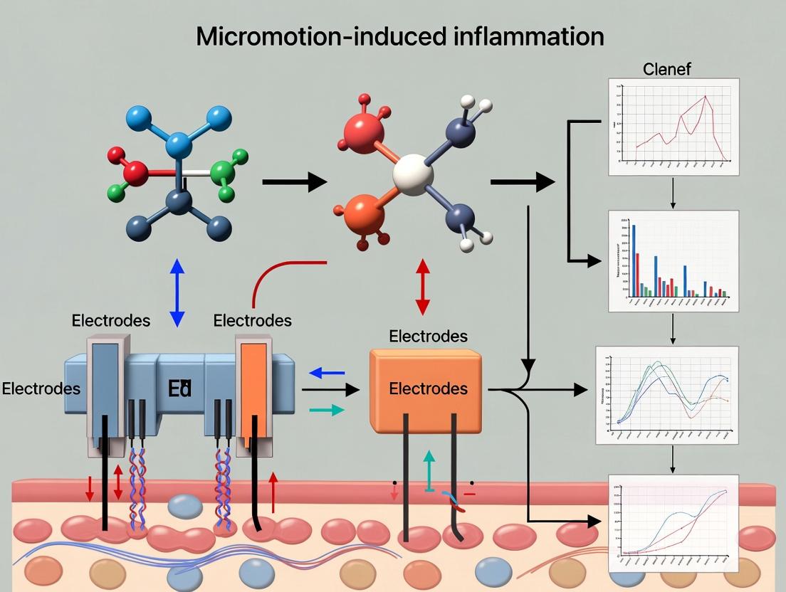
Understanding the Inflammatory Cascade: The Biological Basis of Micromotion in Bioelectronics
Micromotion refers to the small-scale, relative movement between an implanted biomedical device (e.g., a neural electrode, a bone screw, a glucose sensor) and the surrounding host tissue. This movement occurs at the micron to sub-millimeter scale and is driven by physiological processes such as breathing, muscle contractions, vascular pulsation, and general body movement. In the context of bioelectronics, this persistent mechanical mismatch and friction at the tissue-device interface is a primary trigger for chronic inflammatory response, leading to fibrotic encapsulation, increased electrical impedance, and ultimate device failure or signal degradation.
FAQs on Micromotion in Bioelectronics
Q1: What are the primary physiological sources of micromotion? A1: The main sources are:
- Macro-scale body movement: Walking, stretching.
- Visceral movement: Breathing, peristalsis.
- Pulsatile movement: Vascular pulsation from heartbeats.
- Muscle contractions: Both voluntary and involuntary.
Q2: How does micromotion lead to inflammation and device failure? A2: Micromotion creates a sustained injury cycle:
- Mechanical abrasion damages the delicate foreign body response (FBR) capsule and adjacent cells.
- This repeated damage activates immune cells (e.g., macrophages), causing them to release pro-inflammatory cytokines.
- The sustained inflammatory signaling leads to the recruitment of more immune cells and fibroblasts.
- Fibroblasts deposit dense, collagen-rich fibrotic tissue, isolating the device and impairing its function (e.g., electrical signal transmission for electrodes or analyte diffusion for sensors).
Q3: Can we completely eliminate micromotion in implants? A3: No, micromotion is inevitable. The human body is a dynamic mechanical environment. Absolute immobilization of an implant is biologically impossible without causing severe tissue damage or necrosis. The research focus is therefore on mitigating its effects through material design, mechanical buffering, and pharmacological strategies, rather than achieving complete elimination.
Q4: What are the key experimental metrics for quantifying micromotion effects? A4: Researchers quantify the outcome using both histological and functional metrics.
Table 1: Key Quantitative Metrics for Assessing Micromotion-Induced Inflammation
| Metric Category | Specific Measurement | Typical Method/Assay | Significance |
|---|---|---|---|
| Histological | Fibrotic Capsule Thickness (µm) | H&E staining, microscopy | Direct measure of insulation barrier. |
| Histological | Macrophage Density (cells/mm²) | IHC for CD68/CD206 | Indicates level of inflammatory activity. |
| Histological | Collagen Density (%) | Masson's Trichrome, Picrosirius Red | Maturity and density of fibrotic scar. |
| Functional | Electrode Impedance (kΩ) | Electrochemical Impedance Spectroscopy | Signal quality loss at neural interface. |
| Functional | Signal-to-Noise Ratio (dB) | In vivo electrophysiology recording | Functional performance of recording electrode. |
| Biochemical | TNF-α, IL-1β Concentration (pg/mL) | ELISA of peri-implant fluid | Level of pro-inflammatory signaling. |
Troubleshooting Guide: Common Experimental Challenges
Issue: High variability in fibrotic capsule measurements around identical implants.
- Potential Cause: Inconsistent sectioning plane or angle through the implant site.
- Solution: Use histological guides or implant markers to ensure cross-sections are taken through the implant's central axis. Measure capsule thickness at multiple, standardized clock positions (e.g., 12, 3, 6, 9 o'clock) and report the average and standard deviation.
Issue: Inconsistent cytokine profile data from tissue homogenates.
- Potential Cause: Dilution effect from homogenizing whole tissue, masking the critical local concentration at the interface.
- Solution: Employ a minimally invasive microdialysis catheter placed adjacent to the implant in vivo to collect interstitial fluid over time, or perform lavage of the implant pocket upon explantation for more localized analysis.
Issue: Rapid rise and stabilization of electrode impedance post-implantation.
- Potential Cause: This is the classic signature of the foreign body response. Initial stabilization may not indicate a stable interface but a completed fibrotic seal.
- Solution: Impedance must be correlated with histology. Use spectroscopic impedance (EIS) to model the interface. A purely resistive high-frequency impedance indicates mature fibrosis, while changes in low-frequency impedance may relate to ongoing cellular activity.
Key Experimental Protocol: Evaluating the Foreign Body Response to a Moving Implant
Title: In Vivo Assessment of Micromotion-Induced Fibrosis
Objective: To quantitatively compare the chronic inflammatory and fibrotic response to a static versus a mechanically actuated implant in a subcutaneous rodent model.
Materials (The Scientist's Toolkit):
Table 2: Essential Research Reagents & Materials
| Item | Function in Experiment |
|---|---|
| Polyimide or Silicone-based Implant | Biocompatible, flexible substrate mimicking a bioelectronic device. |
| Miniature Actuator/Piezoelectric Motor | To induce controlled, cyclical micromotion (e.g., 150µm displacement) in the test group. |
| Titanium or Stainless Steel Casing | Rigid, bioinert housing for the actuator and control implant. |
| Anti-CD68 & Anti-CD206 Antibodies | For immunohistochemical identification of total macrophages and M2 phenotype, respectively. |
| Picrosirius Red Stain | For specific visualization and birefringence analysis of collagen types I and III. |
| ELISA Kits for TNF-α and IL-1β | To quantify key pro-inflammatory cytokines from peri-implant lavage samples. |
| Electrochemical Impedance Spectrometer | For functional assessment of electrode-coated implants (if applicable). |
Methodology:
- Implant Fabrication: Fabricate two groups of sterile implants: (A) Static Control: Fixed within a rigid casing. (B) Micromotion Group: Mounted on an actuator programmed to induce a defined displacement (e.g., 100µm) at a physiological frequency (e.g., 1 Hz) for set periods daily.
- Surgical Implantation: Aseptically implant devices in the subcutaneous dorsum of anesthetized rats (n≥5 per group). Ensure the control device is secured to underlying fascia to minimize unintended movement.
- In Vivo Actuation: Activate the motion regimen for the test group post-surgery and maintain for the study duration (e.g., 4 weeks).
- Terminal Analysis (Week 4): a. Lavage: Gently inject and retrieve 0.5 mL of saline into the implant pocket for ELISA. b. Explantation: Carefully explant devices with surrounding tissue intact. c. Histology: Fix tissue in 4% PFA, process, section, and stain with H&E, Picrosirius Red, and for macrophage markers. d. Quantification: Perform blinded analysis of capsule thickness, cell density, and collagen area fraction using image analysis software (e.g., ImageJ).
- Statistical Analysis: Use Student's t-test or ANOVA to compare means between static and motion groups (p < 0.05 considered significant).
Visualizing Key Concepts
Diagram 1: Micromotion-Inflammation-Fibrosis Pathway
Diagram 2: Experimental Workflow for Micromotion Study
Troubleshooting Guides & FAQs
Q1: In our in vitro cell stretch model, we observe inconsistent inflammatory cytokine (IL-1β, TNF-α) release between experiments. What are the primary variables to control? A: Inconsistency often stems from poor control of mechanical parameters or cell state. Key variables to standardize are:
- Substrate Stiffness: Use hydrogels (e.g., polyacrylamide, PDMS) with validated, consistent Young's modulus. Batch-to-batch variation is a common culprit.
- Strain Magnitude & Rate: Calibrate your stretch apparatus (e.g., Flexcell) regularly. Ensure strain is uniform across the membrane. A sudden high-rate strain triggers different pathways (e.g., Piezo1) than chronic low-rate strain.
- Cell Confluency & Passage Number: Always use cells at the same passage and density (recommended 80-90% confluency for stretch experiments).
- Serum Starvation: Inconsistent serum levels before stimulation can alter baseline signaling. Implement a standardized serum-reduction protocol (e.g., 0.5% FBS for 12-16h) prior to mechanical stimulation.
Q2: When implanting a bioelectronic device in our murine model, how do we distinguish micromotion-induced inflammation from the normal foreign body response (FBR) in histology? A: This requires multiplexed spatial and temporal analysis.
- Temporal Cue: Micromotion-driven inflammation is chronic and oscillatory. It persists beyond the standard FBR timeline (which may peak at ~2 weeks and fibrose). Monitor cytokine levels and immune cell presence at multiple late timepoints (e.g., 4, 8, 12 weeks).
- Spatial Cue: Normal FBR creates a concentric, layered capsule. Micromotion-induced inflammation shows disrupted, irregular capsule morphology with persistent, mixed leukocyte infiltrates (neutrophils, macrophages) adjacent to the moving device interface, not just at the static surface. Use multiplex immunohistochemistry (IHC) for pan-immune (CD45), macrophage (F4/80), and neutrophil (Ly6G) markers.
Q3: Our assays for key mechanosensors (YAP/TAZ, NF-κB nuclear translocation) show high background in control, static cells. How can we improve signal-to-noise ratio? A: High background indicates inadequate quiescence or non-mechanical stress.
- Optimize Fixation & Permeabilization: For YAP/TAZ, use 4% PFA for 15 min at RT, followed by 0.2% Triton X-100 for 10 min. Over-permeabilization increases background.
- Include Pharmacological Controls: Treat control cells with an inhibitor to establish baseline. Use Verteporfin (YAP/TAZ inhibitor, 1µM for 4h) or BAY-11-7082 (NF-κB inhibitor, 5µM for 2h). The difference between inhibited and uninhibited static cells is your true "mechanical-off" state.
- Quantify, Don't Just Qualify: Use high-content imaging and measure nuclear/cytoplasmic fluorescence intensity ratio. Set thresholds based on inhibitor-treated controls.
Q4: Which is the most relevant readout for early micromotion-induced inflammatory signaling: calcium flux, cytokine secretion, or phosphorylation events? A: The hierarchy and timing are critical for troubleshooting experimental design.
| Readout | Typical Onset | Key Advantage | Key Limitation | Best For |
|---|---|---|---|---|
| Calcium Flux (e.g., Fluo-4 AM) | Milliseconds to Seconds | Captures initial ion channel (Piezo) activation. | Transient; can be noisy; not specific to inflammation. | Identifying the proximal mechanosensing event. |
| Phosphorylation (e.g., p-IκBα, p-FAK, p-ERK) | Minutes to 1 Hour | Directly shows pathway activation; highly specific. | Requires phospho-specific antibodies; may not translate to functional output. | Mapping the immediate signaling cascade (e.g., NF-κB, MAPK). |
| Cytokine Secretion (e.g., IL-6, TNF-α via ELISA) | Hours to Days | Functional, downstream output; clinically relevant. | Significant delay from stimulus; subject to autocrine/paracrine amplification. | Confirming a pro-inflammatory functional outcome. |
Experimental Protocol: Assessing Piezo1/NF-κB Axis in Macrophages under Cyclic Strain
- Objective: To link mechanical strain to inflammatory priming via the Piezo1 ion channel.
- Materials: RAW 264.7 or primary bone marrow-derived macrophages (BMDMs), flexible-bottom 6-well plates (e.g., BioFlex), cyclic stretch system, Piezo1 agonist (Yoda1), antagonist (GsMTx4), ELISA kits for TNF-α/IL-6.
- Method:
- Seed macrophages at 8x10^5 cells/well in complete medium. Adhere for 6h.
- Pre-treatment: Add 5µM GsMTx4 or vehicle (DMSO) 1 hour prior to strain.
- Stimulation: Apply 10% cyclic tensile strain at 0.5 Hz for 6 hours. Include static controls.
- Positive Control: Parallel wells of static cells treated with 10µM Yoda1 for 6 hours.
- Analysis: Collect supernatant for ELISA. Lyse cells for Western blot (p-IκBα, total IκBα) or RNA for qPCR (Il6, Tnf).
Key Research Reagent Solutions
| Item | Function & Relevance |
|---|---|
| GsMTx4 (Spider Venom Peptide) | Selective inhibitor of cationic mechanosensitive ion channels (e.g., Piezo1). Used to block mechanically-induced calcium influx. |
| Yoda1 | Small molecule agonist of Piezo1. Serves as a non-mechanical positive control to mimic channel activation. |
| Cytoskeleton-Disrupting Agents (Latrunculin A, Cytochalasin D) | Disrupts actin filaments. Used to decouple external force from intracellular transmission to the nucleus. |
| Verteporfin | Disrupts YAP-TEAD interaction. Critical control for confirming YAP/TAZ-mediated mechanotranscription. |
| Tensegrity-Mimicking Hydrogels (e.g., Polyacrylamide of tunable stiffness) | Provides a physiologically relevant 2D/3D substrate to study the effect of stiffness (a static mechanical cue) independent of dynamic strain. |
| Phospho-Specific Antibodies (e.g., p-IκBα (Ser32/36), p-FAK (Tyr397), p-ERK1/2 (Thr202/Tyr204)) | Essential for detecting rapid, force-induced activation of key signaling nodes via Western blot or ICC. |
Diagram: Core Mechano-Inflammatory Signaling Axis
Diagram: Experimental Workflow for In Vitro Validation
Troubleshooting Guides & FAQs for Micromotion-Induced Inflammation Studies
FAQ: General Concepts & Experimental Design
Q1: What is the primary link between device micromotion and the pro-fibrotic foreign body response (FBR)? A1: Repetitive mechanical stress from micromotion activates specific mechanotransduction pathways (e.g., via Piezo1/2 channels, integrin signaling) in macrophages and fibroblasts. This sustains a pro-inflammatory (M1) and later a pro-fibrotic (M2) macrophage phenotype, and directly activates myofibroblasts, leading to excessive collagen deposition and fibrous capsule formation.
Q2: How can I reliably measure micromotion at the tissue-implant interface in small animal models? A2: The most current methodologies combine in vivo imaging with ex vivo analysis:
- In Vivo: Micro-CT or Ultrasound Speckle Tracking for gross movement (>50 µm).
- Ex Vivo: High-resolution techniques like Digital Image Correlation (DIC) of histological sections or using fluorescent beads embedded in the implant vicinity and tracking their displacement via confocal microscopy post-explantation.
Q3: What are the key markers to distinguish between general inflammation and micromotion-specific inflammation? A3: While overlap exists, a sustained elevation of specific markers indicates mechano-activation. Key markers include:
- Macrophages: Sustained YAP/TAZ nuclear localization, COX-2, ARG1 (in late-stage M2).
- Fibroblasts: Alpha-Smooth Muscle Actin (α-SMA), ED-A Fibronectin, Phosphorylated FAK.
- ECM: Aligned and cross-linked Collagen I fibers (visible via Second Harmonic Generation imaging).
FAQ: Technical & Analytical Issues
Q4: My fibrosis capsule thickness data is highly variable. What are common sources of error? A4: Variability often stems from inconsistent sampling. Follow this protocol:
- Sectioning: Serially section the entire implant site. Analyze every 10th section to ensure representativeness.
- Measurement: Use image analysis software (e.g., QuPath, ImageJ) to measure capsule thickness at 12 equidistant points around the implant circumference per section.
- Reporting: Report both average thickness and the range (min-max) to accurately convey variability.
Q5: How do I accurately quantify macrophage polarization in vivo from tissue sections? A5: Rely on multiplex immunofluorescence (mIF) over single markers. A recommended panel:
- Pan-Macrophage: CD68 or F4/80
- M1-like: iNOS or MHC-II
- M2-like: CD206 or ARG1 Quantify using spectral unmixing or sequential fluorescence, reporting the percentage of dual-positive cells for each phenotype relative to total macrophages.
Q6: My implantable sensor's signal degrades within days, suggesting rapid FBR. How can I test if micromotion is the culprit? A6: Implement a two-pronged protocol:
- Stabilization Control: Create an experimental group where the implant is surgically fixed (e.g., using a titanium bracket or biodegradable glue) to minimize interfacial motion.
- Assessment: Compare the control to the standard implant group using:
- Functional: Sensor performance lifetime (e.g., sensitivity drift).
- Histological: Capsule thickness and cellular density at 1, 2, and 4 weeks. A significant improvement in the stabilized group confirms micromotion's role.
Detailed Experimental Protocol: Assessing Micromotion-Driven Pathways
Protocol: Evaluating YAP/TAZ Mechanotransduction in Peri-Implant Macrophages. Objective: To quantify nuclear translocation of YAP/TAZ in macrophages as a readout of micromotion-induced mechano-activation. Materials: See "Research Reagent Solutions" table. Method:
- Implantation & Explanation: Implant your device in the target tissue (e.g., subcutaneous, intramuscular). Explain devices with a 2-3 mm margin of surrounding tissue at time points (e.g., 3, 7, 14 days).
- Tissue Processing: Fix tissue in 4% PFA for 24h at 4°C. Cryoprotect in 30% sucrose, embed in OCT, and section at 10 µm thickness.
- Multiplex Immunofluorescence: a. Block with 5% species-appropriate serum + 0.3% Triton X-100. b. Incubate with primary antibody cocktail (e.g., Anti-F4/80, Anti-YAP/TAZ) overnight at 4°C. c. Incubate with fluorescent secondary antibodies and DAPI for 2h at RT.
- Imaging & Analysis: Acquire high-resolution z-stack images via confocal microscopy. Use analysis software to: a. Create a mask from the DAPI channel to identify nuclei. b. Create a mask from the F4/80 channel to identify macrophages. c. Measure mean YAP/TAZ fluorescence intensity in the nuclear region (DAPI mask) of F4/80+ cells versus the cytosolic region (cell mask minus nuclear mask). d. Calculate Nuclear-to-Cytosolic (N:C) ratio for at least 100 cells per sample.
Research Reagent Solutions Table
| Item | Function/Application | Example (Specific Brand/Type) |
|---|---|---|
| Piezo1 Agonist | Chemically induce mechanosensitive channel opening to mimic micromotion signaling in vitro. | Yoda1 |
| FAK Inhibitor | Inhibit integrin-mediated focal adhesion kinase signaling to disrupt mechanotransduction. | PF-573228 |
| M2 Macrophage Inducer | Polarize macrophages to an anti-inflammatory/pro-fibrotic phenotype for in vitro co-culture studies. | IL-4 / IL-13 Cytokine Cocktail |
| Collagen Hybridizing Peptide (CHP) | Fluorescently tag denatured/disrupted collagen to visualize micro-damage from micromotion. | 3Helix F-CHP |
| Biodegradable Hydrogel | Used as a soft, conformal coating to dampen interfacial micromotion; control material. | GelMA (Gelatin Methacryloyl) |
| Sustained-Release Corticosteroid Pellet | Local anti-inflammatory control to differentiate inflammation sources. | Dexamethasone (slow-release, implanted adjacent to device) |
Table 1: Impact of Implant Stiffness & Fixation on Fibrotic Outcomes (Rodent Model, 4 Weeks)
| Implant Type | Young's Modulus | Fixation Method | Avg. Capsule Thickness (µm) | % α-SMA+ Area | Dominant Macrophage Phenotype |
|---|---|---|---|---|---|
| Silicone (PDMS) | 1.5 MPa | Unsecured | 145 ± 35 | 22.1 ± 4.5 | M2 (CD206+) |
| Porous Polyethylene | 150 MPa | Unsecured | 220 ± 50 | 38.7 ± 6.2 | Mixed (M1/M2) |
| Soft Hydrogel (PEG) | 15 kPa | Unsecured | 85 ± 20 | 10.5 ± 2.1 | M2 (CD206+) |
| Silicone (PDMS) | 1.5 MPa | Suture-Fixed | 95 ± 25 | 12.8 ± 3.0 | M2 (CD206+) |
Table 2: Key Molecular Markers in Micromotion-Accelerated FBR
| Pathway | Target Molecule | Up/Down Regulation (vs. Static Implant) | Detection Method | Typical Timepoint (Post-Implant) |
|---|---|---|---|---|
| Mechanotransduction | Nuclear YAP/TAZ | Up 3-5x | Immunofluorescence (N:C Ratio) | Day 3-7 |
| Pro-fibrotic Signaling | Phospho-FAK (Tyr397) | Up 2-3x | Western Blot / IHC | Day 7-14 |
| ECM Remodeling | LOXL2 | Up 4x | qPCR | Day 14-28 |
| M2 Polarization | ARG1 Expression | Up earlier & sustained | qPCR / IHC | Day 7-28 |
Diagrams
Title: Signaling Pathway from Micromotion to Fibrosis
Title: Workflow for Micromotion-FBR Experiment
Title: Factors Influencing Macrophage Polarization in FBR
Technical Support Center: Troubleshooting Micromotion-Induced Inflammation
Frequently Asked Questions (FAQs)
Q1: My chronically implanted bioelectronic sensor shows a progressive decline in signal fidelity over weeks. What is the likely cause and how can I confirm it? A1: Signal degradation is a classic consequence of the foreign body response (FBR) and chronic inflammation. The buildup of a fibrous capsule (composed primarily of collagen and myofibroblasts) physically distances the electrode from the target tissue, increasing impedance and reducing signal-to-noise ratio. To confirm, perform:
- Electrochemical Impedance Spectroscopy (EIS): A steady rise in low-frequency impedance (~1-100 Hz) is indicative of fibrous tissue encapsulation.
- Histology: Explant the device and surrounding tissue. Stain with H&E for general morphology, Masson's Trichrome for collagen, and immunohistochemistry for α-SMA (myofibroblasts) and CD68 (macrophages).
Q2: My flexible neural implant has failed due to mechanical fracture at the tissue-device interface. Could chronic inflammation be a factor? A2: Absolutely. Chronic inflammation leads to a hostile biochemical environment. Key factors include:
- Phagocytic Attack: Persistent macrophages and foreign body giant cells release reactive oxygen species (ROS) and acidic lysosomal enzymes that degrade polymer substrates and thin metal traces.
- Continuous Mechanical Stress: The contracting fibrous capsule applies cyclic mechanical stress (micromotion) at the interface, accelerating material fatigue.
- Solution: Utilize accelerated aging tests in simulated inflammatory media (e.g., hydrogen peroxide/Fe²⁺ solution for ROS, low-pH buffer) combined with cyclic mechanical strain to model this failure mode.
Q3: We observe significant neuronal loss and glial scarring beyond the immediate implant site. Is this related to the implant's micromotion? A3: Yes. Persistent micromotion perpetuates the inflammatory cascade, transforming acute inflammation into a chronic state. This leads to sustained release of pro-inflammatory cytokines (IL-1β, TNF-α) and neurotoxic molecules from activated microglia and astrocytes, causing bystander tissue damage.
- Protocol for Assessment: Perform multi-label immunofluorescence on tissue sections for:
- Neurons (NeuN)
- Astrocytes (GFAP)
- Activated Microglia (Iba1 + CD68)
- Apoptosis (Cleaved Caspase-3). Quantify cell densities at defined distances (e.g., 50µm, 100µm, 200µm) from the implant interface.
Q4: How can I quantitatively differentiate between the normal healing phase and detrimental chronic inflammation in my animal model? A4: Monitor temporal cytokine profiles and cellular composition. A resolution peak followed by a return to baseline indicates normal healing. A sustained or secondary elevated plateau indicates chronic inflammation.
Table 1: Key Differentiators Between Acute Healing and Chronic Inflammation
| Parameter | Acute/Healing Phase (Days 3-14) | Chronic Inflammation Phase (>Week 4) |
|---|---|---|
| Macrophage Phenotype | Mixed M1 (pro-inflammatory) and M2 (pro-healing) | Predominantly M1, Foreign Body Giant Cells |
| Cytokine Profile | Transient peak of IL-1β, TNF-α, IL-6 | Sustained elevated levels of IL-1β, TNF-α; Presence of TGF-β |
| Fibrous Capsule | Developing, cellular, vascularized | Dense, collagenous, avascular, contractile (α-SMA+) |
| Tissue Integrity | Localized, repairing | Progressive bystander damage/apoptosis |
Experimental Protocols
Protocol 1: In Vivo Assessment of the Foreign Body Response Objective: To histologically characterize the chronic inflammatory response to an implanted bioelectronic device.
- Implantation: Aseptically implant your device into the target tissue (e.g., brain, subcutaneous, muscle) of an animal model (e.g., rodent).
- Time Points: Euthanize animals and explant the device with surrounding tissue at critical time points (e.g., 1, 2, 4, 8, 12 weeks).
- Fixation: Immediately fix tissue in 4% paraformaldehyde for 48 hours.
- Sectioning: Embed in paraffin or OCT compound. Section tissue perpendicular to the device interface (5-10 µm thickness).
- Staining:
- H&E: General morphology and cellular infiltration.
- Masson's Trichrome: Collagen deposition (capsule thickness).
- Immunofluorescence: Stain for macrophages (CD68/Iba1), myofibroblasts (α-SMA), astrocytes (GFAP), and neurons (NeuN).
- Analysis: Use image analysis software to quantify capsule thickness, cellular density, and fluorescence intensity at defined distances.
Protocol 2: Electrochemical Impedance Spectroscopy (EIS) for In Vivo Monitoring Objective: To non-invasively track the progression of fibrous encapsulation.
- Setup: Connect your implanted electrode to a potentiostat capable of EIS measurements. Use a standard 3-electrode configuration (working = implant, counter, reference).
- Measurement Parameters: Apply a sinusoidal voltage perturbation (10 mV amplitude) across a frequency range of 1 Hz to 100 kHz. Perform measurements periodically (e.g., daily for week 1, then weekly).
- Data Modeling: Fit the obtained Nyquist or Bode plots to an equivalent circuit model. The model
R_s(C_dl(R_ct(Z_W)))in series withC_fibrosis(R_fibrosis)is often used, where the low-frequency impedance increase is attributed to theR_fibrosis(fibrosis resistance) andC_fibrosis(fibrosis capacitance) elements. - Correlation: Correlate increases in low-frequency impedance magnitude with post-mortem histological capsule thickness.
Diagrams
Title: Micromotion-Induced Chronic Inflammation Consequences Pathway
Title: In Vivo FBR Assessment Experimental Workflow
The Scientist's Toolkit: Research Reagent Solutions
Table 2: Essential Materials for Investigating Implant-Induced Chronic Inflammation
| Item | Function & Application |
|---|---|
| Flexible Polymer Substrates (e.g., Polyimide, Parylene C) | Provides a soft, conformable interface to minimize mechanical mismatch and initial micromotion-induced damage. |
| Anti-inflammatory Coatings (e.g., Dexamethasone, IL-4, IL-10) | Localized, controlled release coatings to modulate the host immune response, promoting an M2 healing phenotype. |
| Hydrogel Barriers (e.g., Alginate, PEG) | Acts as a physical and biochemical buffer between the device and tissue, absorbing micromotion and delivering therapeutic agents. |
| Conductive Polymers (e.g., PEDOT:PSS) | Lowers interfacial impedance, improving signal acquisition despite mild encapsulation; can be doped with anti-inflammatory drugs. |
| ROS-Scavenging Materials (e.g., Cerium Oxide Nanoparticles, Selenium) | Incorporated into coatings or materials to neutralize reactive oxygen species released by activated macrophages, protecting both tissue and device. |
| Simulated Inflammatory Media (e.g., H₂O₂/Fe²⁺, Low pH Buffer) | For in vitro accelerated aging tests to predict long-term stability of materials in a hostile inflammatory environment. |
| Multiplex Cytokine ELISA/LEGENDplex Assays | For quantifying the precise profile of pro- and anti-inflammatory cytokines in tissue homogenates or from cell culture around explants. |
Troubleshooting Guides and FAQs
FAQ 1: Inconsistent Histological Inflammation Scores Between Animals in the Same Implant Group
- Q: Why am I seeing high variability in capsule thickness and immune cell counts between subjects with identical implants?
- A: This is frequently caused by uncontrolled micromotion. Ensure surgical fixation is highly consistent and consider using a rigid fixation control group. Monitor animal activity; standardize housing (single vs. group) post-op. Verify implant sterilization to rule out subclinical infection.
FAQ 2: Poor Antibody Penetration in Dense Fibrous Capsules for Immunofluorescence
- Q: My immunofluorescence staining for macrophages (e.g., CD68) is weak or patchy in thick, collagen-rich capsules.
- A: This is an antigen masking issue. Implement antigen retrieval optimized for formalin-fixed tissue (e.g., citrate buffer heat-induced epitope retrieval). Increase Triton X-100 concentration in blocking buffer (e.g., to 0.5%). Consider using enzymatic digestion (e.g., proteinase K, but test for target antigen compatibility). Use longer antibody incubation times (overnight at 4°C).
FAQ 3: Difficulty Distinguishing Pro-inflammatory (M1) vs. Pro-healing (M2) Macrophages In Situ
- Q: Standard markers like CD86 and CD206 show co-localization, making macrophage polarization state ambiguous.
- A: Single markers are often insufficient. Recommend a multi-marker panel (e.g., iNOS for M1, Arg1 for M2) combined with cytokine staining (e.g., TNF-α, IL-10). Validate with RNAscope for key transcripts. Use spectral imaging or sequential staining to prevent antibody crossover.
FAQ 4: Tissue Shrinkage/Artifacts Around the Implant Site During Processing
- Q: The tissue-implant interface appears gapped or distorted in histology sections, making capsule measurement unreliable.
- A: This is likely a processing artifact. Use a slower, graded dehydration series (e.g., 70%, 80%, 95%, 100% ethanol). For polymer implants, consider resin-based embedding instead of paraffin. If explanting, perform careful perfusion fixation in situ before removing the implant.
FAQ 5: High Background in Luminescence-Based In Vivo Imaging (e.g., IVIS) for Inflammation
- Q: My NF-κB or MMP luciferase reporter mice show high background signal at the implant site, obscuring the specific signal.
- A: This is often due to surgical trauma or background luciferase expression. Include a sham-surgery control. Ensure substrate (D-luciferin) injection is consistent in dose, route, and timing before imaging. Use a spectral unmixing tool if available to separate signals. Allow 7-10 days post-surgery for acute surgical inflammation to subside before baseline imaging.
Key Experimental Protocols
Protocol 1: Standardized Histomorphometric Analysis of the Foreign Body Capsule
- Sample Preparation: Explant tissue with implant en bloc. Fix in 10% neutral buffered formalin for 48 hours.
- Sectioning: Decalcify if necessary. Process and paraffin-embed. Section serially at 5 µm thickness through the entire implant site. Perform H&E and Masson's Trichrome staining on every 10th section.
- Imaging: Digitize slides at 20x magnification. Use image analysis software (e.g., QuPath, ImageJ).
- Quantification: Measure capsule thickness at 8-12 radially equidistant points around the implant perimeter per section. Average across 3 non-adjacent sections.
- Cell Counting: Using IHC for CD45 (pan-leukocyte), count positive nuclei within the capsule in five standardized 0.1 mm² regions. Report cells/mm².
Protocol 2: Multiplex Immunofluorescence (mIF) for Spatial Profiling of the Inflammatory Interface
- Staining: Use a commercial multiplex kit (e.g., Akoya Biosciences OPAL, Roche mIF) on FFPE sections.
- Panel Design: Include primary antibodies: CD68 (macrophages), αSMA (myofibroblasts), CD3 (T-cells), Collagen I, and a nuclear marker (DAPI). Validate each antibody individually first.
- Process: Perform sequential rounds of antibody application, tyramide signal amplification (if using OPAL), and microwave-mediated antibody stripping.
- Analysis: Acquire images on a multispectral microscope. Use spectral unmixing software. Quantify cell densities and distances between different cell phenotypes relative to the implant surface.
Data Presentation
Table 1: Comparative Capsule Thickness in Rodent Models at 4 Weeks Post-Implantation
| Implant Material | Animal Model (n=6) | Mean Capsule Thickness (µm) ± SD | Key Histological Feature |
|---|---|---|---|
| Medical-Grade Silicone | C57BL/6 Mouse | 125.3 ± 18.7 | Dense, aligned collagen, moderate macrophages |
| Polyethylene (Positive Control) | Sprague Dawley Rat | 310.5 ± 45.2 | Hypercellular, disorganized matrix, giant cells |
| Porous Titanium | C57BL/6 Mouse | 85.1 ± 12.4 | Fibrovascular ingrowth, minimal lymphocyte presence |
| Flexible Polyimide (Unfixed) | Lewis Rat | 220.8 ± 75.4* | Highly variable thickness, mixed inflammation |
| Flexible Polyimide (Rigidly Fixed) | Lewis Rat | 95.6 ± 20.1 | Thin, organized layer |
*Indicates statistically significant higher variance (p<0.05, Levene's test) compared to fixed group.
Table 2: Efficacy of Anti-Inflammatory Drug Interventions on Micromotion-Induced Inflammation
| Treatment (Daily) | Model (Micromotion Induced) | Capsule Thickness Reduction vs. Vehicle | M1/M2 Macrophage Ratio (IHC) at Interface |
|---|---|---|---|
| Systemic Dexamethasone (1 mg/kg) | Rat S.C. Model | 40%* | 0.5 (Strong M2 shift) |
| Local Doxcycline Release (Coating) | Mouse Muscle Model | 25% | 1.2 (Moderate M2 shift) |
| Anti-TNF-α mAb (10 mg/kg, 2x/wk) | Rat S.C. Model | 30% | 1.8 (Mild M2 shift) |
| Vehicle Control | Rat S.C. Model | -- | 4.5 (M1 Dominant) |
- p<0.05, * p<0.01, ** p<0.001; mAb: monoclonal antibody.
Visualizations
Title: Micromotion-Driven Fibrous Capsule Formation Pathway
Title: Experimental Workflow for Interface Analysis
The Scientist's Toolkit: Research Reagent Solutions
| Item | Function & Application |
|---|---|
| Anti-CD68 Antibody (clone FA-11) | Pan-macrophage marker for identifying total macrophage infiltration in rodent tissue (IHC/IF). |
| Anti-αSMA-Cy3 Conjugate | Directly conjugated antibody for labeling activated myofibroblasts, reducing staining time and multiplexing complexity. |
| Masson's Trichrome Stain Kit | Differentiates collagen (blue) from muscle/cytoplasm (red), essential for quantifying fibrous encapsulation. |
| Lectin (Griffonia Simplicifolia) FITC | Binds to vascular endothelium; used to quantify angiogenesis within the peri-implant capsule. |
| D-Luciferin, Potassium Salt | Substrate for in vivo bioluminescence imaging in reporter mice (e.g., NF-κB-luc) to track inflammation kinetics. |
| OPAL 7-Color Automation Kit | Enables multiplex immunofluorescence (mIF) on a single FFPE section for spatial phenotyping of the immune response. |
| RNAscope Probe: Mm-Tnf | Allows single-molecule RNA in situ hybridization to visualize pro-inflammatory cytokine expression in specific cells. |
| Slow-Release Dexamethasone Pellet | Provides sustained systemic anti-inflammatory delivery to study pharmacological modulation of the FBR. |
Engineering Solutions: Material and Device Strategies to Dampen Micromotion Effects
Technical Support & Troubleshooting Center
Troubleshooting Guides
Issue: Delamination of Conductive Traces from Elastomeric Substrate
- Symptoms: Sudden increase in electrical impedance, complete loss of signal, visible peeling under microscope.
- Probable Cause: Poor adhesion due to surface contamination, insufficient curing of adhesion layer, or modulus mismatch causing stress concentration.
- Solution: Implement rigorous oxygen plasma treatment (50W, 1 min) followed by (3-aminopropyl)triethoxysilane (APTES) vapor priming. Ensure the Young's modulus of the conductive composite (e.g., PDMS-Au nanoparticle composite) is within one order of magnitude of the substrate. Perform tape test (ASTM D3359) on sample batches.
Issue: Unstable Electrochemical Impedance in Chronic In Vivo Recordings
- Symptoms: Drifting baseline impedance, increased noise, inconsistent stimulation efficacy over time (weeks).
- Probable Cause: Fibrotic capsule formation altering local dielectric environment, or moisture ingress through microcracks.
- Solution: Redesign device with a lower, tissue-matching modulus (<10 kPa) to reduce mechanical mismatch. Apply a bioactive coating (e.g., peptide-functionalized poly(ethylene glycol)) to mitigate foreign body response. Use accelerated aging tests (37°C, 95% relative humidity) to validate encapsulation pre-implantation.
Issue: Inconsistent Performance of Stretchable Interconnects Under Cyclic Strain
- Symptoms: Resistance changes non-linearly with strain, failure occurs below expected strain threshold (e.g., <30%).
- Probable Cause: Improper geometric design of serpentine or horseshoe patterns leading to localized peak stress.
- Solution: Optimize the "island-bridge" layout using finite element analysis (FEA) simulation. Ensure the radius of curvature in meanders is >500 µm. Validate with a minimum of 10,000 cyclic strain tests (to 15% strain) prior to biological experiments.
Frequently Asked Questions (FAQs)
Q1: What is the target Young's modulus for devices intended for neural interfaces, and why? A1: For direct neural interfacing, especially with the cortex or peripheral nerves, the target modulus range is 1-100 kPa. This range closely matches the modulus of brain tissue (~1-3 kPa) and minimizes shear forces that trigger glial scarring and micromotion-induced inflammation, a key focus of our thesis research.
Q2: My hydrogel-based electrode swells and loses conductivity in physiological buffer. How can I stabilize it? A2: Use a double-network hydrogel strategy. Create a primary network of conductive polymer (e.g., PEDOT:PSS) within a secondary, cross-linked network of a non-swelling polymer like polyacrylamide. Tune the ionic crosslink density (e.g., using Ca²⁺ ions) to control swelling ratio below 10%.
Q3: Which adhesion promoters are most effective for bonding silicone elastomers to inorganic materials (e.g., chips, sensors)? A3: For permanent, biocompatible bonds, a two-step process is recommended. First, treat both surfaces with oxygen plasma. Then, apply a thin primer layer of: 1) APTES for silanol bonding to oxides, or 2) a commercially available silicone primer (e.g., MED-1511 from NuSil). Cure under pressure at 80°C for 2 hours.
Q4: How do I accurately measure the modulus of my soft composite material? A4: Use a combination of techniques. Atomic Force Microscopy (AFM) in force spectroscopy mode is best for localized, surface measurements of very soft materials (<100 kPa). For bulk material properties, perform tensile tests using a micro-mechanical tester with a low-force load cell (<5N). Always test in conditions mimicking the biological environment (37°C, hydrated).
Key Quantitative Data
Table 1: Modulus of Biological Tissues and Common Electronic Materials
| Material/Tissue | Young's Modulus (Approx. Range) | Key Characteristics/Implications |
|---|---|---|
| Brain Tissue | 0.5 - 3 kPa | Extremely soft; requires ultra-compliant interfaces. |
| Peripheral Nerve | 50 - 500 kPa | Stiffer than brain; allows for slightly more rigid cuffs. |
| Cardiac Tissue | 10 - 100 kPa | Dynamic, cyclic straining necessitates high fatigue resistance. |
| PDMS (Sylgard 184) | 0.5 MPa - 3 MPa | Easily tunable but often 2-3 orders stiffer than brain. |
| Polyimide (Kapton) | 2.5 GPa | Classic flexible PCB material; modulus mismatch is severe. |
| Ecoflex (00-30) | 30 - 60 kPa | Off-the-shelf elastomer well-suited for soft interfaces. |
| Hydrogel (PAAm) | 1 - 100 kPa | Highly hydratable; can match tissue modulus precisely. |
Table 2: Performance Metrics of Stretchable Conductor Compositions
| Conductor Composition | Conductivity (S/cm) | Max Strain at Failure | Critical Strain for R Increase >10% | Key Trade-off |
|---|---|---|---|---|
| EGaln Liquid Metal | ~3.4 x 10⁴ | >500% | ~250% | Low viscosity leads to leakage. |
| PDMS + Flake Silver | ~5,000 | ~120% | ~50% | Conductivity drops sharply after yield. |
| SEBS + PEDOT:PSS | ~300 | >200% | ~100% | Lower absolute conductivity. |
| Au Nanomesh on PU | ~1.1 x 10⁵ | ~160% | ~80% | Complex, expensive fabrication. |
Experimental Protocols
Protocol: Fabrication of a Soft, Stretchable Microelectrode Array (MEA) for Epicortical Recording
- Substrate Preparation: Mix and degas a soft silicone (e.g., Ecoflex 00-30, 1:1 ratio). Spin-coat onto a sacrificial glass slide at 500 RPM for 60s to achieve a ~150 µm film. Cure at 60°C for 30 minutes.
- Laser Patterning of Conductive Traces: Apply a pressure-sensitive adhesive mask with serpentine trace patterns. Deposit a 20 nm Cr adhesion layer followed by a 200 nm Au layer via electron-beam evaporation. Lift-off in acetone to define the circuit.
- Encapsulation: Spin-coat a second layer of the same soft silicone (300 µm) over the traces, leaving only electrode sites and contact pads exposed. Cure fully.
- Electrode Site Functionalization: Treat exposed Au sites with oxygen plasma. Electrochemically deposit PEDOT:PSS using chronopotentiometry at 0.5 mA/cm² for 30 seconds in an aqueous PEDOT:PSS dispersion.
- Release & Characterization: Carefully release the device from the glass slide. Measure electrochemical impedance spectroscopy (EIS) in 1x PBS (100 Hz - 100 kHz). Perform cyclic stretching test (10,000 cycles to 15% strain) while monitoring resistance.
Protocol: Accelerated Aging Test for Hydration Barrier Efficacy
- Sample Preparation: Fabricate test devices with your full encapsulation stack. Include defined metal lines for resistance monitoring.
- Conditioning: Place samples in a controlled climate chamber at 37°C and 95% Relative Humidity (RH).
- Monitoring: At defined intervals (0, 24, 48, 96, 168 hours), remove samples and immediately measure the line resistance and perform EIS in PBS.
- Failure Criterion: Define a threshold (e.g., 20% increase in line resistance or a 50% drop in impedance phase angle at 1 kHz) as encapsulation failure.
- Analysis: Plot resistance/impedance versus time. Use the time-to-failure to compare different encapsulation materials or thicknesses.
Diagrams
Dot Script 1: Micromotion-Induced Inflammatory Cascade
Dot Script 2: Soft Electronics Development Workflow
The Scientist's Toolkit: Research Reagent Solutions
Table 3: Essential Materials for Soft Bioelectronics Research
| Item | Example Product/Chemical | Function & Rationale |
|---|---|---|
| Soft Elastomer | Ecoflex 00-30 (Smooth-On) | Platinum-cure silicone with modulus (~60 kPa) close to many soft tissues. Easy to use. |
| Conductive Hydrogel Precursor | Polyacrylamide (PAAm), PEDOT:PSS | Forms soft (kPa range), ionically conductive networks for tissue-like electrodes. |
| Adhesion Promoter | (3-Aminopropyl)triethoxysilane (APTES) | Silane coupling agent that forms strong bonds between inorganic surfaces and polymers. |
| Liquid Metal | Eutectic Gallium-Indium (EGaIn) | Highly conductive, intrinsically stretchable material for interconnects. |
| Bioactive Coating | CD47 Peptide or CXCL12 | "Self" peptide or chemokine to mitigate foreign body response and fibrosis. |
| Degradable Encapsulant | Poly(lactic-co-glycolic acid) (PLGA) | Provides temporary barrier function for resorbable electronics. |
| Stretchable Dielectric | Styrene-Ethylene-Butylene-Styrene (SEBS) | Thermoplastic elastomer with stable insulation properties under strain. |
Technical Support Center: Troubleshooting & FAQs
Thesis Context: This support content is designed for researchers developing novel bioelectronic interfaces to mitigate micromotion-induced inflammation and fibrotic encapsulation, thereby improving long-term device performance and biocompatibility.
Frequently Asked Questions (FAQs)
Q1: My conductive polymer (PEDOT:PSS) coating shows poor adhesion and delaminates from the metal electrode during cyclic mechanical strain testing. What could be the cause? A: Poor adhesion is a common issue when simulating micromotion. Primary causes include insufficient surface pretreatment, incorrect dopant or crosslinker concentration, and mismatch in mechanical modulus. Ensure you:
- Pre-treat the metal surface: Use an O₂ plasma cleaner (100W, 5 min) or apply a silane coupling agent (e.g., (3-Aminopropyl)triethoxysilane, 2% v/v in ethanol) to create active binding sites.
- Optimize crosslinking: For PEDOT:PSS, add 1-3% v/v of (3-glycidyloxypropyl)trimethoxysilane (GOPS) as a crosslinker and cure at 140°C for 15-30 minutes to enhance adhesion and stability in aqueous environments.
- Consider an adhesion interlayer: Apply a thin layer of polyurethane or polydopamine (<100 nm) on the electrode prior to polymer deposition.
Q2: The bioactivity of my peptide-coated hydrogel seems to degrade rapidly in vitro. How can I stabilize the bioactive signals? A: Rapid degradation often indicates poor immobilization chemistry or susceptibility to enzymatic cleavage.
- Check your conjugation chemistry: For RGD peptides, ensure you are using a stable covalent bond. Sulfosuccinimidyl 4-(N-maleimidomethyl)cyclohexane-1-carboxylate (Sulfo-SMCC) is a common heterobifunctional crosslinker for amine-to-thiol coupling. Always perform a post-immobilization wash with a mild detergent (0.1% SDS) to remove physisorbed peptides.
- Use protease-resistant sequences: Opt for D-amino acid peptides or cyclized sequences (e.g., c[RGDfK]) to reduce degradation by serum proteases.
- Test concentration: Use surface characterization like X-ray Photoelectron Spectroscopy (XPS) to confirm peptide density. Optimal bioactivity often occurs within a specific density range (see Table 1).
Q3: My hydrogel-based interface exhibits a significant increase in impedance (> 50 kΩ at 1 kHz) after 7 days of implantation in a rodent model. What steps should I take? A: Increased impedance typically points to fouling, fibrosis, or dehydration of the hydrogel.
- Characterize the tissue response: Histologically section and stain (H&E, Masson's Trichrome) the explanted tissue to differentiate between inflammation (cellular infiltration) and collagenous capsule formation.
- Modify the hydrogel: Incorporate anti-inflammatory agents (e.g., dexamethasone, ~ 5 µM load) or non-fouling polymers like poly(ethylene glycol) (PEG) into the network to suppress the foreign body response.
- Ensure hydration: If using a non-swelling hydrogel, consider increasing its hydrophilicity or designing a protective seal to prevent fluid loss in vivo.
Q4: During electrophysiological recording, my conductive polymer film shows increased noise. How can I improve its electrochemical performance? A: High noise suggests increased interfacial impedance or inhomogeneous charge transport.
- Measure the Electrochemical Impedance Spectroscopy (EIS): Compare the impedance spectrum (from 1 Hz to 100 kHz) of the film before and after testing. A rise across all frequencies indicates poor contact or delamination. A low-frequency rise suggests poor ionic penetration.
- Re-evaluate your deposition method: For electrodeposited polymers, ensure a consistent potential/current density. For spin-coated films, check for uniformity. Incorporate high-surface-area nanomaterials like carbon nanotubes (0.1-0.5% wt) to lower impedance.
- Check dopant integrity: Ensure your dopant ions (e.g., LiClO₄, Tosylate) are properly integrated and not leaching out. Post-treatment with ethylene glycol for PEDOT:PSS can enhance conductivity.
Troubleshooting Guides
Issue: Inconsistent Polymerization of Conductive Hydrogels.
- Symptoms: Variable conductivity, uneven color/texture, gelation failure.
- Protocol & Solution:
- Degas monomers: Dissolve your monomers (e.g., 3,4-ethylenedioxythiophene - EDOT) and crosslinkers in deionized water. Bubble with nitrogen or argon for 15 minutes to remove dissolved oxygen, which can inhibit polymerization.
- Standardize initiator handling: For redox initiators like Ammonium Persulfate (APS), prepare a fresh stock solution for each experiment. Keep it shielded from light and on ice.
- Control temperature precisely: Perform polymerization in a temperature-controlled water bath (±0.5°C). For common poly(acrylamide) hybrids, 25°C is standard.
- Monitor pH: Use a buffer (e.g., 0.1 M phosphate buffer, pH 7.4) to maintain consistent reaction kinetics.
Issue: Poor Cell Attachment on Bioactive Coating.
- Symptoms: Cells remain rounded, low viability, easy detachment during media changes.
- Protocol & Solution:
- Validate coating activity with a positive control: Coat a separate well with a commercial ECM product (e.g., Matrigel or collagen I). If cells attach there but not on your coating, the issue is with your coating.
- Quantify ligand density: Use a fluorescence-based quantification kit (e.g., for amine-containing peptides) to ensure your coating density is within the effective range (typically 1-10 pmol/cm² for RGD).
- Check sterility and solvent residues: Ensure all coating steps are performed aseptically. If organic solvents were used (e.g., for polymer coatings), expose the coated surface to UV light and rinse extensively with sterile PBS to remove residual solvent.
Data Presentation
Table 1: Performance Comparison of Interface Materials for Mitigating Micromotion Effects
| Material System | Typical Impedance at 1 kHz (Ω) | Adhesion Strength (MPa) | Fibrotic Capsule Thickness in vivo (µm, 4 weeks) | Key Advantage | Primary Challenge |
|---|---|---|---|---|---|
| Platinum/IrOx | 5 - 50 kΩ | N/A | 80 - 150 | Stable, low impedance | Inflammatory foreign body response |
| PEDOT:PSS (with GOPS) | 0.5 - 3 kΩ | 0.8 - 1.5 | 40 - 100 | High charge capacity, soft | Long-term hydration stability |
| Conductive Hydrogel (PAAm/PEDOT) | 2 - 10 kΩ | 0.1 - 0.5 (to substrate) | 20 - 60 | Extreme softness, high water content | Mechanical durability, delamination risk |
| Bioactive Coating (e.g., Laminin peptide on polymer) | Varies with substrate | 0.5 - 2 (coating-to-substrate) | 30 - 80 | Promotes specific cellular integration | Bioactivity half-life, stability |
Table 2: Common Reagent Solutions for Anti-Fibrotic Coatings
| Reagent | Typical Concentration/Formula | Function in Experiment | Key Consideration |
|---|---|---|---|
| Poly(dimethylsiloxane) (PDMS) | Sylgard 184, 10:1 base:curing agent | Flexible substrate for simulating soft tissue modulus. | Requires surface activation (plasma, chemical) for bonding. |
| (3-Glycidyloxypropyl)trimethoxysilane (GOPS) | 1-3% (v/v) in PEDOT:PSS dispersion | Crosslinker for PEDOT:PSS, improves adhesion and stability in water. | Critical for in vivo application. Cure temperature/time must be optimized. |
| Sulfo-SMCC | 0.5 - 2 mM in PBS (pH 7.2-7.4) | Heterobifunctional crosslinker for covalent peptide (thiol) to surface/polymer (amine) conjugation. | Hydrolyzes in water; prepare immediately before use. |
| c[RGDfK] Peptide | 0.1 - 1.0 mg/mL in sterile water | Integrin-binding sequence to promote specific cell adhesion and reduce pro-inflammatory macrophage activation. | Use cyclic form for stability. Confirm density via fluorescence tag or ELISA. |
| Dexamethasone | 1 - 10 µM loaded in hydrogel/matrix | Synthetic glucocorticoid to suppress local inflammatory response and fibroblast proliferation. | Controlled release profile (e.g., via degradable microspheres) is crucial to avoid systemic effects. |
Experimental Protocols
Protocol 1: Fabrication and Characterization of a Micromotion-Resistant Conductive Hydrogel Coating. Objective: To synthesize an interpenetrating network hydrogel of polyacrylamide and PEDOT on a neural electrode and characterize its electromechanical stability. Materials: Gold electrode, EDOT, acrylamide, N,N'-methylenebisacrylamide (BIS), APS, tetramethylethylenediamine (TEMED), phosphate buffered saline (PBS). Methodology:
- Surface Priming: Clean gold electrode with piranha solution (Caution: Highly corrosive). Rinse with DI water and dry under N₂.
- Pre-gel Solution: Prepare a degassed aqueous solution of 20% (w/v) acrylamide, 0.3% (w/v) BIS crosslinker, and 0.1M EDOT.
- Initiation & Electrodeposition: Add APS (0.5% w/v) and TEMED (0.1% v/v) to the pre-gel solution. Immediately immerse the electrode and apply a constant potential of +1.0 V (vs. Ag/AgCl) for 30-60 seconds. This co-deposits PEDOT and initiates acrylamide gelation.
- Curing: Transfer the coated electrode to a 37°C oven for 1 hour to complete hydrogel crosslinking.
- Characterization:
- Impedance: Perform EIS in PBS (1 Hz - 100 kHz).
- Adhesion Test: Perform a standard tape test (ASTM D3359) or use a micro-peel tester.
- Cyclic Strain: Mount in a tensile tester and subject to 10,000 cycles of 10% strain while monitoring resistance.
Protocol 2: Immobilization of Bioactive Peptides on a Hydrogel Substrate. Objective: To covalently tether c[RGDfK] peptides to an amine-functionalized hydrogel surface. Materials: Polyacrylamide hydrogel with surface amine groups, Sulfo-SMCC, c[RGDfK] peptide with a terminal cysteine, Dulbecco's Phosphate Buffered Saline (DPBS, pH 7.4), Zeba Spin Desalting Columns. Methodology:
- Activate the Hydrogel Surface:
- Prepare a 2 mM solution of Sulfo-SMCC in DPBS.
- Incubate the hydrogel in the Sulfo-SMCC solution for 1 hour at room temperature on a rocker.
- Rinse thoroughly with DPBS to remove unreacted crosslinker.
- Prepare the Peptide:
- Dissolve c[RGDfC] peptide in DPBS at 1 mg/mL.
- Purify the peptide solution using a desalting column to remove any reducing agents if present.
- Conjugation:
- Incubate the SMCC-activated hydrogel with the purified peptide solution overnight at 4°C on a rocker.
- Quenching and Washing:
- Quench any remaining maleimide groups by incubating with a 10 mM L-cysteine solution for 30 minutes.
- Wash sequentially with DPBS, 0.1% SDS in DPBS (to remove physisorbed peptide), and finally DPBS again.
- Validation: Use fluorescence microscopy (if peptide is tagged) or a colorimetric amine assay to confirm surface modification.
Visualizations
Title: Micromotion-Induced Failure & Material Solutions Pathway
Title: Workflow for Developing Novel Bioelectronic Interfaces
Welcome to the Technical Support Center for research on mechanical decoupling strategies in bioelectronics. This resource, framed within a broader thesis on mitigating micromotion-induced inflammation, provides troubleshooting guides and FAQs to assist researchers and drug development professionals.
Frequently Asked Questions & Troubleshooting
Q1: My floating electrode array is exhibiting unstable impedance readings in vivo. What could be the cause? A: Fluctuating impedance is often due to poor tissue integration or fluid ingress. Ensure your encapsulation layer (e.g., Parylene-C, silicone) is pinhole-free. Perform pre-implantation impedance spectroscopy in PBS at 37°C for 72 hours to establish a baseline. A steady increase suggests encapsulation failure, while large oscillations may indicate poor electrode-tissue contact.
Q2: The compliant serpentine interconnects in my design have fractured after cyclic testing. How can I improve their durability? A: Fracture typically occurs at stress concentration points. Redesign the interconnect with wider radii at the bend apex (≥ 300 µm). Consider using a neutral mechanical plane design by embedding the metal trace within a bilayer of polyimide (PI, ~5 µm) and silicone (PDMS, ~20 µm). Use a validated fatigue test protocol (see Experimental Protocol 1 below).
Q3: I observe persistent fibrotic encapsulation around my suspended microneedle device despite the decoupling design. What factors should I re-evaluate? A: Suspended designs reduce strain transfer but not the initial foreign body response. Re-evaluate: 1) Feature Size: Ensure cross-sectional dimensions are < 50 µm where possible. 2) Surface Topography: Incorporate subcellular (1-10 µm) textured patterns. 3) Drug Elution: Consider coating with an anti-inflammatory agent (e.g., dexamethasone). Measure the actual micromotion at the implant site; your suspension may be insufficient for the local strain magnitude.
Q4: How do I electrically and mechanically validate the decoupling performance of my complete system before in vivo use? A: Follow a multi-modal validation protocol:
- Mechanical: Use a micro-indenter or piezoelectric stage to apply controlled, cyclical displacement (e.g., 50-200 µm at 1 Hz) while monitoring strain on the device substrate via digital image correlation (DIC).
- Electrical: During mechanical cycling, perform continuous impedance measurement at 1 kHz and cyclic voltammetry (CV) scans every 100 cycles to track charge storage capacity (CSC) and charge injection limit (CIL) stability. A decoupled system should show variation of < 5% in these electrical metrics.
Q5: My wireless module disconnects when implanted. Could this be related to the compliant interconnect? A: Yes. RF performance is highly sensitive to antenna geometry and surrounding material. A stretching interconnect can detune the antenna. 1) Characterize the S11 parameter of your antenna in vitro under simulated stretching (0-15% strain). 2) Consider using a magnetically coupled LC tank circuit for power/data transfer, which is less sensitive to geometric deformation than radiative antennas.
Experimental Protocols
Protocol 1: Accelerated Fatigue Testing of Compliant Interconnects
Objective: To determine the mean cycles to failure of thin-film metallic traces on elastomeric substrates.
- Fabrication: Spin-coat PDMS on a glass carrier. Pattern a bilayer of PI (1.2 µm)/Au (300 nm)/PI (1.2 µm) via photolithography and etch to form serpentines.
- Mounting: Fix the substrate ends to a linear motorized stage (e.g., Zaber) and a fixed clamp. Connect traces to a digital multimeter for continuous resistance monitoring.
- Testing: Apply uniaxial tensile strain (e.g., 10-20% strain) at 2 Hz. Define failure as a 20% increase in baseline resistance.
- Analysis: Record cycles to failure for N≥10 samples. Use Weibull analysis to predict reliability.
Protocol 2:In VivoAssessment of Micromotion-Induced Inflammation
Objective: To correlate device-tissue relative motion with histopathological markers.
- Surgical Implantation: Implant test (decoupled) and control (rigid) devices in subcutaneous or neural tissue of a rodent model (e.g., Sprague-Dawley rat, n=6 per group).
- Motion Tracking: Suture 100 µm fluorescent beads to the device surface and adjacent tissue. Use intravital microscopy through a chronic window to track bead displacement over 7-14 days during natural movement.
- Endpoint Analysis: Perfuse-fixate at endpoint. Explant tissue, section, and stain for inflammation markers (CD68/IBA1 for macrophages, α-SMA for myofibroblasts, Collagen I/III). Quantify capsule thickness and cell density.
- Correlation: Perform linear regression between the measured daily micromotion amplitude and the resulting fibrotic capsule thickness.
Summarized Quantitative Data
Table 1: Performance Comparison of Decoupling Strategies
| Strategy | Typical Strain Reduction vs. Rigid | Chronic CSC Drop (< 30 days) | Typical Encapsulation Thickness (vs. Tissue) | Key Failure Mode |
|---|---|---|---|---|
| Floating Electrode | 60-80% | < 15% | 2-3x | Encapsulation delamination, fluid ingress |
| Compliant Serpentine | 85-95% | < 10% | 1.5-2x | Metal trace fatigue fracture |
| Suspended Design | 90-99% | < 5% | 1-2x | Anchor failure, biological overgrowth |
Table 2: Material Properties for Decoupling Components
| Material | Young's Modulus | Function in Decoupling | Key Consideration |
|---|---|---|---|
| PDMS (Sylgard 184) | 0.5 - 2 MPa | Soft substrate, encapsulant | Permeable to gases, can absorb small molecules |
| Parylene-C | 2.8 - 4 GPa | Biostable encapsulation barrier | Stiff; use in thin layers (<5 µm) for flexibility |
| Polyimide (PI-2611) | 8.5 GPa | Flexible dielectric, trace carrier | Excellent fatigue life in thin films (<5 µm) |
| Gold (Au) Trace | 79 GPa | Conductive interconnect | Use thin (<500 nm), wide designs on neutral plane |
| Liquid Crystal Polymer | 2 - 10 GPa | Flexible circuit substrate | Low moisture absorption, good RF properties |
The Scientist's Toolkit: Research Reagent Solutions
Table 3: Essential Materials for Decoupling Experimentation
| Item | Function | Example Product/Specification |
|---|---|---|
| Micro-Loaded Silicone | Conductive adhesive for anisotropic connections | CHASE BLX Silicone, Ag-loaded |
| Parylene-C Deposition System | For conformal, pinhole-free bio-inert coating | SCS Labcoater Series, ~1 µm thickness |
| Fluorescent Microbeads | For in vivo micromotion tracking | Polystyrene beads, 100 µm, FluoSpheres |
| Cyto-Compatible Strain Jig | For in vitro cyclic testing of devices | Custom or commercial (Bose ElectroForce), with saline bath |
| Neural Recording Simulant | Ionic solution for in vitro electrical testing | PBS or Hanks' solution, 37°C, pH 7.4 |
| Anti-fibrotic Coatings | To reduce FBR independent of mechanics | Dexamethasone-loaded PLGA, ~5 µg/mm² |
| Digital Image Correlation (DIC) Software | To map strain on devices during testing | GOM Correlate, open-source Ncorr |
| Impedance Spectroscopy Analyzer | For continuous electrochemical validation | PalmSens4, Biologic VSP-300 |
Visualizations
Title: Decoupling Strategies Disrupt the Micromotion-Inflammation Pathway
Title: Experimental Workflow for Validating Decoupling Devices
Technical Support Center: Troubleshooting & FAQs
FAQ 1: My engineered nano-grating substrates show inconsistent cell alignment. What could be the cause?
- Answer: Inconsistent alignment is often due to topographical defects or surface chemistry variability.
- Issue A: Pattern Fidelity. Use Atomic Force Microscopy (AFM) to verify feature dimensions (ridge width, groove depth, pitch). Deviations >10% from design specifications can significantly reduce contact guidance. See Table 1 for acceptable tolerances.
- Issue B: Contamination. Organic residues from fabrication (e.g., photoresist) or packaging create a non-uniform surface energy landscape, masking topographic cues. Perform a rigorous cleaning protocol (see Protocol 1).
- Issue C: Cell Seeding Density. Too high a density causes cell-cell interactions to override substrate guidance. Optimize seeding density for your cell type (typically 5,000 - 20,000 cells/cm² for initial attachment studies).
FAQ 2: The biofunctional peptide coating on my micro-pillar array is delaminating during cell culture. How can I improve adhesion?
- Answer: Delamination indicates poor binding between the coating and the underlying substrate.
- Solution A: Surface Activation. Ensure the polymer (e.g., PDMS, PCL) is properly activated. For PDMS, use oxygen plasma treatment (100W, 30 sec) immediately before coating. Delay >5 minutes significantly reduces surface hydroxyl groups available for conjugation.
- Solution B: Crosslinker Choice. For covalent grafting of RGD or other peptides, use a heterobifunctional crosslinker like Sulfo-SANPAH. It reacts with surface amines (after plasma-induced amination) upon UV activation, leaving an NHS ester for peptide coupling. Standard adsorption is insufficient for long-term culture.
- Solution C: Coating Validation. Use a fluorescently-tagged scrambled peptide control and perform a post-culture fluorescence assay to quantify retained coating.
FAQ 3: How do I quantify the inflammatory response (pro-inflammatory cytokine release) of macrophages on my textured surfaces?
- Answer: Use a standardized cytokine multiplex assay (e.g., Luminex) of conditioned media.
- Critical Step: Normalization. Cytokine concentration (pg/mL) must be normalized to total cellular protein (using a BCA assay) or cell number (via DNA quantification) from the same sample. This accounts for differences in cell adhesion/proliferation across topographies. See Table 2 for key cytokines.
- Protocol 2: Macrophage Inflammatory Profiling. 1) Seed THP-1 derived macrophages or primary cells onto test substrates (n≥4). 2) After 24-48h, collect conditioned media. 3) Centrifuge media (1000×g, 10 min) to remove debris. 4) Analyze supernatant immediately or store at -80°C. 5) Lyse cells for parallel protein/DNA quantification. 6) Run multiplex assay per manufacturer's instructions and normalize data.
Experimental Protocols
Protocol 1: Substrate Cleaning & Activation for Polymer Surfaces
- Sonication: Immerse substrates in 70% ethanol and sonicate for 15 minutes.
- Rinse: Rinse three times with sterile deionized water.
- Plasma Activation: Place substrates in a plasma cleaner. Evacuate chamber to <0.2 mbar. Introduce oxygen gas at 0.4 mbar. Apply RF power (100W) for 45-60 seconds.
- Immediate Functionalization: Within 5 minutes, incubate substrates in the desired biofunctionalization solution (e.g., 0.1 mg/mL laminin in PBS for 1 hour at 37°C).
Protocol 2: Macrophage Inflammatory Profiling on Engineered Surfaces (See detailed steps in FAQ 3 answer above.)
Data Presentation
Table 1: Acceptable Tolerances for Common Topographical Features
| Feature Type | Target Dimension | Acceptable Tolerance | Verification Tool |
|---|---|---|---|
| Nano-grating Pitch | 800 nm | ± 80 nm | AFM, SEM |
| Nano-grating Depth | 200 nm | ± 20 nm | AFM, Profilometer |
| Micro-pillar Diameter | 2 µm | ± 0.2 µm | SEM |
| Micro-pillar Height | 5 µm | ± 0.5 µm | Confocal, SEM |
| Pore Diameter (Scaffold) | 150 µm | ± 25 µm | Micro-CT, SEM |
Table 2: Key Pro-inflammatory Cytokines for Micromotion Response Analysis
| Cytokine | Primary Source | Function in FBR | Typical Assay Range (pg/mL) |
|---|---|---|---|
| TNF-α | M1 Macrophages | Early activator, promotes inflammation. | 10 - 5000 |
| IL-1β | M1 Macrophages, NLRP3 Inflammasome | Pyroptosis, chronic inflammation. | 5 - 2000 |
| IL-6 | Macrophages, Fibroblasts | Acute phase, B-cell differentiation. | 20 - 10000 |
| IL-8 (CXCL8) | Many cell types | Neutrophil chemotaxis & activation. | 50 - 5000 |
The Scientist's Toolkit: Research Reagent Solutions
| Item | Function | Example Product/Catalog # |
|---|---|---|
| Sulfo-SANPAH | Heterobifunctional crosslinker for covalent peptide immobilization on surfaces. | Thermo Fisher, 22589 |
| Poly-L-lysine-g-PEG | Creates a non-fouling background; prevents non-specific protein/cell adhesion. | SuSoS, PLL(20)-g[3.5]-PEG(2) |
| CellRox Deep Red | Fluorescent probe for measuring intracellular reactive oxygen species (ROS). | Thermo Fisher, C10422 |
| LIVE/DEAD Viability/Cytotoxicity Kit | Simultaneously stains live (calcein-AM, green) and dead (EthD-1, red) cells. | Thermo Fisher, L3224 |
| Human Cytokine 10-Plex Panel | Multiplex bead-based ELISA for key inflammatory markers (IL-1β, IL-6, TNF-α, etc.). | Thermo Fisher, EPX010-10265-901 |
| OsteoAssay Surface | Commercially available tissue culture plate with nano-hydroxyapatite coating for mineralization studies. | Corning, 3988 |
Visualizations
Surface Engineering Mitigates Micromotion Inflammation
Experimental Workflow for ECM-Mimetic Surfaces
The Promise of Bioresorbable and Transient Electronics
Technical Support Center: Troubleshooting & FAQs
Troubleshooting Guides
Issue: Premature Device Degradation During In Vivo Testing
- Problem: Device dissolves or loses function before the end of the intended monitoring period.
- Root Cause Analysis: Common causes include elevated local inflammation (lowering pH), greater-than-anticipated mechanical stress (micromotion), or deviations in local hydration from model assumptions.
- Solution Protocol:
- Pre-Implant Calibration: Conduct accelerated degradation testing in PBS at pH 5.0 and 7.4 at 37°C to establish a baseline. Compare kinetics.
- Post-Explant Analysis: Use SEM imaging to examine device surface for crack propagation versus uniform dissolution.
- Material Adjustment: If micromotion is suspected (cracking), consider increasing the molecular weight of your polymer (e.g., PLGA) or adding a plasticizer like polyethylene glycol (PEG). If uniform accelerated dissolution is observed, apply a thin, additional encapsulation layer of a slower-degrading polymer like polycaprolactone (PCL).
Issue: Unstable Electrical Output in Transient Sensors
- Problem: Signal drift or loss in bioresorbable pressure or strain sensors.
- Root Cause Analysis: Often related to poor interfacial adhesion between the dissolvable conductive layer (e.g., Mg, Zn, Mo) and the polymer substrate, exacerbated by micromotion.
- Solution Protocol:
- Interface Engineering: Implement an oxygen plasma treatment (50W, 1 minute) on the polymer substrate immediately prior to metal deposition.
- Layer Design: Introduce a thin, adhesive intermediate layer (e.g., 5-10 nm of silicon dioxide, SiO₂, deposited via ALD).
- In-Situ Validation: Use a cyclic bending test (1,000 cycles at 1Hz, 1% strain) in a simulated body fluid bath while monitoring resistance to validate stability pre-implantation.
Issue: Excessive Foreign Body Response (FBR) Defeating Device Purpose
- Problem: Severe inflammation and fibrous capsule formation occur, isolating the device and compromising its function or resorption profile.
- Root Cause Analysis: Standard device geometry and stiff materials provoke a classic FBR. Micromotion aggravates this by causing chronic tissue irritation.
- Solution Protocol (Aligned with Anti-FBR Thesis):
- Geometry Optimization: Redesign device edges to be sub-micron thickness (< 1 µm) using lithography to minimize physical disruption.
- Surface Functionalization: Covalently bond anti-inflammatory molecules (e.g., dexamethasone phosphate) to the device surface using silane or carbodiimide chemistry.
- Mechanical Mimicry: Formulate substrate materials (e.g., PLGA/PEG blends) to match the elastic modulus of the target tissue (see Table 1).
Frequently Asked Questions (FAQs)
Q1: How do I accurately measure the dissolution rate of my bioresorbable electronic device in vitro? A: Follow a standardized immersion protocol. Use a controlled bath (PBS, 37°C, pH monitored). Measure mass loss gravimetrically at intervals and characterize the effluent via ICP-MS for metal ions or HPLC for polymer fragments. Always run parallel control samples in pH-buffered solutions at 5.0 and 7.4.
Q2: What are the best practices for securing a flexible, transient device to moving tissue to minimize micromotion? A: Avoid non-degradable sutures. Use a biocompatible, degradable surgical adhesive (e.g., fibrin glue or a cyanoacrylate-based bioadhesive). Alternatively, design a porous mesh interface that allows for tissue integration. Ensure the adhesive's degradation profile is faster than the device's to not impede resorption.
Q3: Which signaling pathways are most relevant to inflammation induced by chronic micromotion, and how can device design modulate them? A: The primary pathways are the NLRP3 inflammasome activation and the TGF-β/Smad pathway driving fibrosis. Device design can modulate these by:
- Releasing specific anti-inflammatory agents (e.g., MCC950 to inhibit NLRP3).
- Incorporating microtopography that reduces macrophage adhesion and fusion into foreign body giant cells.
- Using materials that buffer local acidic microenvironments.
Q4: My Mg-based conductive traces dissolve too quickly. What are my options? A: You can:
- Alloy the Mg: Use Mg-Zn or Mg-W alloys to fine-tune the corrosion rate.
- Apply a Controlled Barrier: Use a nanoscale, conformal coating of SiO₂ or silk fibroin via ALD or spin-coating. Thickness directly controls dissolution delay (see Table 2).
- Increase Trace Thickness: As a linear variable, thickness provides a straightforward, predictable extension of functional lifetime.
Data Presentation
Table 1: Elastic Modulus of Common Materials vs. Biological Tissues
| Material / Tissue Type | Elastic Modulus (Approx.) | Relevance to Micromotion |
|---|---|---|
| Silicon | 160-180 GPa | Stiff, provokes strong FBR with motion. |
| PLGA (85:15) | 1.5 - 2.0 GPa | Moderately flexible, but often still mismatched. |
| PCL | 0.2 - 0.4 GPa | Softer, good for neural interfaces. |
| Myocardium (Heart) | 0.1 - 0.5 MPa | Target for mechanical matching. |
| Skin | 4 - 20 MPa | Target for mechanical matching. |
| Brain | 1 - 3 kPa | Target for mechanical matching. |
Table 2: Dissolution Delay of 50nm Mg Films with Different Encapsulants
| Encapsulation Layer (Thickness) | Avg. Functional Lifetime in PBS (37°C, pH 7.4) | Lifetime Extension vs. Bare Mg |
|---|---|---|
| None (Bare Mg) | 4 ± 0.5 hours | -- |
| SiO₂ (50 nm via ALD) | 28 ± 3 hours | 7x |
| Silk Fibroin (1 µm spin-coated) | 48 ± 6 hours | 12x |
| PLGA (2 µm spin-coated) | 120 ± 10 hours | 30x |
Experimental Protocols
Protocol 1: Assessing Micromotion-Induced Inflammation in a Subcutaneous Model
- Objective: To quantify the relationship between device mobility and capsule thickness/inflammatory markers.
- Materials: Test devices (varying stiffness), control devices, animal model, histological supplies, ELISA kits for TNF-α and IL-1β.
- Method:
- Implantation: Implant devices subcutaneously in two cohorts: one with devices loosely placed (high motion) and one with devices tightly secured (low motion).
- Explanation & Analysis: At 1, 2, and 4-week endpoints:
- Explant the device-tissue complex.
- Fix in 4% PFA, section, and stain with H&E and Masson's Trichrome.
- Measure fibrous capsule thickness using image analysis software (≥10 measurements/section).
- Homogenize adjacent tissue and perform ELISA for pro-inflammatory cytokines.
- Data Correlation: Statistically correlate capsule thickness and cytokine levels with the "motion" condition and device mechanical properties.
Protocol 2: In Situ Electrical Performance Monitoring During Degradation
- Objective: To record the real-time functional decline of a transient circuit element.
- Materials: Device with a defined resistor or transistor, electrochemical workstation or source measure unit (SMU), constant immersion bath (simulated body fluid, 37°C).
- Method:
- Setup: Immerse the device, keeping electrical connections dry. Connect to an SMU.
- Measurement: Program a continuous, low-frequency monitoring routine (e.g., measure resistance or threshold voltage every 60 seconds).
- Data Collection: Plot parameter (e.g., resistance) versus time. The point of sudden divergence or open circuit defines the functional lifetime. Correlate with visual/physical degradation.
Visualizations
Diagram Title: Signaling Pathways in Micromotion-Induced Fibrosis
Diagram Title: Anti-FBR Transient Electronics Research Workflow
The Scientist's Toolkit: Key Research Reagent Solutions
| Item | Function & Relevance to Thesis |
|---|---|
| PLGA (Poly(lactic-co-glycolic acid)) | Primary substrate/encapsulant. Degradation rate tunable via LA:GA ratio. Matches resorption to healing timeline. |
| Magnesium (Mg) Foil/Target | Bioresorbable conductive material. Used for electrodes and interconnects. Degrades into biocompatible ions. |
| Silicon Dioxide (SiO₂) ALD Precursor | Provides thin, conformal barrier layers. Precisely controls dissolution kinetics of metals like Mg. |
| Fibrin Glue | Bioresorbable surgical adhesive. Secures devices to tissue, minimizing initial micromotion, then dissolves. |
| Dexamethasone-Phosphate | Anti-inflammatory drug. Can be incorporated into polymers for local, sustained release to suppress FBR. |
| MCC950 (CP-456773) | Selective NLRP3 inflammasome inhibitor. Key experimental tool for probing inflammation pathways from micromotion. |
| Simulated Body Fluid (SBF) | Standardized in vitro testing solution. Provides ionic consistency for reproducible degradation studies. |
From Bench to Body: Testing, Modeling, and Refining Anti-Micromotion Designs
Technical Support Center
Troubleshooting Guides & FAQs
FAQ 1: My strain chamber is not producing consistent or repeatable strain profiles. What should I check?
- A: Inconsistent strain is often due to mechanical or calibration issues. Follow this checklist:
- Actuator Calibration: Recalibrate the linear actuator or pneumatic drive using the manufacturer's software. Ensure the displacement sensor (e.g., LVDT) is zeroed.
- Membrane Attachment: Verify the culture membrane is uniformly and securely clamped without wrinkles or slack.
- Fluid Level: Check that the culture medium volume is consistent across runs. Variable hydrostatic pressure can alter the effective strain on cells.
- Software Parameters: Confirm the waveform (sinusoidal, square, trapezoidal), frequency (e.g., 1 Hz), and amplitude (e.g., 5% strain) are correctly input and that the controller is not missing steps.
FAQ 2: I observe high rates of cell detachment in my dynamic culture experiment after applying cyclic strain. How can I improve cell adhesion?
- A: Cell detachment indicates insufficient adhesion strength to withstand shear or tensile forces.
- Surface Coating: Optimize your extracellular matrix (ECM) coating protocol. Use a higher concentration of fibronectin (e.g., 10 µg/mL instead of 5 µg/mL) or collagen I. Extend the coating time to 2 hours at 37°C.
- Seeding Protocol: Allow cells to adhere fully under static conditions before initiating strain. Increase the pre-strain incubation period to 24-48 hours.
- Strain Ramping: Implement a "ramp-up" protocol. Start with a low strain magnitude (e.g., 2%) for 6 hours, then gradually increase to your target strain over 24 hours.
FAQ 3: My control static cultures are showing metabolic or morphological changes compared to cells in standard plates. What is the cause?
- A: This points to issues with the static control environment within the strain chamber system.
- Fluid Flow Artifacts: Ensure the "static" control is truly static. Any perfusion or rocking must be stopped. Use a separate, sealed compartment if the main system has flow.
- Gas Exchange: Verify that the static control has equivalent surface area-to-volume ratio for CO₂/O₂ exchange as the dynamic group. Hypoxia can cause metabolic shifts.
- Material Cytotoxicity: Perform a material compatibility test. Condition the chamber materials with medium for 24 hours, then apply this conditioned medium to cells in a standard plate to check for leachates.
FAQ 4: How do I distinguish between inflammation caused by micromotion versus inflammation from the biomaterial itself?
- A: This requires a carefully designed experimental matrix. Use the following protocol:
- Group 1 (Material Control): Cells seeded on the biomaterial, static culture.
- Group 2 (Motion Control): Cells seeded on a standard, biocompatible coated surface (e.g., tissue-culture plastic with collagen), subjected to cyclic strain.
- Group 3 (Combined Effect): Cells seeded on the biomaterial, subjected to cyclic strain. Measure pro-inflammatory cytokines (IL-1β, IL-6, TNF-α) and pathway markers (NF-κB nuclear translocation) at 6, 24, and 48 hours. Synergistic effects in Group 3 indicate micromotion exacerbates material-induced inflammation.
FAQ 5: My biosensor readings (e.g., TEER, impedance) are noisy during mechanical stimulation. How can I obtain cleaner data?
- A: Noise is typically electromagnetic or mechanical interference.
- Shielding: Use shielded cables for all electrical sensors and ensure the chamber body is grounded.
- Sampling Synchronization: Program your data acquisition system to pause sampling for 100 ms during the reversal of motion if using a high-frequency actuator.
- Sensor Isolation: Physically decouple the sensor (e.g., STX2 electrode for TEER) from the moving membrane using a flexible, sealed port or by taking measurements during brief, programmed pauses in strain.
Table 1: Common Strain Parameters & Cellular Outcomes
| Strain Magnitude (%) | Frequency (Hz) | Duration (hours) | Cell Type | Key Inflammatory Outcome (vs. Static) | Citation Year |
|---|---|---|---|---|---|
| 1-5% | 0.5 - 1 | 24 - 72 | Macrophages (RAW 264.7) | ↑ IL-6 (2.5x), ↑ TNF-α (3.1x) | 2023 |
| 10% | 1 | 48 | Fibroblasts (NIH/3T3) | ↑ COX-2 expression, ↑ PGE2 release | 2022 |
| 15% (Compressive) | 0.3 | 168 | Osteoblasts (MG-63) | ↑ IL-1β (4.8x), ↑ RANKL/OPG ratio | 2024 |
| 2% (Shear + Tensile) | 0.2 | 24 | Endothelial (HUVEC) | ↑ ICAM-1 expression, ↑ NF-κB activation | 2023 |
Table 2: Troubleshooting Summary Table
| Problem | Likely Cause | Immediate Action | Long-term Solution |
|---|---|---|---|
| Cell Death in Strain Zone | Excessive shear stress | Reduce strain frequency by 50% | Redesign membrane deflection geometry |
| No Cellular Response | Sub-physiological strain | Verify applied strain via video analysis | Calibrate actuator with a strain gauge |
| Bacterial/Fungal Contamination | Seal failure during motion | Check O-rings/gaskets, add antifungals | Implement sterile, closed-loop system |
| Inconsistent results across chambers | Manufacturing tolerance | Use chambers from same production lot | Implement pre-experiment QC strain test |
Experimental Protocols
Protocol 1: Standardized Macrophage Inflammatory Response to Cyclic Tensile Strain Objective: To quantify the pro-inflammatory cytokine release from macrophages in response to defined micromotion.
- Cell Seeding: Differentiate THP-1 cells to macrophages using 100 nM PMA for 48 hours on a silicone membrane coated with 10 µg/mL fibronectin. Seed at 50,000 cells/cm².
- Rest Period: Culture cells statically for 24 hours in complete RPMI-1640 medium.
- Strain Application: Mount chamber on a commercially available cyclic strain system (e.g., FlexCell, STREX). Apply a sinusoidal tensile strain profile: 5% magnitude, 1 Hz frequency.
- Controls: Include static control chambers (identical setup, no strain) and a material control (cells on membrane without coating).
- Sample Collection: At 24h and 48h, collect supernatant and lyse cells for RNA/protein.
- Analysis: Quantify IL-1β, IL-6, TNF-α via ELISA. Analyze NF-κB pathway activation via western blot for p65 nuclear translocation.
Protocol 2: Co-culture Model for Micromotion-Induced Fibrosis Objective: To simulate the peri-implant fibrotic capsule formation driven by mechanical strain.
- Setup: Use a transwell-based strain chamber. Seed fibroblasts (e.g., NIH/3T3, 30,000 cells/cm²) on the flexible bottom membrane. Seed macrophages (e.g., RAW 264.7, 10,000 cells/cm²) in the upper chamber.
- Conditioning: Culture for 24 hours to allow cell attachment and paracrine communication establishment.
- Dynamic Culture: Apply 10% cyclic tensile strain at 0.5 Hz for 72 hours.
- Endpoint Assays:
- Immunofluorescence: Fix and stain for α-SMA (myofibroblast marker), Collagen I, and CD68 (macrophage marker).
- Gene Expression: Perform qPCR on fibroblasts for ACTA2, COL1A1, TGF-β1.
- Cytokine Array: Analyze co-culture supernatant for TGF-β, PDGF, IL-4, IL-13.
The Scientist's Toolkit: Research Reagent Solutions
| Item | Function & Rationale |
|---|---|
| Flexcell FX-6000T System | A widely cited tension system. Provides computer-controlled cyclic strain to 6-well culture plates. Essential for standardized, high-throughput micromotion studies. |
| Bioflex Silicone Rubber Plates | Collagen I-coated, flexible-bottomed culture plates designed for strain systems. Ensure uniform strain application and optical clarity for imaging. |
| Poly-dimethylsiloxane (PDMS) Sylgard 184 | A two-part elastomer for custom strain chamber fabrication. Allows tuning of stiffness (by varying base:curing agent ratio) to match target tissue mechanics. |
| Fibronectin, Human Plasma | Critical ECM protein coating. Enhances integrin-mediated cell adhesion, preventing detachment under shear and providing physiological mechanotransduction cues. |
| CellROX Green Oxidative Stress Reagent | A fluorogenic probe for detecting reactive oxygen species (ROS) in live cells. Links mechanical strain to oxidative stress, a key inflammation driver. |
| NF-κB (p65) Transcription Factor Assay Kit (Colorimetric) | Measures NF-κB binding activity in nuclear extracts. A direct quantitative readout of a primary inflammatory pathway activated by micromotion. |
| LIVE/DEAD Viability/Cytotoxicity Kit | Simultaneously stains live (calcein-AM, green) and dead (EthD-1, red) cells. Crucial for assessing the cytotoxic threshold of applied mechanical strain protocols. |
Diagrams
Title: Micromotion-Induced Inflammation & Fibrosis Pathway
Title: In Vitro Micromotion Assay Workflow
Finite Element Analysis (FEA) and Computational Modeling of the Implant-Tissue Interface
Technical Support Center
Troubleshooting Guides & FAQs
Q1: My FEA model shows unrealistic stress concentrations at the electrode-tissue boundary, leading to singularities. How can I resolve this? A: Stress singularities are common at sharp geometric corners and material discontinuities. Implement a cohesive zone model (CZM) at the interface. Use a bi-linear traction-separation law to define the interfacial behavior. In your pre-processor (e.g., Abaqus, COMSOL), apply a fine, structured mesh at the interface and progressively coarsen it away. Ensure mesh refinement until the results converge (less than 2% change in max principal stress). Replace perfectly sharp corners with a microfabrication-realistic fillet radius (e.g., 5-10 µm).
Q2: How do I accurately model the time-dependent, viscoelastic response of neural tissue in a micromotion simulation? A: Neural tissue exhibits non-linear, time-dependent behavior. Use a quasi-linear viscoelastic (QLV) or a hyper-viscoelastic constitutive model (e.g., a Prony series expansion of a hyperelastic Ogden or Mooney-Rivlin model). Obtain relaxation modulus data from recent rheological studies (see Table 1). Run a two-step analysis: First, a static step for implant insertion/initial contact, followed by a viscoelastic step applying cyclic displacement (e.g., 50 µm at 1 Hz) to simulate physiological micromotion over 10^4 cycles.
Q3: My computational model of cytokine diffusion predicts an unrealistically large pro-inflammatory zone. What parameters are most sensitive?
A: The diffusion coefficient (D) and the cellular uptake/decay rate (k) are highly sensitive. Ensure your diffusion-reaction equation parameters are cell-type specific and sourced from recent peer-reviewed studies (see Table 2). Validate your model by comparing the predicted concentration profile at 24h against in vitro data from a transwell assay. Calibrate k using a least-squares optimization loop to match experimental ELISA measurements at discrete time points.
Q4: I am getting convergence errors when simulating delamination at the implant coating interface. What solver settings should I adjust? A: This is typical for contact and fracture problems. Switch to an implicit dynamic solver (Abaqus/Standard) for stable quasi-static solutions. Increase the maximum number of increments to 10,000. For the contact definition, use a "small sliding" formulation initially and increase the stiffness scaling factor for the normal behavior to 0.1. For the CZM, reduce the initial time increment to 1e-8 and use the "line search" stabilization option. Monitor the status variable (SDEG) to track damage progression.
Q5: How can I model the feedback loop where micromotion-induced stress alters cell phenotype, which in turn modifies local tissue material properties? A: Implement a coupled multiphysics framework (see Diagram 1: Multiphysics Feedback Loop). Use a User-Defined Field (UDF) or MATLAB/COMSOL LiveLink. At each computational time step:
- Calculate mechanical stimulus (e.g., strain energy density) at each node in the tissue domain.
- Map this stimulus to a local change in inflammatory cell density (e.g., macrophages M1 phenotype) using a phenomenological transfer function from in vitro data.
- Update the local elastic modulus (E) and permeability (k) of the tissue based on the new inflammatory cell density (e.g., Enew = E0 * (1 + α * [M1])).
- Re-run the mechanical analysis for the next increment with updated properties.
Experimental Protocols for Model Validation
Protocol 1: Quantifying Interfacial Micromotion in a Rodent Model.
- Objective: To measure in vivo micromotion magnitude and frequency for input into FEA models.
- Materials: Piezoelectric micro-actuator integrated implant, high-speed digital image correlation (DIC) system, bone anchor, telemetry unit.
- Method:
- Implant a custom bioelectronic device with a fluorescent speckle pattern on its surface into the target tissue (e.g., sciatic nerve).
- Securely anchor the device's proximal end to adjacent bone to minimize bulk motion.
- Post-recovery, use a translucent chronic window and high-speed camera (1000 fps) to record DIC videos during natural locomotion (treadmill).
- Extract 2D displacement fields using DIC software (e.g., GOM Correlate). Filter frequencies >100 Hz.
- Plot micromotion amplitude (µm) vs. gait cycle phase. Output: Mean peak-to-peak displacement and dominant frequency.
Protocol 2: Correlating Computational Stress with Histological Inflammation.
- Objective: To validate FEA-predicted stress contours against post-mortem tissue analysis.
- Materials: Explanted tissue with implant, cryostat, antibodies for Iba1 (macrophages), TNF-α, DAPI, confocal microscope.
- Method:
- Run FEA simulation for a specific implant geometry under load conditions from Protocol 1.
- Sacrifice animal and explant the tissue-implant construct. Flash-freeze in OCT.
- Section tissue longitudinally (10 µm slices). Perform immunofluorescence staining for Iba1 and TNF-α.
- Image sections using confocal microscopy. Quantify fluorescence intensity (FI) as a proxy for inflammatory activity in 100 µm concentric zones from the implant surface.
- Correlate the spatial map of FI with the FEA-generated map of Von Mises stress or strain energy density using spatial regression analysis.
Data Tables
Table 1: Viscoelastic Prony Series Parameters for Neural Tissue (37°C)
| Material | G∞ (Shear Modulus, kPa) | g₁ | τ₁ (s) | g₂ | τ₂ (s) | Source (Year) |
|---|---|---|---|---|---|---|
| Brain (Grey Matter) | 0.5 | 0.45 | 0.5 | 0.35 | 50 | Budday et al. (2020) |
| Peripheral Nerve | 12.0 | 0.30 | 1.2 | 0.25 | 80 | García-Grajales et al. (2022) |
| Fibrous Capsule (Chronic) | 250.0 | 0.15 | 10.0 | 0.10 | 500 | Previous FEA Calibration |
Table 2: Key Parameters for Cytokine Diffusion-Reaction Models
| Cytokine | Diffusion Coefficient (D) in Tissue (µm²/s) | Cellular Uptake/Decay Rate (k) (1/s) | Typical Peak Concentration in vivo (pg/mL) | Key Producing Cell (in context) |
|---|---|---|---|---|
| TNF-α | 110 ± 15 | 1.5 x 10⁻³ | 200-500 | Activated M1 Macrophage |
| IL-1β | 95 ± 10 | 2.1 x 10⁻³ | 100-300 | Inflammasome-activated Myeloid |
| IL-10 | 120 ± 20 | 0.9 x 10⁻³ | 50-150 | Regulatory M2 Macrophage |
Diagrams
Title: Micromotion to Inflammation Feedback Loop
Title: FEA Model Setup Workflow for Implant Interface
The Scientist's Toolkit: Research Reagent Solutions
| Item | Function in Research | Example Product/Specification |
|---|---|---|
| Poly(dimethylsiloxane) (PDMS) | Elastomeric Substrate: Used for creating flexible, biocompatible implant coatings and in vitro cell-stretch devices to simulate micromotion. | Sylgard 184, 10:1 base:curing agent ratio for ~1 MPa modulus. |
| Piezoelectric Microactuators | Micromotion Generation: Provides precise, high-frequency mechanical displacement to implants in benchtop or in vivo validation setups. | PI P-611.2 NanoCube XYZ stage, 100 µm travel, sub-nm resolution. |
| Fluorescent Microbeads (0.5 µm) | Digital Image Correlation (DIC): Applied to implant or tissue surface to create a speckle pattern for high-resolution displacement tracking. | TetraSpeck microspheres, 4-color emission for multi-plane tracking. |
| Recombinant Cytokines & Neutralizing Antibodies | Model Calibration: Used in diffusion-reaction experiments to establish source terms and decay rates for computational models. | Recombinant Mouse TNF-α (Carrier-free), Anti-Mouse TNF-α mAb (Clone XT3.11). |
| Phalloidin-iFluor 488 | Cytoskeletal Visualization: Stains F-actin to visualize cellular deformation and alignment in response to simulated interfacial stress. | Abcam ab176753, 1:1000 dilution in IF buffer. |
| Pressure-Controlled Cell Stretcher | In Vitro Mechanostimulation: Applies cyclic strain to cell monolayers on flexible membranes to mimic the implant-tissue mechanical environment. | Flexcell FX-6000T system, <1% to 20% strain, 0.01-5 Hz. |
Accelerated Lifetime Testing and Fatigue Analysis for Flexible Components
Technical Support Center: Troubleshooting & FAQs
Frequently Asked Questions
Q1: During accelerated bending fatigue tests of my flexible electrode, the electrical impedance increases erratically after 50,000 cycles, not following a smooth degradation curve. What could be the cause? A: Erratic impedance jumps are a classic indicator of interfacial delamination or micro-crack propagation reaching a conductive pathway. First, pause the test and perform microscopic inspection (SEM/optical) of the bend region. Likely causes are: 1) Adhesive failure between the conductive layer and substrate due to differing moduli, 2) Fatigue accumulation at a pre-existing defect acting as a stress concentrator. Protocol Check: Ensure your test fixture aligns the neutral bending axis precisely with the component's geometric center. Misalignment induces unintended tensile/compressive stresses.
Q2: My accelerated test in PBS at 37°C shows catastrophic failure (open circuit) much earlier than predicted by Arrhenius-based lifetime models. Are these models invalid for flexible bioelectronics? A: This discrepancy is common and points to a failure mode shift. The Arrhenius model primarily accelerates bulk material degradation (e.g., polymer oxidation). In saline bio-environments, synergistic effects dominate: ion ingress, electrochemical corrosion at micro-cracks, and swelling-induced stress. Your test is likely accelerating a different, more dominant mechanism. Recommendation: Implement a multi-stress model combining temperature, mechanical cycling frequency, and electrochemical potential. See Table 1 for acceleration factor comparison.
Q3: How do I differentiate between material fatigue failure and inflammation-induced degradation when analyzing explanted flexible components from an in vivo study? A: This requires failure analysis triangulation:
- Surface Analysis: Use EDS/XPS on the failure site. Elevated biological elements (Ca, P, S) suggest biofilm or protein fouling/inflammation involvement.
- Fractography: Fatigue cracks show characteristic striations (beach marks) from cyclic loading. Inflammation-related degradation may show uniform erosion or pitting.
- Location Correlation: Map failure sites against histology. Co-location with dense macrophage/foreign body giant cell presence indicates inflammation-induced failure. Protocol: Always establish a pre-implantation mechanical/electrical baseline for each device to isolate in vivo effects.
Q4: What is the appropriate control experiment for isolating the effect of micromotion from the general biofouling effect in a subdermal implant fatigue study? A: A two-tier control strategy is essential:
- Control A (Static Fouling): Implant a functionally identical but rigidly encapsulated device at the same site. This isolates pure biofouling/fluid ingress effects.
- Control B (Static Environment): Perform identical accelerated bending on devices in a warm, sterile PBS bath without biological elements. Compare the failure distributions of these controls with your experimental cohort (flexible device under in vivo micromotion). See the experimental workflow diagram.
Q5: When designing an ALT protocol for a novel flexible neural probe, how do I select the right cycle frequency and strain amplitude to be physiologically relevant? A: You must characterize the in vivo micromotion environment first.
- Frequency: Literature data suggests major physiological motions (breathing, heartbeat, tremor) occur at 0.1-10 Hz. Use the high end (10 Hz) for conservative acceleration.
- Strain Amplitude: This is critical. Use finite element analysis (FEA) modeling based on implant geometry and tissue modulus to translate known tissue displacement (e.g., 50-200 µm for brain pulsation) into local strain on your component. Use this calculated strain (often 0.1%-1%) as your test amplitude. Exceeding it will invalidate the test by introducing non-physiological failure modes.
Experimental Protocols & Data
Protocol 1: Synergistic Stress Accelerated Lifetime Test (SSALT) for Flexible Bioelectronics Objective: To simulate combined mechanical fatigue and inflammatory environment.
- Setup: Use a commercial or custom-built multi-axis tester with an environmental chamber.
- Sample Preparation: Encapsulate flexible device, leaving only the active region exposed. Solder connections to a continuous monitoring system for impedance, resistance, or functionality.
- Test Media: Use protein-supplemented PBS (e.g., 10 mg/mL BSA or Fibrinogen) or stimulated macrophage-conditioned medium to simulate inflammatory factors.
- Parameters: Cycle at 5-10 Hz with strain (ε) defined by FEA. Maintain 37°C. Include a potentiostatic control if relevant.
- Monitoring: Record electrical parameters in situ. Sample devices at predetermined intervals (e.g., 10k, 50k, 100k cycles) for SEM/EDS surface analysis.
- Endpoint: Functional failure (e.g., resistance increase >200% or loss of signal transmission).
Protocol 2: Post-Explantation Failure Analysis Workflow
- Gentle Rinse: Rinse explant in gentle stream of deionized water to remove loose biological material.
- Chemical Fixation: Immerse in 4% glutaraldehyde (in PBS) for 4 hours to preserve adherent biological layers.
- Critical Point Drying: To avoid mechanical distortion from surface tension during drying.
- Multi-Modal Imaging:
- Optical Microscopy: Document overall condition.
- SEM/EDS: High-resolution imaging of failure sites with elemental mapping.
- Profilometry: Measure crack depth or erosion pits.
- Data Correlation: Overlay failure site maps with histology slides from surrounding tissue.
Table 1: Comparison of Acceleration Factors for Different Failure Mechanisms
| Failure Mechanism | Accelerating Stress(s) | Common Model | Key Limitation for Flexible Bioelectronics |
|---|---|---|---|
| Polymer Bulk Degradation | Temperature (T) | Arrhenius Equation | Ignores mechanical stress, ion diffusion |
| Fatigue (Crack Growth) | Cyclic Strain (ε), Frequency (f) | Coffin-Manson, Paris' Law | May overlook environmental synergy |
| Corrosion / Electrolysis | Voltage, Temperature, [Cl⁻] | Electrochemical Kinetics | Requires potentiostatic control |
| Delamination | Humidity (H), Temperature, Strain | Peck Model (modified) | Complex interface interactions |
| Synergistic (In Vivo-like) | T + ε + f + Electrolyte | Multi-Stress Model | Complex to parameterize, but most realistic |
Table 2: Research Reagent & Materials Toolkit
| Item | Function & Rationale |
|---|---|
| Polydimethylsiloxane (PDMS) | Common flexible substrate/encapsulant. Biocompatible, tunable modulus. Key for mimicking soft device mechanics. |
| Parylene-C | Conformal, pin-hole free vapor-deposited barrier coating. Critical for moisture and ion ingress protection in ALT. |
| Simulated Body Fluid (SBF) | Ionically matches human plasma. Standard for in vitro biostability and corrosion testing. |
| Lipopolysaccharide (LPS) / IFN-γ | Used to stimulate macrophages in culture to produce an inflammatory-conditioned medium for cell-culture-based ALT. |
| Fluoroelastomers (e.g., PVDF-HFP) | High-performance flexible encapsulant with superior long-term chemical resistance in harsh environments vs. PDMS. |
| Cyclic Olefin Copolymer (COC) | Rigid, high-moisture-barrier polymer used for creating Control A (static fouling control) devices. |
Visualizations
Title: Micromotion-Inflammation-Failure Pathway in Bioelectronics
Title: Experimental Design to Isolate Micromotion Effects
Optimizing Implant Geometry, Fixation, and Surgical Placement
Technical Support Center & FAQs
Q1: During in vivo testing, we observe elevated pro-inflammatory cytokine levels (e.g., IL-1β, TNF-α) around our neural electrode after 1 week. Could this be due to excessive micromotion?
A: Yes, chronic micromotion (>50-100 µm) is a primary driver of the foreign body response (FBR). Mechanical strain on the peri-implant tissue activates mechanosensitive pathways in immune and stromal cells, leading to a sustained inflammatory state.
- Troubleshooting Steps:
- Quantify Micromotion: Use in situ imaging (e.g., synchronized micro-CT or intravital microscopy with fiducial markers) to measure implant displacement relative to bone or stable tissue. Correlate displacement magnitude with histology.
- Check Fixation: Review your fixation method. For cortical bone anchors, ensure screw engagement passes both near and far cortical layers. For soft tissue, assess suture tension and integration with fascial layers.
- Analyze Geometry: Sharp edges or large cross-sectional shifts in implant geometry create stress concentrations. Consider smoothing transitions or adding a flexible, strain-isolating proximal segment.
Q2: Our flexible, thin-film subdermal bioelectronics device is failing due to encapsulation and fibrosis, impairing function. How can surgical placement be optimized to minimize this?
A: Placement within specific anatomical planes can drastically reduce mechanical stress. The goal is to align the device's neutral mechanical plane with that of the surrounding tissue.
- Troubleshooting Protocol:
- Identify the Correct Fascial Plane: For subdermal devices, place the implant in the loose areolar tissue just above the deep fascia, not in the dense subcutaneous fat. This plane allows for natural gliding.
- Surgical Technique: Use blunt dissection to create a pocket exactly sized to the device. Avoid electrocautery near the pocket edges to reduce necrotic tissue. Anchor the device at one key point using a bioinert suture (e.g., 6-0 polypropylene) to a stable fascial layer to prevent gross migration, while allowing micro-conformability.
- Validation: Perform a follow-up ultrasound scan at 48 hours to check for seroma or hematoma, which exacerbate fibrosis.
Q3: What are the critical implant geometry parameters to reduce shear stress at the tissue interface, and how are they quantified?
A: The key parameters are the effective modulus gradient and the surface curvature. The following table summarizes target values from recent literature:
Table 1: Key Implant Geometry Parameters for Minimizing Shear Stress
| Parameter | Target Value / Ideal Characteristic | Measurement Method | Rationale |
|---|---|---|---|
| Effective Modulus (at interface) | < 1 MPa, matching target tissue | Nanoindentation on implant-tissue cross-section | Minimizes modulus mismatch, reducing stress concentration. |
| Edge Curvature Radius | > 50 µm | Scanning electron microscopy (SEM) | Rounded edges distribute stress more evenly than sharp edges. |
| Aspect Ratio (L/W for leads) | < 10:1 | Design specification | High aspect ratio leads are more prone to buckling and inducing shear. |
| Surface Topography (Lateral Feature Spacing) | 10-20 µm | Atomic force microscopy (AFM) | This scale can direct fibroblast alignment and reduce dense capsule formation. |
Q4: We suspect macrophage activation via mechanotransduction pathways due to micromotion. What is a key signaling pathway to investigate, and what is a standard assay protocol?
A: The Yes-associated protein (YAP)/Transcriptional co-activator with PDZ-binding motif (TAZ) pathway in macrophages is a central mechanotransduction hub linking mechanical cues to pro-inflammatory gene expression.
Diagram Title: YAP/TAZ Mechanosensing in Macrophage Activation
Experimental Protocol: Immunofluorescence for YAP/TAZ Localization in Peri-Implant Tissue
- Sample Preparation: Extract the implant-tissue interface at day 7 post-op. Fix in 4% PFA for 24h, embed in OCT, cryosection at 10 µm.
- Staining:
- Block with 5% BSA/0.3% Triton X-100 for 1h.
- Incubate with primary antibodies: Anti-YAP/TAZ (1:200, Cell Signaling #8418) and Anti-F4/80 (macrophage marker, 1:100, BioRad #MCA497) overnight at 4°C.
- Wash with PBS. Incubate with secondary antibodies (Alexa Fluor 488 anti-rabbit, Alexa Fluor 594 anti-rat, 1:500) and DAPI for 1h.
- Imaging & Analysis: Image with confocal microscopy. Quantify the nuclear-to-cytoplasmic fluorescence intensity ratio of YAP/TAZ specifically within F4/80+ cells across 5 fields of view per sample (n≥3). A high ratio indicates mechano-activation.
The Scientist's Toolkit: Key Research Reagents & Materials
Table 2: Essential Reagents for Micromotion & Inflammation Studies
| Item | Function / Application | Example Product / Model |
|---|---|---|
| Polyimide-based Flexible Electrode Arrays | Low-modulus substrate for neural interfaces; reduces mechanical mismatch. | NeuroNexus μECoG arrays, custom fabrication. |
| Medical-Grade Silicone Elastomer (PDMS) | Encapsulation and strain relief layer; tunable modulus. | Dow Silastic MDX4-4210. |
| Bioinert Mesh | Provides macro-scale tissue integration to limit gross movement. | Sefar PET-1000/40 (Polyethylene Terephthalate). |
| Fiducial Markers (Zirconia beads) | For precise in vivo tracking of micromotion via imaging. | 50 µm ZrO₂ beads (e.g., Cospheric). |
| Cytokine Multiplex Assay | Quantify panel of inflammatory cytokines from tissue homogenate. | Luminex Mouse ProcartaPlex Panel. |
| Mechanical Testing System | Measure ex vivo pull-out force or implant bending stiffness. | Instron 5848 MicroTester. |
| Anti-YAP/TAZ Antibody | Key reagent for detecting mechanotransduction activity. | Cell Signaling Technology #8418. |
Integrating Anti-inflammatory Drug Elution with Mechanical Design
Technical Support Center
Troubleshooting Guides & FAQs
Q1: Our drug-eluting coating shows rapid, uncontrolled burst release in the first 24 hours, depleting the reservoir. How can we achieve a more sustained, linear release profile?
A: This is a common issue related to coating microstructure and polymer-drug interaction. Implement a multi-layer coating strategy.
- Protocol: Use an ultrasonic spray coater to apply sequential layers:
- Base Layer: A pure Parylene-C layer (2-3 µm) for insulation and adhesion.
- Drug-Loaded Layer: Mix your anti-inflammatory drug (e.g., Dexamethasone) with a biodegradable polymer like PLGA (75:25 LA:GA) at a 20% w/w drug load. Dissolve in dimethyl sulfoxide (DMSO) at 10% w/v. Spray to achieve a 10 µm coating.
- Rate-Limiting Membrane: Apply a thin, top coat of pure PLGA (1-2 µm) to act as a diffusion barrier.
- Data: The following table compares release kinetics from different coating architectures:
| Coating Architecture | Burst Release (0-24h) | Linear Release Duration | Total Drug Eluted |
|---|---|---|---|
| Single-Layer (PLGA+Dex) | 65% ± 8% | 7 days | 98% by Day 10 |
| Dual-Layer (PLGA+Dex / Pure PLGA) | 22% ± 5% | 28 days | 95% by Day 35 |
| Triple-Layer (Parylene / PLGA+Dex / Pure PLGA) | 15% ± 3% | >42 days | Ongoing at Day 42 |
Q2: Micromotion during in vivo testing causes delamination of our drug-eluting film from the bioelectronic device substrate. How can we improve adhesion?
A: Delamination indicates insufficient interfacial bonding and mismatch in mechanical compliance.
- Protocol: Surface Functionalization & Mechanical Testing.
- Surface Activation: For titanium or platinum substrates, use oxygen plasma treatment (100W, 5 min) to create hydroxyl groups.
- Primer Application: Immediately apply a silane-based primer (e.g., (3-Aminopropyl)triethoxysilane, APTES) via vapor deposition (75°C, 2 hrs). This creates a chemical bridge.
- Coating Application: Apply your polymer-drug coating on the primed surface using the spray protocol above.
- Adhesion Test: Perform tape tests (ASTM D3359) and cyclic bending tests (10,000 cycles at 2% strain) to validate.
Q3: The incorporated anti-inflammatory drug appears to lose its bioactivity after the coating fabrication process. How do we verify drug stability and efficacy post-elution?
A: High-temperature processing or solvent exposure can degrade sensitive drug molecules.
- Protocol: Post-Process Drug Activity Assay.
- Elution Collection: Elute the coated device in phosphate-buffered saline (PBS) at 37°C under gentle agitation. Collect aliquots at set time points.
- HPLC Analysis: Use High-Performance Liquid Chromatography (HPLC) to quantify the concentration of the intact drug molecule and detect degradation peaks.
- Cell-Based Bioassay: Treat LPS-stimulated RAW 264.7 macrophage cells with your eluate. After 24h, measure the production of key pro-inflammatory cytokines (TNF-α, IL-6) using an ELISA kit to confirm the drug's functional anti-inflammatory effect.
- Data:
| Process Step | Drug Recovery (HPLC) | Bioactivity (Inhibition of TNF-α vs. Fresh Drug) |
|---|---|---|
| Solvent Casting (Chloroform) | 92% ± 4% | 88% ± 7% |
| Ultrasonic Spray (DMSO) | 95% ± 3% | 91% ± 5% |
| Electrospinning (High Voltage) | 85% ± 6% | 78% ± 9% |
Q4: How do we quantitatively correlate the reduction in local inflammation with the improvement in bioelectronic signal fidelity?
A: This requires a multi-modal in vivo experimental setup.
- Protocol: Integrated In Vivo Assessment.
- Implant: Device with integrated drug-eluting coating and a control (non-eluting) into the target tissue.
- Signal Measurement: Continuously record electrophysiological signal amplitude and signal-to-noise ratio (SNR).
- Histological Analysis: At terminal time points (e.g., 2, 4, 8 weeks), explant tissue. Stain sections with H&E and for immune cell markers (CD68 for macrophages, CD3 for T-cells).
- Quantification: Use image analysis software to calculate the fibrous capsule thickness and immune cell density within 100 µm of the device interface.
- Data:
| Implant Type (Week 4) | Fibrous Capsule Thickness | Macrophage Density (cells/µm²) | Signal SNR (dB) |
|---|---|---|---|
| Control (No Drug) | 85.2 µm ± 12.3 | 0.15 ± 0.04 | 8.5 ± 1.2 |
| Dexamethasone-Eluting | 22.7 µm ± 5.6 | 0.03 ± 0.01 | 14.8 ± 0.9 |
The Scientist's Toolkit: Research Reagent Solutions
| Item | Function & Rationale |
|---|---|
| PLGA (75:25 LA:GA) | Biodegradable copolymer. Erosion time of weeks to months, allows for sustained drug release. The LA:GA ratio tunes degradation rate. |
| Dexamethasone Sodium Phosphate | Potent synthetic glucocorticoid. Inhibits NF-κB pathway, reducing expression of multiple pro-inflammatory cytokines (TNF-α, IL-1β, IL-6). |
| Parylene-C | USP Class VI biocompatible polymer. Provides a conformal, pin-hole free insulating barrier and excellent substrate adhesion. |
| APTES Silane | Adhesion promoter. Forms covalent bonds with metal oxide surfaces and provides amine groups for polymer attachment. |
| RAW 264.7 Cell Line | Murine macrophage cell line. Standard in vitro model for assessing anti-inflammatory drug efficacy via cytokine release assays. |
Diagram 1: Signaling pathway of micromotion-induced inflammation and drug inhibition.
Diagram 2: Integrated experimental workflow for device development.
Diagram 3: Logical relationship of integrating mechanical and pharmacological strategies.
Evaluating Success: Comparative Analysis and Validation Frameworks for Clinical Translation
Troubleshooting Guides & FAQs
Q1: During a chronic neural probe implantation study, we observe a sudden, sustained increase in electrode impedance after day 7. What are the primary potential causes and how can we isolate them? A1: A post-acute impedance spike is commonly linked to inflammatory cell encapsulation or biofilm formation.
- Isolation Protocol:
- Perform Cyclic Voltammetry (CV): Run a CV sweep on the suspect electrode. A decrease in charge storage capacity (CSC) alongside the impedance increase strongly indicates active, conductive cellular encapsulation (e.g., microglia/macrophages).
- Bench-top Saline Test: Carefully extract and sterilize the device. Test impedance in sterile PBS. If impedance remains high, the issue is likely non-biological (e.g., electrode corrosion or delamination).
- Histology Correlation: Perfuse-fix the subject and process brain tissue for immunohistochemistry (IHC). Stain for GFAP (astrocytes), IBA1 (microglia), and Neuronal Nuclei (NeuN). Correlate the glial scar density at the electrode track with the electrical recording site.
- Microbial Culture: Aseptically plate explanted device components on agar plates to check for biofilm.
Q2: Our cytokine multiplex assay from peri-implant tissue shows high IL-1β and TNF-α, but the recorded neural signal quality remains stable. How do we reconcile strong inflammatory markers with functional longevity? A2: Acute, transient cytokine release does not always equate to catastrophic failure. Functional longevity depends on the phenotype of the inflammatory response.
- Diagnostic Steps:
- Time-Point Analysis: Compare cytokine levels at day 3 (acute peak) vs. day 14 (chronic phase). A decline suggests resolution.
- Phenotype Staining: Perform IHC for CD86 (M1, pro-inflammatory macrophage marker) and CD206 (M2, anti-inflammatory/healing marker). A mixed or shifting phenotype may permit continued function.
- Analyze Signal Metrics: Quantitatively assess signal-to-noise ratio (SNR) and single-unit yield over time. Stability indicates that the inflammatory cascade has not led to significant neuronal loss or dense fibrotic encapsulation at the recording interface.
Q3: What is the gold-standard protocol for longitudinal, in vivo impedance spectroscopy to monitor the device-tissue interface? A3: The protocol balances data richness with minimal tissue perturbation.
- Experimental Protocol:
- Equipment: Use a recording system capable of electrochemical impedance spectroscopy (EIS), e.g., Intan RHD with EIS board, or a standalone potentiostat.
- Parameters: Apply a sinusoidal waveform (10 mV RMS) across a frequency range of 10 Hz to 32 kHz. Log the data at 1 Hz intervals.
- Timing: Perform measurements at the same time daily, preferably before any recording session, to ensure consistent tissue state.
- Modeling: Fit the resulting Nyquist plot to a modified Randles equivalent circuit model. Track changes in specific components:
- Rs: Solution/medium resistance.
- Rct: Charge transfer resistance (interface health).
- Cdl: Double-layer capacitance.
- Zw: Warburg element (diffusion-related).
Q4: When correlating histology with electrical metrics, what are the critical region-of-interest (ROI) dimensions for quantitative analysis around the implant? A4: Standardized ROIs ensure comparable metrics across studies.
- Standardized Protocol:
- Core ROI: A 50 µm radius from the implant edge. Quantify neuronal density (NeuN+ cells) and microglial density (IBA1+ cells) here.
- Inner Periphery: A 50-100 µm annulus from the implant edge. Quantify astrocyte activation (GFAP intensity) and macrophage infiltration (CD68+ cells).
- Outer Periphery: A 100-200 µm annulus. Assess for broader glial activation and any vascular changes.
- Normalization: Always normalize cell counts in each ROI to the equivalent area in a contralateral, unimplanted control region from the same animal.
Data Presentation
Table 1: Correlation of Electrical, Molecular, and Histological Metrics Over Time
| Time Post-Implant | Mean Impedance (1 kHz) | Key Cytokine Elevation (vs. control) | Neuronal Density (50µm ROI) | Astrocyte Scar Thickness | Primary Failure Mode Indicated |
|---|---|---|---|---|---|
| Day 3 | +20% | IL-1β (10x), TNF-α (8x) | -15% | 25 µm | Acute Neuroinflammation |
| Day 7 | +80% | IL-6 (5x), IL-1β (4x) | -20% | 45 µm | Encapsulation Initiation |
| Day 21 (Stable) | +60% | TGF-β (3x), IL-1ra (6x) | -25% | 65 µm | Chronic Glial Scar |
| Day 21 (Failed) | +300% | IL-1β (15x), TNF-α (12x) | -70% | 120 µm | Severe Neuronal Loss |
Table 2: Equivalent Circuit Model Parameters from In-Vivo EIS
| Interface Condition | Rs (Ω) | Rct (kΩ) | Cdl (nF) | Zw (kΩ·s-1/2) | Typical Nyquist Plot Shape |
|---|---|---|---|---|---|
| Baseline (in PBS) | 500 ± 50 | 12 ± 2 | 3.1 ± 0.5 | ~0 | Semicircle |
| Day 1 (in vivo) | 800 ± 100 | 50 ± 10 | 1.8 ± 0.3 | 5 ± 1 | Depressed Semicircle + Tail |
| Day 7 (Cellular Encapsulation) | 950 ± 150 | 250 ± 50 | 0.5 ± 0.2 | 15 ± 3 | Large Semicircle + Prominent Tail |
| Device Delamination | 550 ± 100 | 500 ± 100 | 0.1 ± 0.05 | ~0 | Very Large, Irregular Semicircle |
The Scientist's Toolkit: Key Research Reagent Solutions
| Item | Function & Relevance to Micromotion Studies |
|---|---|
| Ibuprofen (or other NSAID) | Non-steroidal anti-inflammatory drug administered via drinking water to systemically dampen the initial prostaglandin-driven inflammatory response, testing its effect on acute impedance rise. |
| Minocycline | A microglial activation inhibitor. Used to dissect the specific role of microglia (vs. astrocytes) in the formation of the inflammatory scar and its impact on signal attenuation. |
| Dexamethasone-Eluting Coating | A potent glucocorticoid. Locally eluted from the implant surface to create a highly localized immunosuppressive environment, directly testing the effect of suppressing the full immune cascade on functional longevity. |
| Fluorescent Dextran (e.g., 70kDa FITC-Dextran) | A vascular permeability tracer. Injected IV prior to perfusion to assess blood-brain barrier disruption around the implant site, a key indicator of neuroinflammatory severity. |
| Cell Viability Assay (e.g., MTT, Calcein AM) | For in-vitro studies with neural cell lines or glial cultures subjected to simulated micromotion. Quantifies live/dead cell ratio in response to mechanical perturbation. |
Experimental Protocols
Protocol 1: Multi-Modal Assessment of the Device-Tissue Interface
- Objective: To concurrently measure electrical performance, inflammatory biomarker levels, and histological outcomes from a single implant.
- Method:
- Surgery & Grouping: Implant microelectrode arrays (e.g., Michigan or Utah style) in rodent model (n≥8 per group). Include sham surgery controls.
- Longitudinal Electrical Testing: Daily EIS (10Hz-32kHz) and weekly neural recording (SNR, unit yield) in vivo.
- Terminal Perfusion & Tissue Harvest: At predetermined timepoints (e.g., 3, 7, 28 days), administer FITC-Dextran via tail vein. 10 minutes later, deeply anesthetize and perfuse with 4% PFA.
- Micro-dissection: Using a biopsy punch, extract a 2mm diameter tissue cylinder centered on the implant track.
- Biomarker Quantification: Homogenize half the sample for multiplex ELISA (IL-1β, TNF-α, IL-6, IL-4, IL-10, TGF-β).
- Histology: Cryosection the other half. Perform sequential IHC stains (GFAP, IBA1, NeuN, CD68) on serial sections. Image with confocal microscopy.
Protocol 2: In-Vitro Micromotion Simulation for Inflammation Induction
- Objective: To establish a causal link between controlled micromotion and glial inflammatory activation in a reduced system.
- Method:
- Cell Culture: Seed primary rat astrocytes or microglial cell line (e.g., BV2) onto flexible, collagen-coated silicone membrane plates.
- Device Mock-up: Place a sterile, inert polymer post (mimicking implant stiffness) into the well, contacting the membrane.
- Motion Regime: Use a bioreactor or calibrated piezoelectric system to apply cyclic, lateral displacement to the post (e.g., 10-50µm, 1 Hz, for 24-72 hrs). Include static controls.
- Endpoint Analysis:
- Molecular: Collect supernatant for cytokine ELISA (e.g., IL-6, MCP-1). Lyse cells for qPCR analysis of inflammatory genes (iNOS, Arg1, GFAP).
- Morphological: Fix and stain for actin (phalloidin) and nuclei (DAPI). Analyze cell alignment and process extension toward the post.
- Viability: Perform Calcein AM/EthD-1 live/dead assay.
Visualizations
Title: Micromotion-Induced Inflammatory Cascade
Title: Multi-Modal Experimental Workflow
Title: Randles Circuit Model of Electrode Interface
Technical Support Center
Troubleshooting Guides & FAQs
Q1: During in vivo testing of my rigid microelectrode array, I observe a persistent inflammatory signal (e.g., elevated GFAP/Iba1) beyond 4 weeks. What are the primary troubleshooting steps? A: This indicates chronic foreign body response (FBR). Follow these steps:
- Verify Micromotion: Confirm secure skull fixation (for neural implants) or tissue anchoring. Use high-speed videography or motion capture markers in situ to measure relative movement at the implant-tissue interface.
- Analyze the Fibrotic Capsule: Perform explant histology with Masson's Trichrome stain. Measure capsule thickness at multiple points (>50µm suggests significant FBR).
- Check Material Integrity: Inspect the explant for microfractures (rigid implants) or delamination of coatings using SEM. Failure can expose new, non-biofunctionalized surfaces.
- Review Surgical Protocol: Ensure sterile technique and minimal meningeal damage (for CNS). Consider prophylactic, localized anti-inflammatory drug elution during future implants.
Q2: My soft, transient conductive polymer film is degrading too quickly in vitro, losing >80% conductivity within 3 days, unlike the predicted 2-week stability. What could be the cause? A: Accelerated degradation typically points to environmental factors.
- Test Buffer Ionic Composition: High chloride ion concentrations can accelerate oxidative degradation of polymers like PEDOT:PSS. Compare your cell culture medium or PBS recipe to the protocol's specified solution.
- Measure Local pH: Use a micro-pH probe. Acidic shifts (pH <6) or alkaline shifts (pH >8) can hydrolyze degradable links (e.g., ester bonds in PLGA substrates).
- Check for Enzymatic Activity: If using serum-containing media or in vivo models, test in a serum-free, buffered saline control. Proteases (e.g., matrix metalloproteinases) may be degrading protein-based components.
- Calibrate Temperature: Ensure incubator or bath is at precisely 37°C. A 2-3°C increase can double degradation rates for some hydrolytically-cleaved polymers.
Q3: Signal-to-noise ratio (SNR) deteriorates progressively in my permanent soft hydrogel electrode over 1 month. How do I diagnose the issue? A: Progressive SNR loss in stable soft implants often relates to surface fouling or mechanical failure.
- Perform Electrochemical Impedance Spectroscopy (EIS): Track at 1 kHz weekly. A steady increase in impedance suggests protein adsorption or cellular encapsulation. A sharp increase may indicate conductive layer fracture.
- Inspect for Swelling/Hydration Changes: Soft hydrogels can swell/deswell with ionic changes. Measure dimensions in situ via optical coherence tomography. Swelling can increase distance to target cells.
- Validate Mechanical Compliance: Ensure the hydrogel's elastic modulus matches the target tissue (e.g., ~1 kPa for brain). Re-evaluate your polymerization or cross-linking protocol if mismatch occurs, as stress can damage the local tissue.
Q4: When comparing rigid and soft implants side-by-side, what are the key quantitative metrics I should collect to evaluate micromotion-induced inflammation? A: A standardized comparison requires multi-modal data collection, as summarized below.
Table 1: Key Quantitative Metrics for Implant Paradigm Evaluation
| Metric Category | Specific Measurement | Tool/Method | Typical Timeline |
|---|---|---|---|
| Micromotion | Relative displacement (µm) | Micro-CT, Speckle Imaging, Digital Image Correlation | Acute (0-24h), Chronic (1-12 wks) |
| Immune Response | Iba1+ area (%) [Microglia] | Immunohistochemistry & confocal microscopy | 3d, 1, 2, 4, 12 wks |
| GFAP+ intensity [Astrocytes] | Immunohistochemistry & confocal microscopy | 3d, 1, 2, 4, 12 wks | |
| CD68+ cell count [Macrophages] | Immunohistochemistry & confocal microscopy | 3d, 1, 2, 4, 12 wks | |
| Fibrous Encapsulation | Capsule thickness (µm) | H&E, Masson's Trichrome stain | 2, 4, 12 wks |
| Neuronal Health | Neuron density (#/mm²) at interface | NeuN staining | 2, 4, 12 wks |
| Functional Performance | Electrode Impedance at 1 kHz (kΩ) | Electrochemical Impedance Spectroscopy (EIS) | Daily/Weekly |
| Signal-to-Noise Ratio (SNR) (dB) | In vivo recording system | Daily/Weekly | |
| Single-unit yield | Spike sorting software | Daily/Weekly |
Experimental Protocols
Protocol 1: In Vivo Quantification of Peri-Implant Micromotion Objective: To measure relative displacement between a cranial implant and brain tissue. Materials: Mouse/rat model, stereotaxic frame, rigid (e.g., silicon) and soft (e.g., hydrogel) implants, titanium bone screws, surgical tools, fluorescent microbeads (0.5µm), two-photon or confocal microscope. Method:
- Cranial Window & Bead Injection: Create a standard cranial window. Using a microinjector, inject 1-2 µL of fluorescent bead solution (diluted in saline) 500µm deep into the target region.
- Implant Placement: Secure the implant (rigid or soft) according to your design, ensuring it sits atop the pia within the window. Fix rigid implants firmly to skull screws. Allow soft implants to conform.
- Imaging & Perturbation: Under anesthesia, take a baseline 3D image stack of beads relative to the implant edge. Apply a controlled perturbation (e.g., gentle skull tap, controlled breath). Acquire a second 3D stack immediately.
- Analysis: Use particle image velocimetry (PIV) software to calculate the vector field of bead displacement between pre- and post-perturbation stacks. Calculate mean displacement magnitude (µm) for regions 0-100µm and 100-200µm from the implant surface.
Protocol 2: Histological Assessment of the Foreign Body Response Objective: To compare inflammatory capsule thickness and cellular markers. Materials: Explanted tissue with implant, 4% PFA, 30% sucrose, O.C.T. compound, cryostat, primary antibodies (Iba1, GFAP, CD68, NeuN), appropriate secondary antibodies. Method:
- Perfusion & Fixation: At endpoint, transcardially perfuse with PBS followed by 4% PFA. Explant the tissue with the implant carefully in situ. Post-fix for 24h at 4°C.
- Sectioning: Cryoprotect in 30% sucrose. Embed in O.C.T. Section tissue longitudinally through the implant center at 20µm thickness.
- Immunostaining: Perform standard immunofluorescence. Use a multiplexed approach or sequential staining on serial sections.
- Quantification:
- Capsule Thickness: On Masson's Trichrome or CD68-stained sections, measure the dense cellular layer perpendicular to the implant surface at 10+ random locations per section.
- Fluorescence Intensity: For GFAP/Iba1, define a region of interest (ROI) extending 500µm from the implant interface. Measure mean fluorescence intensity normalized to a contralateral control area.
Mandatory Visualizations
The Scientist's Toolkit: Research Reagent Solutions
Table 2: Essential Materials for Implant Inflammation Studies
| Item | Function/Application | Key Considerations |
|---|---|---|
| PEDOT:PSS Conductive Ink | Forms soft, conductive coating for electrodes. | Viscosity affects spin/spray coating; additives (e.g., DMSO, surfactants) enhance stability. |
| PLGA (Poly(lactic-co-glycolic acid)) | Biodegradable substrate for transient implants. | L:G ratio & MW determine degradation rate (weeks to years). |
| PEG-based Hydrogel Precursor | Forms soft, biocompatible, swellable matrix for implants. | Degree of functionalization & cross-link density control modulus & permeability. |
| Iba1 & GFAP Antibodies | Immunostaining for microglia and astrocytes, respectively. | Validate for species; choose clones for multiplexing. |
| Masson's Trichrome Stain Kit | Visualizes collagenous fibrous capsule. | Standardize staining time for consistent capsule thickness measurement. |
| Fluorescent Polyethylene Microbeads (0.5µm) | In vivo fiducial markers for tracking tissue displacement (micromotion). | Choose excitation/emission wavelengths distinct from tissue autofluorescence. |
| Electrochemical Impedance Spectroscope | Monitors electrode integrity and biofouling in real-time. | Use a physiological saline solution (e.g., PBS) as a standard for baseline comparison. |
| Matrix Metalloproteinase (MMP) Sensitive Peptide Linker | Enzyme-responsive element for transient, bioresponsive implants. | Select MMP subtype (e.g., MMP-2/9) based on target inflammatory environment. |
Technical Support Center: Troubleshooting Micromotion-Induced Inflammation
Frequently Asked Questions (FAQs)
Q1: Our chronic neural recording electrode shows a progressive decline in signal-to-noise ratio (SNR) and increased impedance after 2 weeks in vivo. What is the likely cause and how can we confirm it? A: This pattern is highly indicative of the foreign body response (FBR) and glial scar formation. Micromotion at the tissue-device interface exacerbates chronic inflammation, leading to an insulating layer of activated microglia and astrocytes around the electrode. To confirm:
- Perform immunohistochemistry on explanted tissue for Iba1 (microglia/macrophages) and GFAP (astrocytes).
- Measure the local field potential (LFP) power spectrum; a global increase in 1/f noise often correlates with encapsulation.
- Use electrochemical impedance spectroscopy (EIS) fitting to a equivalent circuit model; an increase in the tissue encapsulation resistance (
Rencap) component is a direct sign.
Q2: Our peripheral nerve cuff is causing focal demyelination and reduced compound muscle action potential (CMAP) amplitude in long-term studies. How can we modify our interface to mitigate this? A: This is a classic sign of mechanical mismatch and pressure-induced injury from cuff micromotion. Solutions include:
- Material Switch: Use softer, low-modulus elastomers (e.g., PDMS, Ecoflex) instead of silicone rubber.
- Design Change: Implement a spiral cuff design that self-adapts to nerve swelling and allows fluid exchange.
- Interface Layer: Apply a hydrogel coating (e.g., alginate, hyaluronic acid) as a compliant, bio-lubricating layer between the cuff and epineurium to dampen shear forces.
Q3: For our epicardial pacing lead, we observe elevated capture thresholds and fibrotic encapsulation. What are the best strategies to improve electrical performance chronically? A: Elevated thresholds are driven by fibrotic tissue (collagen deposition) increasing the distance between electrode and cardiomyocytes.
- Drug Elution: Incorporate a dexamethasone-eluting collar on the lead to suppress local inflammation.
- Surface Engineering: Create micro- or nano-scale porous electrode surfaces (e.g., platinum black, PEDOT:PSS coatings) that lower interfacial impedance and may better integrate with tissue.
- Mechanical Tethering: Use a mesh or sutureless design that promotes stable, non-traumatic integration, minimizing repetitive micro-trauma with each heartbeat.
Q4: We see high variability in inflammatory marker expression (TNF-α, IL-1β) across subjects with identical neural implants. What factors should we control for? A: Beyond surgical technique, key variables are:
- Implant Site Micro-Motion: Ensure consistent, minimal tension on the device tether/connector. Use strain relief loops.
- Sterility: Confirm the absence of subclinical bacterial biofilm via post-explant sonication and plating.
- Individual Biological Variability: Implement a within-subject control design (e.g., implant two different materials/geometries in the same animal) and normalize biomarker levels to a baseline serum sample.
Table 1: Chronic Performance Metrics Across Bioelectronic Interfaces
| Interface Type | Primary Failure Mode | Key Quantitative Metric (Acute) | Key Quantitative Metric (Chronic - 4 wks) | Common Mitigation Strategy |
|---|---|---|---|---|
| Cortical Microelectrode | Glial Scarring | Impedance @1 kHz: 50-200 kΩ | Impedance @1 kHz: 500-2000 kΩ | Anti-inflammatory drug elution (Dexamethasone) |
| Single-Unit Yield: >50% | Single-Unit Yield: <20% | Soft polymer substrates (e.g., NeuroGrid) | ||
| Peripheral Nerve Cuff | Focal Demyelination | CMAP Amplitude: 100% baseline | CMAP Amplitude: 40-60% baseline | Spiral/Softer cuff design, hydrogel coatings |
| Conduction Velocity: Normal | Conduction Velocity: Reduced 15-30% | |||
| Epicardial Pacing Lead | Fibrotic Encapsulation | Capture Threshold: <0.5 V | Capture Threshold: >1.5 V | Porous electrode coatings, steroid elution |
| Sensing Amplitude: >5 mV | Sensing Amplitude: <2 mV |
Table 2: Inflammatory Biomarker Timeline Post-Implantation
| Time Post-Implant | Dominant Cell Type | Key Molecular Mediators (Upregulated) | Functional Consequence |
|---|---|---|---|
| 1-3 Days | Neutrophils, M1 Macrophages | TNF-α, IL-1β, ROS | Acute inflammation, device encapsulation initiation |
| 3-7 Days | M1/M2 Macrophages, Fibroblasts | IL-6, TGF-β1, MMPs | Chronic inflammation, ECM remodeling begins |
| 1-4 Weeks | M2 Macrophages, Activated Fibroblasts, Astrocytes (CNS) | TGF-β1, PDGF, Collagen I/III | Fibrotic/Glial scar maturation, increased interfacial impedance |
Experimental Protocols
Protocol 1: Assessing the Foreign Body Response to an Implanted Electrode
- Objective: To quantify chronic inflammation and glial/fibrotic scarring around an implanted bioelectronic device.
- Materials: Test device, rodent model, perfusion setup, cryostat, antibodies (Iba1, GFAP, CD68, Collagen IV), confocal microscope.
- Method:
- Implant device for desired chronic duration (e.g., 2, 4, 12 weeks).
- Transcardially perfuse with PBS followed by 4% paraformaldehyde (PFA).
- Explant the device with surrounding tissue, post-fix in PFA for 24h, then cryoprotect in 30% sucrose.
- Section tissue (20-40 µm thickness) using a cryostat perpendicular to the device axis.
- Perform immunohistochemical staining for target biomarkers (e.g., Iba1 for microglia, GFAP for astrocytes).
- Image using confocal microscopy. Quantify cell density and fluorescence intensity in concentric zones (0-50 µm, 50-100 µm, 100-150 µm) from the device surface using image analysis software (e.g., ImageJ, Imaris).
Protocol 2: Electrochemical Impedance Spectroscopy (EIS) for Interface Monitoring
- Objective: To non-destructively track changes at the electrode-tissue interface indicative of inflammation and scarring.
- Materials: Potentiostat/Gamry, 3-electrode setup (working=implant, counter=Pt wire, reference=Ag/AgCl), PBS or in vivo recording setup.
- Method:
- Connect the implanted electrode as the working electrode in a 3-electrode configuration.
- Set the potentiostat to perform EIS from 100,000 Hz to 0.1 Hz with a 10 mV RMS sinusoidal perturbation.
- Measure in vivo at regular intervals (pre-implant, post-implant, daily/weekly).
- Fit the resulting Nyquist plot to a modified Randles equivalent circuit model that includes a tissue encapsulation resistor (
Rencap) in series with the standard solution resistance (Rs) and double-layer constant phase element (CPE). - Monitor the increase in
Rencapand the decrease inCPEmagnitude over time, which correlate with fibrous tissue growth.
Signaling Pathways in Micromotion-Induced Inflammation
Diagram 1: Micromotion-induced inflammation and device failure pathway.
Experimental Workflow for Interface Evaluation
Diagram 2: Workflow for evaluating bioelectronic interface performance.
The Scientist's Toolkit: Research Reagent Solutions
Table 3: Essential Materials for Mitigating Micromotion Inflammation
| Item | Function & Rationale |
|---|---|
| Dexamethasone Sodium Phosphate | Synthetic glucocorticoid eluted locally to suppress pro-inflammatory cytokine (TNF-α, IL-1β) release and macrophage activation at the implant site. |
| Poly(3,4-ethylenedioxythiophene):Polystyrene sulfonate (PEDOT:PSS) | Conductive polymer coating. Lowers electrochemical impedance, improving charge transfer; softer mechanical profile may reduce inflammatory cues. |
| Polyethylene Glycol (PEG) or Alginate Hydrogels | Used as compliant coatings or device encapsulants. Hydrated layer dampens shear forces from micromotion and can be functionalized with anti-fouling or drug-delivery motifs. |
| Iba1 & CD68 Antibodies | Immunohistochemical markers for identifying and quantifying resident microglia and infiltrating macrophages, key drivers of the early foreign body response. |
| Transforming Growth Factor-Beta 1 (TGF-β1) ELISA Kit | Quantifies levels of this pivotal cytokine that drives the transition from inflammation to fibrotic scar formation around chronic implants. |
| Soft Lithography Materials (SU-8, PDMS) | For fabricating flexible, low-modulus neural probes or cuff electrodes that mechanically mimic neural tissue, reducing strain mismatch. |
Troubleshooting Guides & FAQs
Q1: In our large animal model for a chronically implanted bioelectronic device, we observe persistent fibrotic encapsulation and elevated inflammatory markers (e.g., IL-1β, TNF-α) at the 4-week endpoint. How do we determine if this is primarily driven by micromotion versus a foreign body response (FBR), and what data will regulators expect us to provide?
A: Regulators (FDA, EMA) require a clear mechanistic understanding. You must design experiments to dissect the contribution of micromotion from the baseline FBR.
- Key Evidence Needed:
- Controlled Motion Study: Implant identical devices in a paired model: one with a standard fixation (allowing micromotion) and one with an enhanced, motion-attenuating fixation (e.g., using a porous sleeve or extra sutures). Compare histology and cytokine profiles.
- Micromotion Quantification: Use micro-CT or kinematic analysis post-explantation to measure actual displacement at the tissue-device interface. Correlate displacement magnitude with capsule thickness and cellular infiltration density.
- Time-Course Analysis: Collect data at 1, 2, 4, and 12 weeks. A sustained or increasing inflammatory signal suggests ongoing mechanical irritation, while a peak and decline is more indicative of a classic FBR.
- Regulatory Expectation: You must provide quantitative histomorphometry (capsule thickness, cell counts) and multiplex cytokine data (from local tissue homogenate) from both experimental groups in a directly comparable format. A clear, dose-response-like relationship between motion magnitude and inflammation severity is highly persuasive.
Q2: Our in vitro macrophage activation assay shows a favorable profile, but in vivo results are inconsistent. What is the standard model for testing micromotion-induced inflammation, and what are the critical control groups?
A: The gold standard is a rodent subcutaneous implant model with a controlled actuation system.
- Detailed Protocol:
- Implant Fabrication: Prepare sterile test devices (e.g., 5mm x 5mm x 1mm polymer squares). Attach to a calibrated piezoelectric or mechanical actuator.
- Animal Groups (n=8 minimum per group):
- Group 1 (Negative Control): Sham surgery (incision, no implant).
- Group 2 (FBR Control): Static implant (no induced motion).
- Group 3 (Test): Implant with daily, calibrated micromotion (e.g., 150µm displacement, 1Hz, for 60 minutes).
- Group 4 (Material Control): Implant of a clinically approved material (e.g., medical-grade silicone) with the same micromotion protocol.
- Endpoint Analysis (Day 14):
- Explant peri-implant tissue.
- Histology: H&E for capsule thickness, Masson's Trichrome for collagen, immunohistochemistry for macrophages (CD68), myofibroblasts (α-SMA).
- Molecular: RT-qPCR on tissue for Il1b, Tnfa, Tgfb1, Col1a1.
- Systemic: Serum levels of CRP (in larger species).
- Regulatory Focus: They will scrutinize the actuation parameters (justification of displacement, frequency, duration) and the statistical powering of the study. The positive control (Group 4) is essential to benchmark against known materials.
Q3: What specific biomarkers are considered most predictive and acceptable to regulatory agencies for demonstrating control over micromotion-induced inflammation?
A: Agencies expect a multi-omics panel that captures acute inflammation, chronic fibrosis, and tissue remodeling.
- Primary Biomarkers (Must-Include):
- Histopathology: Fibrotic capsule thickness (µm), cell density at interface.
- Immune Cell Phenotyping: Ratio of M2 (CD206+) to M1 (iNOS+) macrophages via flow cytometry of dissociated capsule tissue.
- Key Cytokines: Local tissue protein levels of IL-1β, TNF-α, IL-6 (acute/pro-inflammatory) and TGF-β1, PDGF (pro-fibrotic).
Table 1: Core Biomarker Panel for Micromotion Studies
| Biomarker Category | Specific Marker | Analytical Method | Expected Trend with Excessive Micromotion |
|---|---|---|---|
| Structural | Fibrotic Capsule Thickness | Histomorphometry | Increase (>150µm is often problematic) |
| Cellular | M1/M2 Macrophage Ratio | Flow Cytometry, IHC | Increase (Higher M1 proportion) |
| Molecular (Protein) | IL-1β, TNF-α | Multiplex Immunoassay | Significant Increase |
| Molecular (Protein) | TGF-β1 | Multiplex Immunoassay | Sustained Increase |
| Molecular (Gene) | COL1A1, ACT4A2 | RT-qPCR | Significant Upregulation |
| Functional | Device Impedance | Electrochemical Testing | Progressive Increase (if applicable) |
Q4: What does the FDA consider as a "valid" preclinical model for a bioelectronic device intended for a moving organ (e.g., a peripheral nerve, heart, or bladder)?
A: The model must recapitulate the mechanical environment. For a peripheral nerve cuff, for example, a rodent sciatic nerve model with joint flexion simulation is superior to a static implant.
- Required Evidence: You must provide data showing your test system induces comparable strain on the implant/tissue interface as predicted in humans. This often requires finite element analysis (FEA) modeling.
- Build a computational model of the human anatomy and device.
- Calculate strain fields under physiological motion.
- Replicate these strain fields in your animal model via controlled actuation.
- Validate by implanting strain gauges or using digital image correlation in vivo.
- Regulatory Pathway Insight: For a PMA application, this mechanistic, model-justification data is critical. For a De Novo request, it establishes the basis for your new device classification and special controls.
The Scientist's Toolkit: Research Reagent Solutions
Table 2: Essential Materials for Micromotion Inflammation Studies
| Item | Function & Rationale |
|---|---|
| Polymeric Test Coupons (e.g., PDMS, Parylene-C, Polyimide) | Standardized material samples for controlled in vivo implantation to isolate material and motion variables. |
| Calibrated Piezoelectric Actuators | To apply precise, quantifiable micromotion (e.g., 50-200 µm) to implants in animal models. |
| Optical Clear Tissue Reagent (SeeDB2, ScaleS) | For deep-tissue imaging to visualize device-tissue interface and immune cell interactions in 3D without distortion. |
| Multiplex Cytokine Panels (e.g., Mouse/Rat ProcartaPlex) | To simultaneously quantify a suite of key inflammatory and fibrotic cytokines from small tissue lysate samples. |
| CD68/iNOS/CD206 Antibodies | For immunohistochemical staining to phenotype M1 (pro-inflammatory) vs. M2 (pro-healing) macrophages in the fibrous capsule. |
| Micro-CT Contrast Agent (e.g., Lugol's Iodine) | To stain and visualize soft tissue encapsulation and its structure around explanted devices in 3D. |
| Fluorescently Tagged Annexin V / PI | To assess level of apoptosis/necrosis in cells at the immediate device interface, a driver of inflammation. |
| Mechanical Testing System (e.g., Bose ElectroForce) | To perform tensile/compressive testing on explanted tissue-device complexes to measure adhesive strength and capsule mechanics. |
Experimental Protocols
Protocol 1: Ex Vivo Micromotion-Induced Macrophage Activation Assay
- Differentiate THP-1 cells into macrophages using 100 ng/mL PMA for 48 hours.
- Seed macrophages onto flexible silicone membranes coated with your device material in a 6-well BioFlex plate.
- Mount plates on a FlexCell FX-6000T strain system.
- Apply a defined mechanical regimen: 10% cyclic strain, 1 Hz, for 1 hour followed by 5 hours rest, repeated for 24-72 hours. Control plates remain static.
- Harvest supernatant for cytokine (IL-1β, TNF-α) analysis via ELISA.
- Lyse cells for RNA extraction and RT-qPCR analysis of IL1B, TNF, ARG1.
- Fix and stain for actin (phalloidin) and nuclei (DAPI) to visualize cytoskeletal remodeling.
Protocol 2: In Vivo Quantification of Tissue-Strain and Inflammation
- Implant a fluorescently tagged (e.g., DiI) device subcutaneously in a rodent model.
- After 7 days, anesthetize the animal and surgically expose the implant window.
- Apply fiduciary markers around the implant site.
- Record high-speed video while inducing physiological movement (e.g., limb flexion) or using an actuator.
- Use Digital Image Correlation (DIC) software to calculate Lagrangian strain tensors in the peri-implant tissue.
- Immediately euthanize and explant the tissue for biomarker analysis (see Q3).
- Correlate local strain magnitude with local biomarker expression levels via spatial mapping.
Signaling Pathways & Workflows
Diagram 1: Micromotion Activates NLRP3 Inflammasome
Diagram 2: Preclinical Evidence Generation Workflow
Technical Support Center: Troubleshooting Micromotion-Induced Inflammation in Bioelectronics Implants
Disclaimer: The following guidance is for research purposes only. Protocols should be adapted and validated by qualified personnel within specific institutional safety guidelines.
Frequently Asked Questions (FAQs) & Troubleshooting
Q1: During in vivo testing of my neural electrode, I observe a persistent fibrotic capsule exceeding 100 µm thickness after 4 weeks. What are the primary experimental variables I should adjust to mitigate this?
A: A fibrotic capsule of this thickness indicates a pronounced foreign body reaction, often exacerbated by chronic micromotion. Your experimental adjustments should focus on the material-tissue interface and mechanical stability.
- Primary Variables to Adjust:
- Implant Surface Topography: Shift from smooth to structured surfaces. Consider implementing controlled micro-scale (1-10 µm) or nano-scale (50-500 nm) roughness or porous architectures to disrupt fibrous collagen alignment and promote beneficial cell adhesion.
- Mechanical Mismatch (Modulus): Re-evaluate the effective Young's modulus of your device at the tissue interface. The use of soft, conductive coatings (e.g., PEDOT:PSS hydrogels, silicone-carbon composite skirts) can buffer strain mismatch.
- Secure Surgical Fixation: Implement a standardized, quantified fixation protocol. Use bone anchor sutures or biodegradable mesh cuffs to reduce peak micromotion amplitudes. Track and log the force applied during fixation.
- Drug-Elution Strategy: Integrate a localized, sustained release of anti-inflammatory agents (e.g., dexamethasone). Verify release kinetics in vitro before implantation.
Q2: My immunofluorescence data shows sustained M1 macrophage (CD86+) polarization at the implant site beyond the acute phase (Day 14). Does this definitively point to micromotion as the cause?
A: Sustained M1 polarization is a key hallmark of chronic inflammation and is strongly correlated with persistent mechanical perturbation. While other factors (material chemistry, infection) can contribute, micromotion is a prime suspect. To confirm:
- Perform a Dual-Marker Analysis: Co-stain for a pan-macrophage marker (e.g., Iba1, CD68) alongside M1 (CD86) and M2 (CD206) markers. Calculate the M1:M2 ratio. A ratio >1.5 at Day 14-28 strongly suggests a non-resolving inflammatory response.
- Correlate with Histomorphometry: Section the implant-tissue interface and measure the % area density of collagen (picrosirius red stain) and capsule thickness at multiple, standardized points. Use the following table to benchmark your findings:
| Time Point | Ideal Benchmark (No/Low Micromotion) | Problem Indicator (Excessive Micromotion) |
|---|---|---|
| Day 7 | M1 > M2; Capsule < 50 µm | M1 >> M2; Capsule > 80 µm |
| Day 14 | M1 ≈ M2; Capsule ~ 50-80 µm | M1 > M2; Capsule > 100 µm |
| Day 28 | M2 ≥ M1; Capsule stable or thinning | M1 sustained; Capsule > 150 µm & dense collagen |
- Implement a Micromotion Control Experiment: If possible, compare your implant against an identically fabricated one placed in a low-strain anatomical site (e.g., subcutaneous in loose fascia) versus a high-strain site (e.g., over a muscle).
Q3: What is the most reliable method to quantify micromotion in a small animal model, and what amplitude is considered the critical threshold for triggering chronic inflammation?
A: Direct in vivo measurement is challenging but critical. The consensus from recent literature indicates a critical threshold.
- Recommended Quantification Methods:
- Micro-CT with Radiopaque Markers: Embed small tantalum beads in your implant. Perform sequential scans post-op and during functional loading. Track bead displacement with 3D image correlation software. Resolution: ~5-10 µm.
- Digital Image Correlation (DIC) on Explanted Tissue: Apply a speckle pattern to the implant surface. After explanation with surrounding tissue intact, apply controlled cyclic load in a bioreactor. Use high-resolution cameras and DIC software to calculate strain fields at the interface.
- Critical Threshold Data: Current evidence suggests sustained interfacial micromotion amplitudes exceeding 50-100 µm shift the healing response from normal wound healing to chronic inflammation and fibrosis. Motions below 30 µm are generally tolerated for softer interfaces.
Experimental Protocols
Protocol 1: Standardized Histological Scoring of the Peri-Implant Foreign Body Response (FBR)
Objective: To quantitatively assess the severity of inflammation and fibrosis at the bioelectronic implant interface.
Materials:
- Explanted tissue with implant carefully removed in situ.
- Optimal Cutting Temperature (OCT) compound or paraffin embedding system.
- Cryostat or microtome.
- Standard histological stain kits (H&E, Picrosirius Red).
- Immunofluorescence antibodies: Anti-CD86 (M1), Anti-CD206 (M2), Anti-CD68 (pan-macrophage), DAPI.
Methodology:
- Fixation & Sectioning: Fix tissue in 4% PFA for 24h. Decalcify if near bone. Embed. Section serially at 5-10 µm thickness through the entire implant site.
- Staining: Perform H&E and Picrosirius Red staining on every 10th section.
- Imaging & Quantification:
- Using brightfield microscopy, measure the fibrotic capsule thickness at four quadrants per section. Report mean ± SD.
- Under polarized light for Picrosirius Red, quantify the % area of birefringent (mature, aligned) collagen within 200 µm of the interface using ImageJ.
- For immunofluorescence, acquire high-resolution z-stack images. Use cell segmentation software to count CD68+/CD86+ and CD68+/CD206+ cells within the capsule. Calculate the M1:M2 ratio.
Protocol 2: In Vitro Characterization of Dynamic Strain on Macrophage Polarization
Objective: To model the effect of micromotion on key immune cells in a controlled environment.
Materials:
- Biocompatible, elastic membrane plates (e.g., silicone culture wells).
- Computer-controlled cyclic stretch bioreactor system.
- Primary human or murine monocyte-derived macrophages.
- Cell culture reagents (differentiation factors: M-CSF/GM-CSF).
- ELISA kits for cytokines (TNF-α, IL-1β, IL-10, TGF-β).
Methodology:
- Cell Seeding & Differentiation: Seed monocytes on collagen-I coated elastic membranes. Differentiate with M-CSF (50 ng/mL) for 7 days to obtain M0 macrophages.
- Application of Cyclic Strain: Apply a defined mechanical regimen to experimental groups (e.g., 5% or 15% strain, 1 Hz, for 6 hours/day). Include static controls.
- Analysis:
- Post-Strain (24h): Collect supernatant for ELISA. Key metric: TNF-α/IL-10 ratio.
- Post-Strain (48h): Fix cells and perform immunocytochemistry for CD86 and CD206. Quantify fluorescence intensity per cell.
- Gene Expression (qPCR): Analyze M1 (iNOS, IL-1β) and M2 (Arg1, IL-10) markers.
Visualizations
Title: Immune Response Pathway Driven by Implant Micromotion
Title: Workflow for Correlating Micromotion with Tissue Response
The Scientist's Toolkit: Key Research Reagent Solutions
| Item | Function & Rationale |
|---|---|
| Soft Conductive Hydrogels (e.g., PEDOT:PSS, PPy/Agarose) | Creates a compliant, ionically conductive interface to buffer strain and lower interfacial impedance. |
| Controlled-Release Coatings (PLGA, Heparin-based) | Enables localized, sustained delivery of anti-inflammatory (dexamethasone) or pro-healing (IL-4) agents. |
| Micropatterned/3D-Porous Substrates | Topographical cues that direct cell adhesion and phenotype, reducing fibrous encapsulation. |
| Biodegradable Mechanical Buffers (e.g., Gelatin, PCL mesh) | Temporary stiff exoskeleton that dissolves, transferring load gradually to soft implant. |
| Fluorescent/Bioluminescent Reporters (NF-κB, TGF-β pathways) | Transgenic models or reporter cell lines to visualize specific inflammatory pathways in real-time. |
| Cyclic Stretch Bioreactors | In vitro systems to apply precise, physiological strain patterns to cells cultured on elastic substrates. |
Conclusion
Micromotion-induced inflammation presents a formidable but surmountable barrier to the long-term success of bioelectronic implants. Success requires a multidisciplinary approach, merging deep biological understanding of the foreign body response with innovative materials science and sophisticated engineering. As outlined, the path forward involves designing devices with inherent mechanical compatibility, employing advanced computational and experimental models to predict in vivo performance, and establishing robust comparative validation frameworks. The future of bioelectronics lies in creating dynamic, adaptive interfaces that not only minimize mechanical mismatch but also actively promote healing and integration. By solving the micromotion challenge, we unlock the potential for stable, lifelong bioelectronic therapies for chronic neurological, cardiac, and metabolic diseases, fundamentally transforming the landscape of medical treatment.