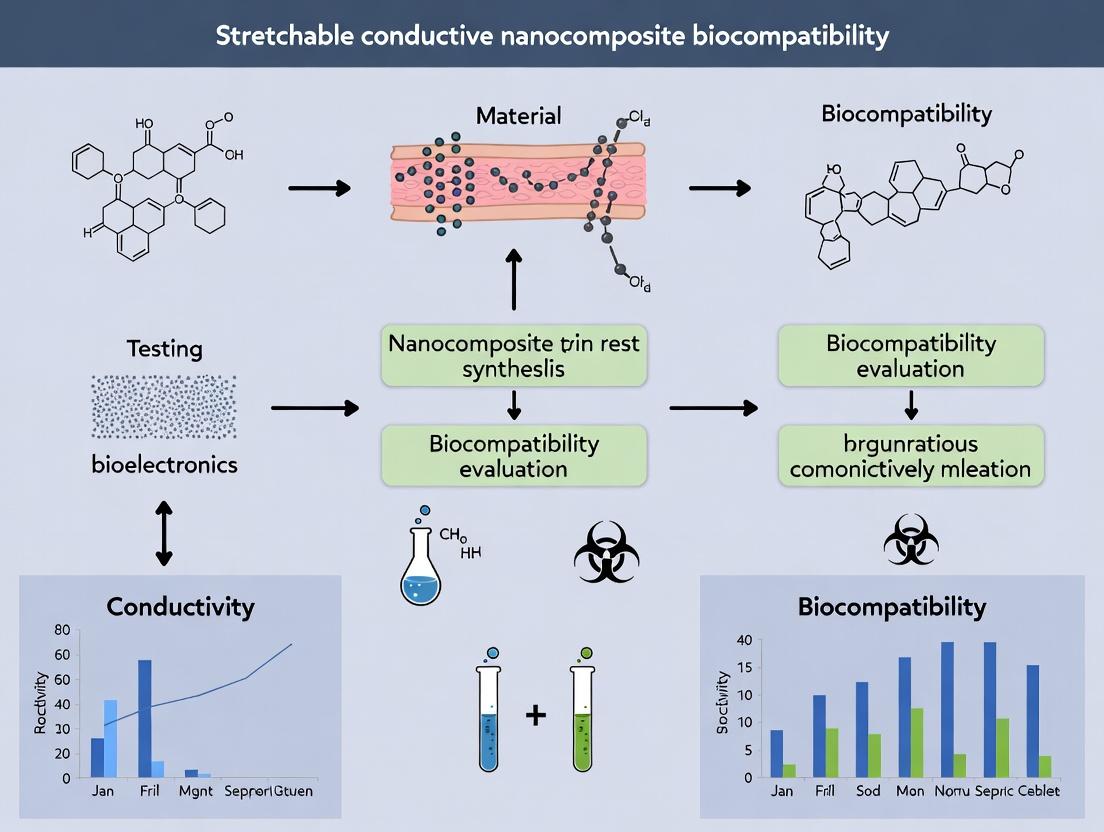
Stretchable Conductive Nanocomposites: A Comprehensive Guide to Biocompatibility for Biomedical Applications
This article provides a detailed examination of the biocompatibility of stretchable conductive nanocomposites, critical materials for next-generation biomedical devices.
Stretchable Conductive Nanocomposites: A Comprehensive Guide to Biocompatibility for Biomedical Applications
Abstract
This article provides a detailed examination of the biocompatibility of stretchable conductive nanocomposites, critical materials for next-generation biomedical devices. Targeting researchers, scientists, and drug development professionals, the content explores the fundamental principles of biocompatibility in dynamic materials, surveys current synthesis and fabrication methodologies, and addresses key challenges in cytotoxicity and long-term stability. It further offers rigorous frameworks for in vitro and in vivo validation, comparing leading material systems like silver nanowire (AgNW), liquid metal, and carbon-based composites. The synthesis of these four core intents serves as a strategic guide for developing safe, effective, and reliable bioelectronic interfaces for applications in wearable monitoring, neural implants, and soft robotics.
The Fundamentals of Biocompatibility in Stretchable Electronics: Definitions, Mechanisms, and Material Components
Defining Biocompatibility for Dynamic, Implantable, and Wearable Interfaces
Within the context of a broader thesis on the biocompatibility of stretchable conductive nanocomposites, this technical guide redefines biocompatibility for dynamic interfaces. Traditional static definitions fail to capture the complex, time-dependent biological interactions of materials that stretch, bend, and flex within a living system. This document provides a framework for assessing biocompatibility as a dynamic, multifactorial performance metric, integrating molecular, cellular, and systemic responses over operational lifetimes.
The ISO 10993 series provides a foundational but incomplete framework for dynamic interfaces. It primarily assesses static, passive materials, whereas stretchable conductive nanocomposites for implants and wearables are active, mechanically dynamic, and often designed for sustained biochemical interaction. Biocompatibility here must be defined as the ability of a dynamic material-device system to perform its intended function with an appropriate host response, throughout its operational lifespan, under relevant mechanical and electrochemical cycling. This necessitates a shift from evaluating inertness to characterizing controlled, predictable interaction.
Core Dimensions of Dynamic Biocompatibility
The biocompatibility of stretchable nanocomposites must be evaluated across three interdependent dimensions:
- Material-Cycle Biocompatibility: The local biological response to the composite material itself, including polymer matrix, conductive nanofillers (e.g., metal nanowires, carbon nanotubes, graphene), and any leachable species, under repeated mechanical strain.
- Interface-Cycle Biocompatibility: The response at the biotic-abiotic interface, considering surface topology changes during movement, electrochemically driven reactions (Faradaic vs. capacitive), and ionic transport.
- Signal-Cycle Biocompatibility: For bioelectronic interfaces, the fidelity of signal transduction (recording/stimulation) without eliciting adverse cellular responses (e.g., electrotoxic gliosis, unsustainable inflammatory activation).
Quantitative Metrics and Key Data
The following tables summarize critical quantitative endpoints for assessing dynamic biocompatibility.
Table 1: In Vitro Cytocompatibility Under Dynamic Conditions
| Metric | Test Method | Acceptable Threshold (Typical) | Key Challenge for Nanocomposites |
|---|---|---|---|
| Cell Viability | Live/Dead assay, MTT/WST-1 on strained substrates | >70% relative to control | Nanoparticle shedding during strain cycles |
| Reactive Oxygen Species (ROS) Generation | DCFH-DA assay, under electrical stimulation | <150% of unstimulated control | Electrochemical byproducts & nanomaterial catalysis |
| Membrane Integrity (LDH Release) | LDH assay in culture medium during cycling | <30% increase over static control | Cyclic strain-induced delamination & sharp edges |
| Inflammatory Cytokine Profile | Multiplex ELISA (e.g., IL-1β, IL-6, TNF-α, IL-10) | Pro-inflammatory cytokines not significantly elevated | Chronic "frustrated" macrophage response to moving surface |
Table 2: In Vivo Performance Metrics for Implantable Interfaces
| Metric | Evaluation Technique | Target Outcome (≥ 30 days) | Relevant Standard/Analog |
|---|---|---|---|
| Foreign Body Response (FBR) Thickness | Histology (H&E) capsule measurement | <150 µm, non-progressive | Compared to medical-grade silicone |
| Chronic Neuronal Loss/Gliosis | IHC (NeuN, GFAP, Iba1) quantification | <50% increase in glial density vs. distal site | Critical for neural interfaces |
| Impedance at 1 kHz | Electrochemical Impedance Spectroscopy (EIS) | Stable or decreasing trend post-acute phase | Indicates stable interface & minimal scar |
| Signal-to-Noise Ratio (SNR) | In vivo electrophysiology recording | Maintained >80% of day 7 baseline | Functional measure of biofouling impact |
Essential Methodologies and Protocols
Protocol: Cyclic Strain Cytocompatibility Assay
Objective: To evaluate the effect of repeated mechanical deformation on cell health and inflammatory response on stretchable nanocomposite substrates.
Materials: Sterilized nanocomposite film, bioreactor or custom strain rig, cell culture reagents, relevant cell line (e.g., fibroblasts, macrophages, neurons).
Procedure:
- Substrate Preparation: Cut nanocomposite to fit culture plates or bioreactor chambers. Sterilize via UV/Ozone or 70% ethanol (validate no degradation).
- Cell Seeding: Seed cells at standard density. Allow attachment for 24-48 hours under static conditions.
- Strain Regime Application: Apply defined uniaxial or biaxial strain (e.g., 10-20% strain, 0.5-1 Hz frequency) using a bioreactor. Include static controls.
- Conditioned Media Collection: At time points (e.g., 24h, 72h, 7d), collect media for soluble factor analysis (LDH, cytokines).
- Endpoint Analysis: After strain period, perform live/dead staining, ROS assays, or cell lysis for gene expression (qPCR for inflammatory markers).
- Characterization Post-Cycling: Analyze film surface via SEM/EDX for cracks, delamination, or nanomaterial release.
Protocol:In VivoElectrophysiological & Histological Co-Evaluation
Objective: To correlate the functional performance of an implantable bioelectronic interface with the histological host response.
Materials: Nanocomposite electrode array, rodent model, stereotaxic frame, electrophysiology system, perfusion and fixation reagents, histological stains/antibodies.
Procedure:
- Surgical Implantation: Aseptically implant the device in the target tissue (e.g., brain cortex, peripheral nerve).
- Chronic Recording/Stimulation: At regular intervals, acquire electrochemical impedance spectra and neural recording/stimulation data under anesthesia.
- Perfusion and Explanation: At terminal time point, transcardially perfuse with PBS followed by 4% PFA. Carefully explant the device with surrounding tissue intact.
- Histological Processing: Cryoprotect and section tissue. Perform serial staining: H&E for general morphology, Masson's Trichrome for collagen, and immunohistochemistry (IHC) for cell types (GFAP, Iba1, NeuN, CD68).
- Correlative Analysis: Register implant location with histological sections. Quantify metrics from Table 2 (capsule thickness, glial density) and correlate directly with impedance and SNR data from the same anatomical locus.
Signaling Pathways in the Dynamic Foreign Body Response
The presence of a moving interface modifies the canonical Foreign Body Response (FBR). Key pathways are outlined below.
The Scientist's Toolkit: Research Reagent Solutions
Table 3: Essential Materials for Dynamic Biocompatibility Research
| Item | Function & Rationale |
|---|---|
| Polydimethylsiloxane (PDMS; Sylgard 184) | Standard elastomeric matrix for stretchable substrates; allows tuning of stiffness; optically transparent. |
| PEDOT:PSS Conductive Polymer | Common conductive hydrogel component; mixed ionic-electronic conductor; enhances interface capacitance. |
| Gold Nanowires / Liquid Metal (EGaIn) | Conductive nanofillers providing percolation network and high stretchability (>100% strain). |
| CellScale BioTester or similar bioreactor | Instrument for applying precise, cyclic mechanical strain to cell-seeded constructs in culture. |
| Multi-electrode Array (MEA) Systems | For functional electrophysiology assessment of neurons on dynamic substrates or in explanted tissue. |
| DCFH-DA / CellROX ROS Detection Kits | Fluorogenic probes for detecting reactive oxygen species generation, a key nanotoxicity metric. |
| Luminex Multiplex Cytokine Assay Panels | Enable simultaneous quantification of a suite of pro- and anti-inflammatory cytokines from small sample volumes. |
| Iba1, GFAP, CD68 Antibodies | Standard immunohistochemistry markers for microglia/macrophages, astrocytes, and phagocytic cells, respectively. |
| Electrochemical Impedance Spectrometer (e.g., Gamry) | Critical for characterizing the electrical stability and charge transfer properties of the biotic-abiotic interface over time. |
An Integrated Assessment Workflow
A comprehensive evaluation requires a staged, integrated approach, as visualized below.
Defining biocompatibility for dynamic interfaces requires a paradigm shift from passive assessment to active, longitudinal performance monitoring. For stretchable conductive nanocomposites, biocompatibility is an emergent property of the material-tissue-system interaction under operational duress. A successful framework integrates quantitative in vitro screening under simulated use conditions with correlated in vivo functional and histological outcomes. This guide provides the foundational metrics, methods, and conceptual models to advance the rigorous development of safe and effective implantable and wearable technologies.
This whitepaper details the core biocompatibility challenges for stretchable conductive nanocomposites (SCNs) intended for chronic biomedical implants and bioelectronic interfaces. The integration of conductive nanofillers (e.g., carbon nanotubes, graphene, metallic nanowires) into elastomeric matrices (e.g., polydimethylsiloxane, polyurethane, hydrogels) creates unique material properties but also introduces significant biological risks. The material-host interface must be meticulously engineered to mitigate mechanical mismatch, nanomaterial leaching, and resultant chronic inflammatory cascades, which can lead to device failure, tissue damage, and systemic toxicity. This guide provides a technical framework for evaluating and addressing these challenges within a comprehensive biocompatibility thesis.
Mechanical Mismatch
Mechanical mismatch occurs when the elastic modulus, stretchability, and viscoelastic properties of the SCN differ substantially from the host tissue (e.g., skin, neural tissue, cardiac muscle). This mismatch creates shear stress at the interface, leading to fibrotic encapsulation, delamination, and signal degradation.
Quantitative Data on Tissue vs. Material Properties
Table 1: Elastic Modulus of Target Tissues and Common SCN Matrices
| Material/Tissue | Typical Elastic Modulus (kPa) | Ultimate Tensile Strain (%) | Key Notes |
|---|---|---|---|
| Brain Tissue | 0.5 - 2 | 10 - 50 | Highly soft, viscoelastic |
| Cardiac Muscle | 10 - 100 | 10 - 15 | Cyclically stressed |
| Skin (Epidermis/Dermis) | 100 - 2,000 | 30 - 115 | Varies with location & age |
| PDMS (Sylgard 184) | 500 - 3,000 | 100 - 150 | Tunable via base:curing agent ratio |
| Polyurethane (Medical Grade) | 50 - 1,000 | 300 - 600 | Wide range available |
| Polyacrylamide Hydrogel | 1 - 100 | > 200 | Highly tunable, often hydrated |
Experimental Protocol:In VitroCyclic Stretch Co-culture Model
Objective: To assess fibroblast activation and inflammatory cytokine release under simulated mechanical mismatch.
- Fabricate SCN substrates with varying elastic moduli (e.g., 10 kPa, 100 kPa, 1 MPa) using the same conductive filler content.
- Seed human dermal fibroblasts (HDFs) or THP-1 derived macrophages at a density of 50,000 cells/cm² on substrates in a bioreactor capable of applying uniaxial or biaxial strain.
- Apply cyclic strain regimens: 0% (control), 5% (physiological mimic), and 15% (mismatch condition) at 1 Hz for 72 hours.
- Collect supernatant at 24h intervals. Analyze for TGF-β1, IL-6, and TNF-α via ELISA.
- Fix cells post-experiment for immunocytochemistry (ICC) staining of α-smooth muscle actin (α-SMA) and pro-collagen I.
- Quantify gene expression via qRT-PCR for fibrosis markers (COL1A1, ACTA2) and inflammatory markers (IL1B, IL6).
Diagram Title: In Vitro Mechanical Mismatch Assay Workflow
Particle Leaching and Degradation
The long-term stability of SCNs is paramount. Abrasion, enzymatic degradation, and oxidative stress can cause the release of nanoscale fillers and polymer debris, posing risks of local cytotoxicity and systemic dissemination.
Quantitative Leaching Data
Table 2: Leaching Profile of Common Nanofillers under Simulated Physiological Conditions
| Nanofiller | Matrix | Test Medium (37°C) | Duration (Days) | Leached Conc. (ppb) | Primary Analytical Method |
|---|---|---|---|---|---|
| Multi-Wall CNTs | PDMS | PBS + 10% FBS | 30 | 15 - 50 | SP-ICP-MS |
| Graphene Oxide (GO) | GelMA Hydrogel | PBS (pH 7.4) | 60 | 100 - 400 | Fluorescence (Labeled GO) |
| Silver Nanowires | Polyurethane | Artificial Sweat | 28 | 200 - 1000 | ICP-OES |
| PEDOT:PSS | PVA Hydrogel | H₂O₂ (10 µM) | 14 | (Sulfur) 500 - 2000 | LC-MS/MS |
Experimental Protocol: Accelerated Aging and Leachate Characterization
Objective: To quantify and characterize particulates/ions leaching from SCNs under accelerated aging.
- Sample Preparation: Sterilize SCN samples (1cm x 1cm, n=5/group). Use samples with intact and deliberately abraded surfaces.
- Leaching Incubation: Immerse samples in 5 mL of simulated biological fluid (e.g., PBS with 0.1% BSA, artificial lysosomal fluid [ALF]) in sealed, low-binding tubes.
- Accelerated Aging: Incubate at 37°C with agitation (60 rpm). For accelerated tests, incubate at 50°C or add 100 µM H₂O₂ to medium. Collect leachate at 1, 7, 14, 30 days.
- Leachate Analysis:
- Size & Concentration: Use Single Particle ICP-MS (SP-ICP-MS) for metallic nanoparticles (Ag, Au). Use Nanoparticle Tracking Analysis (NTA) for non-metallic particles.
- Chemical Speciation: For polymers like PEDOT:PSS, use Liquid Chromatography-Tandem Mass Spectrometry (LC-MS/MS) to identify degradation products.
- In Vitro Toxicity Screen: Apply leachate (diluted 1:10) to macrophage (RAW 264.7) culture for 24h. Assess viability (MTS assay) and ROS production (DCFDA assay).
Chronic Inflammation Signaling Cascade
Persistent foreign body response (FBR) is the ultimate failure mode. Leached particles and mechanical stress activate a complex cascade leading to chronic inflammation, fibrous capsule formation, and device isolation.
Key Signaling Pathways in FBR to SCNs
The pathway involves initial protein adsorption, macrophage adhesion/activation, and fibroblast differentiation.
Diagram Title: Chronic Inflammation & Fibrosis Signaling Pathway
Experimental Protocol:In VivoSubcutaneous Implantation Model (Rat/Mouse)
Objective: To histologically and molecularly grade the FBR to SCNs over time.
- Implant Fabrication: Prepare sterile SCN disks (5mm diameter, 0.5mm thick). Include positive (stiff, non-porous polymer) and negative (medical-grade silicone) controls.
- Surgical Implantation: Anesthetize animals (e.g., Sprague-Dawley rats). Create subcutaneous pockets on the dorsum. Insert one implant per pocket (n=8 per material per time point). Close wound.
- Explanation: Euthanize animals at 1, 4, and 12 weeks. Excise implant with surrounding tissue en bloc.
- Histological Processing: Fix in 4% PFA, dehydrate, embed in paraffin. Section (5µm) and stain with:
- H&E: General morphology and capsule thickness measurement.
- Masson's Trichrome: Collagen deposition (blue).
- Immunohistochemistry (IHC): CD68 (pan-macrophages), iNOS (M1 macrophages), CD206 (M2 macrophages), α-SMA (myofibroblasts).
- Capsule Scoring: Use a standardized foreign body response scoring system (e.g., based on cell density, cell types, vascularization, and collagen alignment).
The Scientist's Toolkit: Research Reagent Solutions
Table 3: Essential Reagents for Biocompatibility Testing of SCNs
| Reagent/Material | Function/Application in SCN Testing | Example Product/Catalog |
|---|---|---|
| Polydimethylsiloxane (PDMS) | Elastomeric matrix; gold standard for soft lithography and modulus tuning. | Dow Sylgard 184 |
| Gelatin Methacryloyl (GelMA) | Photocrosslinkable, biologically active hydrogel matrix; promotes cell adhesion. | Advanced BioMatrix GelMA Kit |
| Multi-Walled Carbon Nanotubes (MWCNTs) | Conductive nanofiller; high aspect ratio, requires functionalization for dispersion. | Nanocyl NC7000 |
| Artificial Lysosomal Fluid (ALF) | Simulates phagolysosomal environment for accelerated degradation/leaching studies. | Prepared per ISO/TR 19057 |
| Reactive Oxygen Species (ROS) Assay Kit | Quantifies oxidative stress in cells exposed to SCN leachates or surfaces. | Abcam ab186027 (DCFDA) |
| TGF-β1 ELISA Kit | Quantifies key pro-fibrotic cytokine released in mechanical mismatch studies. | R&D Systems DB100B |
| CD68 & iNOS Antibodies (for IHC) | Labels total macrophages and M1-polarized macrophages, respectively, in tissue sections. | Abcam ab955 / ab15323 |
| Single Particle ICP-MS Standard (Au, 60nm) | Calibration standard for quantitative analysis of nanoparticle leaching. | NIST RM 8013 |
| Cyclic Stretch Bioreactor Plates | Applies controlled, physiological strain to cell-seeded SCN membranes in vitro. | Flexcell International FX-6000T System |
The development of stretchable conductive nanocomposites for biomedical applications, such as implantable sensors, neural interfaces, and drug-eluting platforms, hinges on the synergistic integration of two core material classes: the polymer matrix and the conductive filler. The overarching thesis of this research field is that biocompatibility is not an intrinsic property of individual components but an emergent property of the composite system, dictated by interfacial chemistry, mechanical mismatch, degradation profiles, and the biological response to leachable substances. This whitepaper provides a technical guide comparing the core polymers and fillers, framing their selection and processing within the imperative of achieving both functional performance and biological safety.
Core Polymer Matrices: Properties, Processing, and Biocompatibility
Polymer matrices provide the foundational mechanical properties, structural integrity, and host environment for conductive fillers.
Polydimethylsiloxane (PDMS)
- Properties: Thermoset elastomer with low Young's modulus (~0.5-3 MPa), high stretchability (≥100%), optical transparency, and high gas permeability. Its hydrophobic surface can be modified via plasma treatment.
- Biocompatibility Context: Widely regarded as biocompatible and bioinert for short- to medium-term implantation. Concerns include the potential leaching of uncrosslinked oligomers and hydrophobic surface promoting protein fouling.
- Key Processing Method:
- Base & Curing Agent Mixing: Typically mixed at a 10:1 weight ratio.
- Degassing: Vacuum desiccation to remove air bubbles.
- Curing: Thermal cure at 65-80°C for 1-2 hours.
- Surface Modification: Optional oxygen plasma treatment (100W, 30-60s) to create a temporary hydrophilic surface.
Styrene-Ethylene-Butylene-Styrene (SEBS)
- Properties: Thermoplastic elastomer with tunable modulus (1-1000 MPa) via styrene content and formulation. Excellent chemical stability and processability (injection molding, extrusion). Often used in gel form with mineral oil.
- Biocompatibility Context: The SEBS polymer itself is considered biostable and non-cytotoxic. Critical consideration: The biocompatibility of the plasticizing oil (e.g., mineral oil, silicone oil) is paramount, as it can leach out and cause inflammatory responses.
- Key Processing Method:
- Dissolution: SEBS pellets are dissolved in a suitable solvent (e.g., toluene, tetrahydrofuran) or mixed with plasticizing oil under vigorous stirring (e.g., 150 rpm, 80°C, 4h).
- Casting/Printing: The gel or solution is cast into molds or direct ink written.
- Solvent Evaporation: If a solvent is used, controlled evaporation (e.g., 40°C, 12h) is required.
Hydrogels
- Properties: Crosslinked polymer networks with high water content, mimicking native tissue modulus (0.1-100 kPa). Ionic conductivity. Mechanical properties are often enhanced with double-network or nanocomposite strategies.
- Biocompatibility Context: Generally exhibit superior biocompatibility and biointegration due to high water content and tissue-like properties. Biodegradability can be engineered. Key risks include inflammatory response to degradation products or residual crosslinkers (e.g., APS/TEMED).
- Key Processing Method (Polyacrylamide Example):
- Solution Preparation: Mix acrylamide monomer (e.g., 40 wt%), bis-acrylamide crosslinker (e.g., 0.1-0.5 wt%), and initiator (Ammonium Persulfate, 0.1 wt%) in deionized water.
- Degassing: Purge with nitrogen gas for 10 minutes.
- Catalyst Addition & Casting: Add catalyst Tetramethylethylenediamine (TEMED, 0.1% v/v), mix quickly, and cast between glass plates with spacers.
- Gelation: Allow to set at room temperature for 30-60 minutes.
Table 1: Comparative Properties of Core Polymer Matrices
| Polymer | Typical Modulus | Stretchability | Key Advantage | Primary Biocompatibility Concern | Common Processing Method |
|---|---|---|---|---|---|
| PDMS | 0.5 - 3 MPa | 100 - 1000% | Reproducibility, Transparency | Leachable oligomers, Protein fouling | Sylgard 184 mixing & thermal cure |
| SEBS | 1 - 1000 MPa* | 500 - 1300%* | Robustness, Processability | Leaching of plasticizer/oil | Dissolution/solvent casting or extrusion |
| Hydrogel | 0.1 - 100 kPa | 200 - 2000%* | Tissue-like, High Hydration | Residual chemicals, Degradation products | Free radical polymerization or ionic crosslinking |
*Highly tunable based on formulation.
Conductive Fillers: Properties, Percolation, and Biological Impact
Fillers impart electrical conductivity. Their interaction with the polymer matrix and biological environment is critical.
Metallic (Silver Flakes/Nanowires, Gold Nanostructures)
- Properties: High intrinsic conductivity (~10⁶ S/m for Ag). Nanowires enable conductivity at low filler loads due to high aspect ratio. Silver offers antimicrobial properties.
- Biocompatibility Context: Silver ions (Ag⁺) are cytotoxic at high concentrations, posing a risk if corrosion or oxidation occurs. Gold is generally considered more biocompatible and stable but is more expensive. Particle size, shape, and surface coating heavily influence cellular response.
Carbon-Based (Carbon Nanotubes, Graphene, Carbon Black)
- Properties: High conductivity, excellent mechanical properties. CNTs and graphene have very high aspect ratios, achieving percolation at very low loading (<1 wt%).
- Biocompatibility Context: Significant debate exists. Pristine CNTs can cause oxidative stress and persistent inflammation. Surface functionalization (e.g., -COOH, -OH) is essential to improve dispersion in polymers and reduce cytotoxic effects. Long-term biodurability is a key research question.
Liquid Metal (Eutectic Gallium-Indium, EGaIn, Galinstan)
- Properties: Conductivity ~3.4 x 10⁶ S/m. Liquid at room temperature, enabling self-healing and extreme stretchability (>1000%) when formed into micro-/nano-droplets within a polymer.
- Biocompatibility Context: Gallium ions (Ga³⁺) can be bioactive and are used in some pharmaceuticals (e.g., gallium nitrate). Indium toxicity is a concern. The formation of a native gallium oxide shell can partially encapsulate the metal, but rupture may lead to release. In vivo data is still emerging.
Table 2: Comparative Properties of Conductive Fillers
| Filler Type | Intrinsic Conductivity | Typical Percolation Threshold | Key Advantage | Primary Biocompatibility Concern | Critical Processing Note |
|---|---|---|---|---|---|
| Metallic (AgNW) | ~1.5-6.3 x 10⁶ S/cm | 0.1-1.5 vol% | High Conductivity, Antimicrobial | Cytotoxicity of ions (Ag⁺) | Dispersion to prevent aggregation |
| Carbon Nanotubes | ~10³-10⁶ S/cm | <0.1-1.0 wt% | High Aspect Ratio, Strength | Persistent inflammation, Oxidative stress | Must be functionalized for dispersion |
| Graphene | ~10⁶ S/cm | 0.1-3.0 vol% | High Conductivity, 2D Geometry | Edge sharpness, Inflammatory response | Exfoliation quality is critical |
| Liquid Metal | ~3.4 x 10⁶ S/cm | 40-80 wt%* | Self-Healing, Extreme Stretchability | Ion release (Ga³⁺, In³⁺), Long-term stability | Shear mixing to form droplets/network |
*Depends heavily on microstructure; a continuous network can form at lower loads.
Experimental Protocol: Cytocompatibility Assessment of a Nanocomposite
Aim: To evaluate the in vitro cytocompatibility of a PDMS-Silver Nanowire (AgNW) nanocomposite according to ISO 10993-5 standards.
Materials: PDMS (Sylgard 184), AgNW dispersion in ethanol, 96-well tissue culture plate, L929 fibroblast cells, Dulbecco's Modified Eagle Medium (DMEM), Fetal Bovine Serum (FBS), Penicillin-Streptomycin, AlamarBlue or MTT reagent, Phosphate Buffered Saline (PBS).
Methodology:
- Composite Fabrication: AgNW dispersion is mixed into uncured PDMS base by planetary mixing (2000 rpm, 2 min). Curing agent is added (10:1 ratio) and mixed. The blend is spin-coated (500 rpm, 60s) onto a culture plate lid and cured (80°C, 1h).
- Extract Preparation (Indirect Test): Composite samples are sterilized (70% ethanol, UV). An extraction medium (serum-supplemented DMEM) is applied at a surface area-to-volume ratio of 3 cm²/mL. Incubate at 37°C for 24h. The supernatant is the "extract."
- Cell Seeding: L929 cells are seeded in a 96-well plate at 10,000 cells/well in 100 µL medium and incubated for 24h.
- Exposure: The medium is replaced with 100 µL of the extract (100% concentration) or serial dilutions (e.g., 50%, 25%). Controls: cells with fresh medium (negative control) and medium with 1% DMSO (positive control). n=6 per group.
- Viability Assay (MTT): After 24h exposure, replace extract with 100 µL medium containing 0.5 mg/mL MTT. Incubate 4h. Remove solution, add 100 µL DMSO to dissolve formazan crystals.
- Analysis: Measure absorbance at 570 nm on a plate reader. Calculate cell viability (%) relative to the negative control. Viability >70% is generally considered non-cytotoxic.
Diagrams
Biocompatibility Assessment Workflow (98 chars)
Biocompatibility Risk and Mitigation Pathway (98 chars)
The Scientist's Toolkit: Essential Research Reagents & Materials
| Item | Function in Research | Key Consideration for Biocompatibility |
|---|---|---|
| Sylgard 184 (PDMS) | Standard elastomer matrix for stretchable devices. | Always fully cure; consider extraction to remove oligomers. |
| SEBS Pellets (e.g., MD1535) | Base polymer for creating tough, thermoplastic gels. | Must pair with a biocompatible plasticizer (e.g., medical-grade silicone oil). |
| Acrylamide/Bis-acrylamide | Monomers for synthesizing polyacrylamide hydrogels. | Residual monomer is neurotoxic; thorough washing (≥72h in PBS) is mandatory. |
| Silver Nanowires (AgNWs) | High-aspect-ratio conductive filler. | Opt for PVP-coated variants; assess ion release via ICP-MS. |
| Carboxylated CNTs | Functionalized carbon filler for improved dispersion. | Carboxylation reduces but does not eliminate cytotoxicity risk. |
| Eutectic Gallium-Indium (EGaIn) | Liquid metal filler for ultra-stretchable composites. | Handle in fume hood; sonicate in polymer to form stable dispersions. |
| AlamarBlue / MTT | Cell viability assay reagents for ISO 10993-5 testing. | Use indirect (extract) method first to avoid interference from materials. |
| L929 Fibroblast Cell Line | Standardized cell line for cytocompatibility screening. | Maintain passages below 20 for consistent response. |
| Medical-Grade Silicone Oil | Biocompatible plasticizer for SEBS gels. | Essential for in vivo applications to prevent inflammatory response to leachates. |
This whitepaper elucidates the fundamental biological interface mechanisms governing the in vivo performance of stretchable conductive nanocomposites. Within a doctoral thesis on next-generation biocompatible electronics (e.g., for neural interfaces or wearable biosensors), understanding these sequential events—protein adsorption, cellular adhesion, and the foreign body response (FBR)—is paramount. The nanocomposite’s surface properties (topography, chemistry, conductivity, modulus) directly dictate these interfacial interactions, ultimately determining the success or failure of the implanted device through fibrous encapsulation or seamless integration.
Protein Adsorption: The Initial Determinant
Within milliseconds of implantation, water and ions interact with the material, followed by rapid, competitive adsorption of proteins from blood and interstitial fluid (Vroman effect). This layer dictates all subsequent biological responses.
Key Factors Influencing Adsorption on Nanocomposites:
- Surface Energy & Wettability: Hydrophobic surfaces typically promote more denatured, dense protein layers.
- Surface Charge: Positively charged surfaces often adsorb more proteins due to electrostatic interactions with negatively charged plasma proteins.
- Nanoscale Topography: Nanoroughness, pores, or conductive filler (e.g., PEDOT:PSS, graphene, silver nanowires) exposure alter protein binding sites and conformation.
- Composition Dynamics: Under cyclic strain, the surface presentation of conductive fillers vs. elastomeric matrix (e.g., PDMS, SEBS) may change, dynamically altering the protein corona.
Quantitative Data on Protein Adsorption:
| Protein (Example) | Molecular Weight (kDa) | Concentration in Plasma (mg/mL) | Typical Adsorbed Layer Thickness on Hydrophobic Surface (nm) | Key Role in Subsequent Adhesion |
|---|---|---|---|---|
| Albumin | 66.5 | 35-50 | ~5-10 | "Passivating"; reduces cell attachment |
| Fibrinogen | 340 | 2-4 | ~10-15 | Primary mediator of platelet adhesion; ligand for integrins |
| Immunoglobulin G (IgG) | 150 | ~10 | ~8-12 | Promotes phagocyte recognition (Fc region) |
| Fibronectin | 440-500 | ~0.3 | ~10-20 | Critical for fibroblast and macrophage adhesion via RGD sequences |
| Vitronectin | 75 | ~0.2-0.4 | ~5-8 | Promotes osteoblast and fibroblast adhesion |
Experimental Protocol: Quartz Crystal Microbalance with Dissipation (QCM-D) for Protein Adsorption Kinetics
- Substrate Preparation: Coat QCM-D sensor chips (Au-coated) with your stretchable nanocomposite via spin-coating or dip-coating. Characterize surface roughness (AFM) and wettability (contact angle).
- Instrument Calibration: Mount coated chip in flow module. Flow phosphate-buffered saline (PBS) at 100 µL/min until stable baseline (frequency, Δf, and dissipation, ΔD) is achieved.
- Protein Solution Introduction: Switch flow to protein solution (e.g., 1 mg/mL fibrinogen in PBS) for 20-30 minutes. Monitor Δf (mass adsorption) and ΔD (viscoelasticity of adlayer).
- Rinsing: Switch back to PBS flow to remove loosely bound proteins. The final Δf/ΔD indicates mass and rigidity of the irreversibly adsorbed layer.
- Data Analysis: Use Sauerbrey or Voigt models to calculate adsorbed mass and layer thickness. Compare adsorption profiles across different nanocomposite formulations.
Cellular Adhesion: The Cellular Foundation
Cells (immune cells, fibroblasts) interact with the adsorbed protein layer via transmembrane integrins, forming focal adhesions. The nanocomposite's mechanical and electrical properties modulate this process.
Signaling Pathways in Integrin-Mediated Adhesion
Diagram: Integrin-Mediated Adhesion Signaling Cascade
Experimental Protocol: Immunofluorescence Staining for Focal Adhesions
- Cell Seeding: Plate fibroblasts (e.g., NIH/3T3) on sterile nanocomposite films in 24-well plates at 10,000 cells/well in complete medium. Culture for 4-24 hrs.
- Fixation: Aspirate medium. Wash with PBS. Fix with 4% paraformaldehyde in PBS for 15 min at RT. Wash 3x with PBS.
- Permeabilization & Blocking: Permeabilize with 0.1% Triton X-100 in PBS for 10 min. Block with 3% BSA in PBS for 1 hr.
- Primary Antibody Incubation: Incubate with mouse anti-paxillin (1:200 in 1% BSA/PBS) for 2 hrs at RT or overnight at 4°C. Wash 3x with PBS.
- Secondary Antibody & Phalloidin Staining: Incubate with Alexa Fluor 488 goat anti-mouse IgG (1:500) and rhodamine-phalloidin (1:200, for F-actin) in 1% BSA/PBS for 1 hr in the dark. Wash 3x.
- Nuclear Staining & Imaging: Incubate with DAPI (1:1000) for 5 min. Wash, mount, and image using a confocal microscope. Quantify focal adhesion number and size using ImageJ software.
The Foreign Body Response: The Ultimate Outcome
The FBR is a continuum of overlapping stages: acute inflammation, chronic inflammation, granulation tissue formation, foreign body giant cell (FBGC) formation, and fibrous encapsulation.
Temporal Progression of the Foreign Body Response
Diagram: Stages and Potential Outcomes of the Foreign Body Response
Key Quantitative Metrics in FBR Assessment:
| FBR Stage | Key Cell Types | Biomarkers for Analysis (Examples) | Measurable Outcome (Typical Range) |
|---|---|---|---|
| Acute Inflammation | Neutrophils, Mast Cells | Myeloperoxidase (MPO), TNF-α | Peak neutrophil density at implant site: 24-48 hrs |
| Chronic Inflammation | Macrophages (M1), Lymphocytes | CD68 (pan-macrophage), iNOS (M1), CD3 (T-cells) | Macrophage density can exceed 50% of cells at 1-2 weeks |
| FBGC Formation | Foreign Body Giant Cells | CD68, CD47/ SIRPα | FBGCs can persist for the implant lifetime |
| Fibrous Encapsulation | Myofibroblasts | α-SMA, Collagen I/III | Capsule thickness: 50-200+ µm; varies with material |
Experimental Protocol: Histological Evaluation of FBR in a Rodent Subcutaneous Model
- Implantation: Sterilize nanocomposite films (e.g., 5x5 mm). Anesthetize rat/mouse. Make a dorsal subcutaneous pocket. Insert one film per pocket. Suture wound.
- Explanation: Euthanize animals at endpoints (3, 7, 14, 28, 56 days). Excise implant with surrounding tissue.
- Fixation & Processing: Fix tissue in 10% neutral buffered formalin for 48 hrs. Process through graded ethanol and xylene, embed in paraffin.
- Sectioning & Staining: Section at 5 µm thickness. Perform:
- H&E Staining: For general morphology and capsule thickness measurement.
- Masson's Trichrome: For collagen (fibrous capsule) visualization.
- Immunohistochemistry: For specific cell types (e.g., anti-CD68 for macrophages, anti-α-SMA for myofibroblasts).
- Analysis: Use digital slide scanners and image analysis software (e.g., QuPath) to quantify capsule thickness, cellular density, and percentage of positive staining cells at the implant-tissue interface.
The Scientist's Toolkit: Essential Research Reagents & Materials
| Item | Function/Application in Bio-Interface Research |
|---|---|
| Quartz Crystal Microbalance with Dissipation (QCM-D) | Real-time, label-free measurement of protein adsorption mass and viscoelastic properties. |
| Surface Plasmon Resonance (SPR) Biosensor | Highly sensitive quantification of protein binding kinetics (ka, kd, KD) on functionalized surfaces. |
| Atomic Force Microscope (AFM) | Nanoscale topographic imaging and measurement of surface modulus (force spectroscopy). |
| Fibronectin from Human Plasma | A key adhesive glycoprotein used to pre-coat surfaces to promote specific integrin-mediated cell attachment. |
| Anti-Paxillin Antibody (mouse monoclonal) | Immunofluorescence staining of focal adhesion complexes to assess cell-material adhesion quality. |
| Rhodamine-Phalloidin | High-affinity F-actin probe for fluorescent labeling of the cell cytoskeleton. |
| Anti-CD68 Antibody (rabbit polyclonal) | Immunohistochemical marker for macrophages in tissue sections during FBR analysis. |
| α-Smooth Muscle Actin (α-SMA) Antibody | Marker for activated myofibroblasts critical for fibrous capsule contraction. |
| Masson's Trichrome Stain Kit | Differentiates collagen (blue) from muscle/cytoplasm (red) in fibrous encapsulation analysis. |
| PEDOT:PSS Aqueous Dispersion | Common conductive polymer component for stretchable nanocomposites. |
| Polydimethylsiloxane (PDMS) Sylgard 184 | Standard silicone elastomer used as a compliant matrix in nanocomposites or as control. |
Essential Regulatory and Standards Landscape (ISO 10993, USP Class VI) for Pre-clinical Evaluation
This whitepaper details the essential regulatory and standards framework governing the preclinical biological safety evaluation of medical devices and materials. For research focused on stretchable conductive nanocomposites intended for applications such as bioelectronic interfaces, implantable sensors, or neuromodulation devices, rigorous biocompatibility assessment is a critical gateway to clinical translation. The selection and execution of appropriate tests, guided by ISO 10993 and supplemented by USP Class VI, provide the foundational evidence required to demonstrate that the novel material, its leachable substances, and its degradation products present no unacceptable biological risk.
Core Standards: ISO 10993 Series
ISO 10993, "Biological evaluation of medical devices," is a harmonized series of standards that provides a systematic, risk-based framework for evaluating the biocompatibility of devices. The process is governed by the principles outlined in ISO 10993-1: "Evaluation and testing within a risk management process."
Key Concept: The Evaluation Matrix (ISO 10993-1) The standard mandates a tailored testing approach based on two primary factors:
- Nature of Body Contact (e.g., surface, external communicating, implant).
- Duration of Contact (e.g., limited (<24h), prolonged (24h-30d), permanent (>30d)).
The matrix specifies which categories of biological effects (e.g., cytotoxicity, sensitization, irritation) must be considered for a given device. For an implanted stretchable nanocomposite electrode (permanent contact with tissue/bone), a comprehensive evaluation is required.
Quantitative Data Summary: Key ISO 10993 Test Requirements for an Implantable Device
Table 1: Core ISO 10993 Tests for a Permanent Implant (e.g., Stretchable Nanocomposite Electrode)
| Test Category (ISO Part) | Test Objective | Key Quantitative Endpoints | Typical Pass/Fail Criteria |
|---|---|---|---|
| Cytotoxicity (10993-5) | Assess cell death, inhibition of cell growth. | Reduction in cell viability (%). | ≥ 70% viability (for elution method) is generally considered non-cytotoxic. |
| Sensitization (10993-10) | Evaluate potential for allergic contact dermatitis. | Magnitude of skin reactions (score 0-4). | Mean score in test group not significantly > negative control. |
| Irritation/Intracutaneous Reactivity (10993-10) | Assess local inflammatory response. | Erythema and edema scores (0-4). | Scores not significantly > control extracts. |
| Systemic Toxicity (10993-11) | Evaluate acute, subacute, or chronic systemic effects. | Mortality, clinical signs, body weight, necropsy findings. | No significant adverse effects vs. control group. |
| Genotoxicity (10993-3) | Detect mutagenic properties of leachables. | Frequency of reverse mutations (Ames), micronuclei, chromosomal aberrations. | No statistically significant increase vs. controls. |
| Implantation (10993-6) | Assess local effects on living tissue at implant site. | Histopathology scoring (inflammation, fibrosis, necrosis; e.g., 0-4 scale). | Response comparable to negative control material at appropriate time points. |
| Hemocompatibility (10993-4) If blood contact | Assess effects on blood/blood components. | Hemolysis (%), platelet adhesion/activation, coagulation times (PTT, PT). | Hemolysis <5%; other parameters within acceptable limits. |
Detailed Experimental Protocol: Cytotoxicity by Elution Method (ISO 10993-5)
- Purpose: To detect the presence of water-soluble leachables toxic to cultured mammalian cells.
- Materials: Test material extract (prepared per ISO 10993-12), L-929 mouse fibroblast cells, complete cell culture medium, multi-well plates, incubator (37°C, 5% CO₂), neutral red or MTT viability assay reagents.
- Procedure:
- Extract Preparation: Sterilize the nanocomposite sample. Incubate in serum-free culture medium (e.g., 0.1 g/mL or 6 cm²/mL) at 37°C for 24±2h.
- Cell Seeding: Seed L-929 cells in a 96-well plate at a density ensuring sub-confluent monolayers after 24h incubation.
- Exposure: After 24h, replace the culture medium in test wells with the material extract. Include a negative control (fresh medium) and a positive control (e.g., latex or ZnCl₂ solution).
- Incubation: Incubate cells with extract for 24±2h.
- Viability Assessment: Perform Neutral Red Uptake (NRU) assay: add neutral red medium, incubate 3h, wash, desorb dye with acidified ethanol, measure absorbance at 540 nm.
- Calculation: Calculate % cell viability = (Abstest / Absnegative control) x 100%.
USP Class VI: The Plastics Standard
United States Pharmacopeia (USP) <88> Class VI is a specific, prescriptive biological test protocol for plastics intended for use in medical products. While ISO 10993 is a comprehensive, risk-managed process, USP Class VI is a defined set of pass/fail tests often requested for materials used in pharmaceutical packaging or as components of devices.
Key Tests: It involves three in vivo assays: (1) Systemic Injection Test (mice), (2) Intracutaneous Test (rabbits), and (3) Implantation Test (rabbits). The material extracts are administered, and biological responses (lethality, weight loss, skin irritation, tissue reaction) are scored against defined thresholds.
Relationship to ISO 10993: USP Class VI can be considered a subset of testing that addresses aspects of systemic toxicity, irritation, and implantation. For device registration in the US, ISO 10993 is the primary framework, but compliance with USP Class VI may be cited as supplementary evidence of material safety.
Strategic Testing Workflow for Nanocomposites
The evaluation of a novel stretchable conductive nanocomposite requires a phased, logical approach integrated with material characterization.
Diagram 1: Biocompatibility Testing Strategy for Nanocomposites
The Scientist's Toolkit: Key Research Reagent Solutions
Table 2: Essential Materials for Biocompatibility Testing of Conductive Nanocomposites
| Reagent/Material | Function in Experiment | Key Application / Rationale |
|---|---|---|
| L-929 Mouse Fibroblast Cell Line | Model cell system for cytotoxicity testing (ISO 10993-5). | Standardized, reproducible cell line sensitive to leachable toxins. |
| Ames Test Strains (e.g., S. typhimurium TA98, TA100) | Bacterial strains for detecting point mutations (ISO 10993-3). | Initial, cost-effective screen for mutagenic potential of extracts. |
| Positive Control Materials (e.g., PE Film, Tin-stabilized PVC, Latex) | Provide a known, consistent response to validate test system sensitivity. | Required by standards to confirm assay is functioning correctly. |
| USP Purified Water & Polar/Semi-polar Solvents | Extraction vehicles to simulate different physiological conditions. | Used to prepare material extracts for testing, as per ISO 10993-12. |
| Histopathological Stains (H&E, Masson's Trichrome) | Stain tissue sections from implantation studies to evaluate cellular response. | Visualize inflammation, fibrosis, capsule formation around implant. |
| Simulated Body Fluids (SBF) | Solution mimicking ionic composition of blood plasma. | Used in in vitro degradation studies to assess ion release and stability. |
| MTT or Neutral Red Dye | Colorimetric reagents for quantifying cell viability and proliferation. | Provide quantitative endpoint for cytotoxicity assays. |
| ELISA Kits (e.g., for TNF-α, IL-1β, IL-6) | Quantify specific inflammatory cytokines released by cells in vitro. | Assess immunogenic potential of nanomaterials beyond standard cytotoxicity. |
Synthesis, Fabrication, and Application Strategies for Bio-Safe Nanocomposites
Within the pursuit of advanced biocompatible stretchable conductive nanocomposites for biomedical applications—such as neural interfaces, wearable biosensors, and drug-eluting scaffolds—the choice of fabrication technique is paramount. These methods directly dictate the microstructural architecture, electrical percolation networks, mechanical compliance, and ultimate biological integration of the composite material. This whitepaper provides an in-depth technical examination of three pivotal fabrication methodologies: In-Situ Polymerization, Solvent Casting, and 3D/4D Bioprinting. Each technique is analyzed for its role in integrating conductive nanofillers (e.g., carbon nanotubes, graphene, silver nanowires) into elastomeric matrices (e.g., PDMS, PU, hydrogels) while preserving or enhancing biocompatibility.
In-Situ Polymerization
In-situ polymerization involves dispersing conductive nanofillers within a monomer solution, followed by polymerization. This technique promotes uniform filler distribution and strong matrix-filler interactions, crucial for stable electrical conductivity under strain.
Detailed Protocol: In-Situ Polymerization of PEDOT:PSS/CNT in Polyurethane
Objective: To synthesize a stretchable, conductive nanocomposite film for epidermal electrophysiological sensing.
Materials:
- Monomer: Pre-polymer (Polyurethane resin, e.g., polyol and diisocyanate mixture).
- Conductive Fillers: Multi-walled carbon nanotubes (MWCNTs, 1-2 wt%), PEDOT:PSS dispersion.
- Dispersion Aid: Sodium dodecyl benzene sulfonate (SDBS) surfactant.
- Solvent: Dimethylformamide (DMF).
- Catalyst: Dibutyltin dilaurate (DBTDL).
- Crosslinker: Glycerol.
- Equipment: Ultrasonic probe sonicator, planetary centrifugal mixer, vacuum oven, glass mold, precision thickness spacers.
Procedure:
- Nanofiller Dispersion: Disperse MWCNTs (1.0 wt% relative to final solid) in DMF containing 0.1% SDBS. Sonicate using a probe sonicator (500 W, 20 kHz) in an ice bath for 30 min (5s on/2s off pulse cycle).
- Monomer-Filler Mixing: Add the PU polyol component to the MWCNT dispersion. Mix under magnetic stirring for 1 hour. Add the PEDOT:PSS dispersion (final ratio 1:1 by weight with PU) and mix for another 30 min.
- In-Situ Polymerization/Crosslinking: Add the PU diisocyanate component at a 1:1 NCO:OH molar ratio to the mixture. Introduce 0.1 wt% DBTDL catalyst and 2 wt% glycerol crosslinker. Mix thoroughly using a centrifugal mixer at 2000 rpm for 2 min to degas and ensure homogeneity.
- Casting & Curing: Pour the mixture into a glass mold with 500 µm spacers. Cure at 80°C in a vacuum oven for 12 hours.
- Post-Processing: Carefully demold the film and condition at 25°C, 60% RH for 24h before characterization.
Key Quality Metrics: Conductivity measured via 4-point probe; uniformity assessed via SEM mapping; cytocompatibility via ISO 10993-5 elution assay with L929 fibroblasts.
The Scientist's Toolkit: In-Situ Polymerization
| Research Reagent / Material | Primary Function in the Process |
|---|---|
| Multi-walled Carbon Nanotubes (MWCNTs) | Primary conductive nanofiller; forms percolation network for electron transport. |
| PEDOT:PSS Aqueous Dispersion | Intrinsically conductive polymer; enhances composite conductivity and interfacial stability. |
| Polyurethane Pre-polymer (Polyol/Diisocyanate) | Elastomeric matrix forming monomers; provides stretchability and mechanical robustness. |
| Dibutyltin Dilaurate (DBTDL) | Organotin catalyst; accelerates urethane linkage formation during polymerization. |
| Dimethylformamide (DMF) | Polar aprotic solvent; dissolves PU components and aids in nanofiller dispersion. |
| SDBS Surfactant | Dispersing agent; reduces surface tension of CNTs, preventing agglomeration in solution. |
Quantitative Performance Data
Table 1: Representative Performance of In-Situ Polymerized Nanocomposites
| Matrix Material | Conductive Filler (Loading) | Electrical Conductivity (S/cm) | Max Tensile Strain (%) | Key Application Context | Ref. (Year) |
|---|---|---|---|---|---|
| Polyurethane (PU) | PEDOT:PSS + CNT (1.5 wt%) | 12.5 | ~350 | Stretchable epidermal electrode | (2023) |
| Polydimethylsiloxane (PDMS) | Silver Flakes + Nanowires (70 wt%) | 4,800 | ~120 | Conductive adhesive for wearables | (2024) |
| Polyacrylamide Hydrogel | Graphene Oxide (2 mg/mL) | 0.05 | ~500 | Strain-sensing scaffold | (2023) |
Solvent Casting
Solvent casting is a foundational technique where a polymer and nanofillers are dissolved/dispersed in a volatile solvent, cast onto a substrate, and the solvent is evaporated to form a film.
Detailed Protocol: Solvent Casting of PDMS/AgNW Nanocomposite
Objective: To fabricate a transparent, conductive, and stretchable film for optoelectronic sensing.
Materials:
- Polymer Matrix: PDMS base and curing agent (Sylgard 184).
- Conductive Filler: Silver nanowires (AgNWs, diameter 30 nm, length 20-30 µm).
- Solvents: Ethanol and toluene.
- Substrate: Glass plate, treated with oxygen plasma for 2 min.
- Equipment: Ultrasonic bath, spin coater, vacuum desiccator, hot plate.
Procedure:
- Solution Preparation: Disperse AgNWs in ethanol (target concentration 2 mg/mL) using a 30-min ultrasonic bath. Separately, dissolve PDMS base in toluene (10% w/v) by magnetic stirring for 2h.
- Nanocomposite Ink Formulation: Slowly add the AgNW dispersion to the PDMS-toluene solution under vigorous stirring. Maintain stirring for 4h to ensure homogeneous mixing. Finally, add the PDMS curing agent at a 10:1 base-to-agent ratio and stir for 10 min.
- Casting: Pour the ink onto the plasma-treated glass substrate. Use a spin coater (500 rpm for 10s, then 1000 rpm for 30s) to achieve a uniform thin film. Alternatively, use a doctor blade with a 300 µm gap.
- Solvent Evaporation & Curing: Place the cast film in a vacuum desiccator for 1h to remove residual solvent and bubbles. Subsequently, transfer to a hot plate and cure at 80°C for 2h.
- Peel-off: Carefully peel the cured nanocomposite film from the glass substrate.
Key Quality Metrics: Sheet resistance (Ω/sq) vs. transmittance (% at 550 nm) trade-off; adhesion strength via peel test; surface roughness via AFM.
Solvent Casting Workflow
Diagram Title: Solvent Casting Nanocomposite Fabrication Workflow
3D/4D Bioprinting
3D bioprinting precisely deposits bioinks—often nanocomposite hydrogels—layer-by-layer to create complex, cell-laden structures. 4D bioprinting introduces a temporal dimension, where printed constructs change shape or functionality post-printing in response to stimuli (e.g., pH, temperature, electrical field).
Detailed Protocol: Extrusion Bioprinting of a Conductive GelMA/Graphene Bioink
Objective: To 3D print a conductive, cell-laden scaffold for cardiac tissue engineering with electrical stimulation capability.
Materials:
- Bioink Base: Gelatin methacryloyl (GelMA, 10% w/v).
- Conductive Filler: Graphene nanoplatelets (GnP, 1 mg/mL).
- Photoinitiator: Lithium phenyl-2,4,6-trimethylbenzoylphosphinate (LAP, 0.25% w/v).
- Cells: Human mesenchymal stem cells (hMSCs), passage 4-5.
- Culture Medium: α-MEM, supplemented.
- Equipment: Extrusion bioprinter (e.g., BIO X), 22G conical nozzle, 405 nm UV light source, sterile petri dish.
Procedure:
- Bioink Preparation: Dissolve GelMA and LAP in PBS at 40°C. Separately, sterilize GnPs under UV light for 1h and disperse in PBS via sonication. Mix the GnP dispersion into the GelMA solution. Filter sterilize the composite through a 0.22 µm syringe filter. Cool to 25°C.
- Cell Harvesting & Encapsulation: Trypsinize and count hMSCs. Centrifuge and resuspend cells in the cooled (~20°C) GelMA/GnP bioink at a density of 5 x 10^6 cells/mL. Gently mix and keep on ice to prevent premature gelation.
- Printing Parameters: Load bioink into a sterile cartridge. Set printer stage temperature to 15°C. Use pressure: 25-30 kPa, speed: 8 mm/s, layer height: 150 µm. Print a 15mm x 15mm grid structure.
- Crosslinking: After each layer is deposited, apply a brief crosslinking step using 405 nm UV light (5 mW/cm² for 15s).
- Post-Printing & Culture: After final layer, perform a final bulk UV crosslink (20s). Transfer the printed construct to a cell culture plate, add warm medium, and incubate at 37°C, 5% CO₂.
- 4D Transformation (Optional): To induce 4D shape morphing, design a bilayered structure with differing GnP concentrations or crosslinking densities. Upon immersion in cell culture medium at 37°C, differential swelling induces controlled curvature over 24h.
Key Quality Metrics: Printability (filament fusion, shape fidelity), post-printing cell viability (Live/Dead assay at 24h), electrical impedance spectroscopy, and contractile function under electrical pacing.
3D/4D Bioprinting Experimental Logic
Diagram Title: 3D to 4D Bioprinting Logic Path for Nanocomposites
Quantitative Performance Data
Table 2: Performance Metrics of Bioprinted Conductive Nanocomposites
| Bioink Formulation | Filler Loading | Cell Type | Post-Print Viability (%) | Conductivity (S/m) | Elastic Modulus (kPa) | Ref. (Year) |
|---|---|---|---|---|---|---|
| GelMA + Graphene | 1 mg/mL | hMSCs | >92% (Day 1) | 0.12 | 35 ± 5 | (2024) |
| Alginate + PEDOT:PSS | 0.3% w/v | C2C12 myoblasts | 88% | 0.08 | 22 ± 3 | (2023) |
| PEGDA + CNT | 0.5 wt% | NIH/3T3 fibroblasts | 85% | 0.25 | 450 ± 50 | (2023) |
Each fabrication method offers distinct advantages and constraints for producing biocompatible stretchable conductive nanocomposites:
- In-Situ Polymerization excels in creating homogeneous dispersions with strong interfacial bonding, leading to excellent electromechanical stability under cyclic loading. It is ideal for thin, robust films for sensors.
- Solvent Casting is a versatile and relatively simple technique suitable for large-area film production. Its main challenges involve solvent residue removal and potential nanofiller sedimentation during slow evaporation.
- 3D/4D Bioprinting provides unmatched spatial control over geometry and composition, enabling complex, cell-embedded constructs. It is the premier technique for creating anatomically relevant, functional tissues, with 4D capabilities adding dynamic responsiveness.
The selection of a fabrication technique must be driven by the target application's requirements for resolution, scalability, mechanical properties, electrical performance, and, most critically, the desired mode of biointegration. Future progress in this field hinges on the synergistic development of novel nanocomposite materials and adaptive fabrication platforms that together satisfy the stringent triad of stretchability, conductivity, and biocompatibility.
Surface Modification and Encapsulation Strategies to Enhance Biocompatibility
Within the broader research on the biocompatibility of stretchable conductive nanocomposites for biomedical applications (e.g., neural interfaces, cardiac patches, wearable biosensors), surface characteristics and bulk encapsulation are paramount. These materials often combine conductive nanoparticles (e.g., silver nanowires, carbon nanotubes, graphene) with elastomeric matrices (e.g., PDMS, SEBS, hydrogels). While offering excellent electromechanical properties, their pristine surfaces can provoke adverse biological responses, including protein fouling, fibroblast encapsulation, and chronic inflammation, ultimately leading to device failure. This guide details current, advanced strategies to engineer the biointerface of such composites to improve host integration and long-term functionality.
Core Surface Modification Strategies
Surface modification aims to alter the outermost layer of the nanocomposite without compromising its bulk conductive and mechanical properties. The goal is to present a biologically favorable interface.
Physical Adsorption & Layer-by-Layer (LbL) Assembly
This method involves the sequential deposition of oppositely charged polyelectrolytes or biomolecules onto the substrate.
Experimental Protocol for LbL on PDMS-based Nanocomposites:
- Substrate Preparation: A PDMS/AgNW composite film is oxygen plasma treated for 2-5 minutes to generate a negatively charged, hydrophilic surface.
- Polyelectrolyte Solutions: Prepare 1 mg/mL solutions of cationic poly(allylamine hydrochloride) (PAH) and anionic poly(sodium 4-styrenesulfonate) (PSS) in 0.5 M NaCl (to increase layer roughness).
- Deposition Cycle:
- Immerse the substrate in PAH solution for 10 minutes.
- Rinse thoroughly in three separate beakers of deionized water (2 min each).
- Immerse the substrate in PSS solution for 10 minutes.
- Rinse again as above.
- This constitutes one bilayer (PAH/PSS). Repeat for 5-10 bilayers.
- Final Layer: To biofunctionalize, the final layer can be a biomolecule like heparin, hyaluronic acid, or collagen. Adsorb by immersion in a 0.1 mg/mL solution for 1 hour.
Chemical Grafting: "Grafting-to" vs. "Grafting-from"
Chemical grafting creates stable covalent bonds between the surface and the functional layer.
Protocol for Silanization & Peptide Grafting (Grafting-to):
- Surface Activation: Plasma treat the nanocomposite as above.
- Silanization: Immediately immerse in a 2% (v/v) solution of (3-Aminopropyl)triethoxysilane (APTES) in anhydrous toluene for 2 hours under inert atmosphere.
- Washing: Rinse with toluene and ethanol to remove physisorbed silane, then cure at 110°C for 15 minutes.
- Peptide Conjugation: Activate the terminal amine groups by reacting with a heterobifunctional crosslinker (e.g., Sulfo-SMCC) in PBS for 1 hour. Rinse and then react with a cysteine-terminated RGD peptide solution (1 mM in PBS) overnight at 4°C to promote cell adhesion.
Protocol for Surface-Initiated Atom Transfer Radical Polymerization (SI-ATRP) (Grafting-from):
- Initiator Immobilization: Following APTES treatment, react the amines with 2-bromoisobutyryl bromide (BiBB) to install ATRP initiators.
- Polymerization: Prepare a degassed mixture of monomer (e.g., poly(ethylene glycol) methacrylate - PEGMA), catalyst (CuBr/PMDETA), and solvent (methanol/water). Inject this into the reaction vessel containing the initiator-functionalized substrate.
- Reaction: Allow polymerization to proceed for 1-4 hours under N₂ atmosphere to grow a dense brush of PEG-like polymer, which resists protein adsorption.
Biomimetic Coatings
These coatings replicate biological structures to "hide" the material from the immune system.
Protocol for Zwitterionic Polymer Brush Coating: Zwitterions (e.g., poly(sulfobetaine methacrylate) - PSBMA) mimic the antifouling properties of cell membranes. Use the SI-ATRP protocol above, substituting SBMA as the monomer.
Protocol for Cell Membrane Mimicry via Lipid Bilayer Deposition:
- Vesicle Preparation: Prepare small unilamellar vesicles (SUVs) from phospholipids (e.g., DOPC:DOPG mixtures) via extrusion through a 50 nm membrane.
- Substrate Conditioning: Ensure the nanocomposite surface is clean and hydrophilic (via plasma).
- Deposition: Incubate the substrate with the SUV suspension (0.5 mg/mL lipid in Tris buffer) for 1-2 hours at room temperature. The vesicles spontaneously rupture and fuse to form a supported lipid bilayer (SLB).
Encapsulation Strategies
Encapsulation involves creating a barrier layer that fully encloses the nanocomposite, isolating it from the biological environment to prevent leakage of nanoparticles or degradation products.
Thin-Film Encapsulation with Biodegradable Polymers
Protocol for Spray-Coating Poly(lactic-co-glycolic acid) (PLGA):
- Solution Preparation: Dissolve PLGA (50:50 LA:GA) in dichloromethane (DCM) to create a 5% (w/v) solution.
- Masking: Mask electrical contact pads with polyimide tape.
- Spray Coating: Use an airbrush sprayer with a 0.2 mm nozzle. Maintain a distance of 15-20 cm from the substrate. Apply multiple thin coats (e.g., 10 passes) with 30-second drying intervals between coats to build a uniform, pinhole-free film of ~10 µm thickness.
- Curing: Dry under vacuum overnight to remove residual solvent.
Hydrogel Encapsulation
Hydrogels provide a soft, hydrating, and often biocompatible barrier.
Protocol for In Situ Gelatin Methacryloyl (GelMA) Encapsulation:
- GelMA Synthesis: Synthesize GelMA following established protocols (reaction of gelatin with methacrylic anhydride).
- Pre-hydrogel Solution: Prepare a 10% (w/v) GelMA solution in PBS containing 0.25% (w/v) photoinitiator (Lithium phenyl-2,4,6-trimethylbenzoylphosphinate - LAP).
- Device Embedding: Place the stretchable electrode on a substrate, pipette the GelMA solution over it to fully embed, and cover with a glass coverslip to control thickness.
- Crosslinking: Expose to 405 nm UV light (5-10 mW/cm²) for 30-60 seconds to form a covalently crosslinked hydrogel encapsulant.
Table 1: Performance Comparison of Surface Modification Strategies on Stretchable Nanocomposites
| Strategy | Technique | Key Metric | Result (Typical Range) | Biological Outcome |
|---|---|---|---|---|
| Physical | PLL/PSS LbL (5 bilayers) | Roughness Increase (AFM) | +15 to +25 nm | Reduced macrophage activation by ~40% vs. bare PDMS |
| Chemical Grafting | PEG Brush (SI-ATRP) | Protein Adsorption (QCM-D) | >90% reduction in fibrinogen adsorption | Fibroblast adhesion reduced by >85% over 7 days |
| Biomimetic | Zwitterionic PSBMA Brush | Hydration Layer Thickness (NMR) | ~2.3 nm | Whole blood fouling reduction: ~95% |
| Biomimetic | Supported Lipid Bilayer | Fluidity (FRAP Recovery) | 70-90% recovery | Inflammatory cytokine (TNF-α) release from monocytes reduced by 70% |
| Encapsulation | PLGA Spray-Coating (10µm) | Barrier Integrity (Impedance in PBS) | Impedance increase >1 MΩ over 30 days | Prevents Ag⁺ ion leakage below 0.1 ppb for 4 weeks |
| Encapsulation | GelMA Hydrogel (10%) | Young's Modulus | 20-50 kPa (matches soft tissue) | Neuronal cell viability on encapsulated electrode >90% at 7 days |
Table 2: Key Reagent Solutions for Biocompatibility Enhancement
| Reagent/Category | Example Product (Supplier Example) | Function in Experiment |
|---|---|---|
| Polyelectrolytes | Poly(allylamine hydrochloride) (PAH) & Poly(sodium 4-styrenesulfonate) (PSS) (Sigma-Aldrich) | Building blocks for LbL assembly; create a controllable, charged nanoscale coating. |
| Silane Coupling Agent | (3-Aminopropyl)triethoxysilane (APTES) (Gelest) | Provides surface amine groups for subsequent covalent conjugation of biomolecules. |
| ATRP Initiator | 2-Bromoisobutyryl bromide (BiBB) (Sigma-Aldrich) | Immobilizes initiator sites on the surface for "grafting-from" polymer brush synthesis. |
| Zwitterionic Monomer | Sulfobetaine methacrylate (SBMA) (Sigma-Aldrich) | Monomer for growing ultra-low fouling polymer brushes via SI-ATRP. |
| Cell-Adhesive Peptide | Cys-Arg-Gly-Asp-Ser (C-RGDS) (Bachem) | Conjugates to surface to provide specific integrin-binding sites for improved cell adhesion. |
| Biodegradable Polymer | Poly(D,L-lactide-co-glycolide) (PLGA 50:50) (Evonik) | Forms a protective, biocompatible, and resorbable barrier layer for encapsulation. |
| Photocrosslinkable Hydrogel | Gelatin Methacryloyl (GelMA) (Advanced BioMatrix) | Forms a soft, hydrated, cell-interactive encapsulation matrix via UV crosslinking. |
| Photoinitiator | Lithium phenyl-2,4,6-trimethylbenzoylphosphinate (LAP) (TCI Chemicals) | Enables rapid, cytocompatible UV crosslinking of GelMA and similar hydrogels. |
Visualized Workflows and Pathways
Surface Modification Decision Pathway
Encapsulation & Modification Workflow
Foreign Body Response vs. Mitigation
The development of chronic neural electrodes for Brain-Machine Interfaces (BMIs) represents a frontier in neuroscience and neuroengineering. The core challenge, and the central thesis of this research, is that long-term functional stability is intrinsically linked to the biocompatibility of the neural interface material. Traditional rigid electrodes (e.g., tungsten, silicon) elicit a foreign body response characterized by glial scarring, neuronal loss, and declining signal quality over weeks to months. This document posits that stretchable conductive nanocomposites—materials engineered to mimic the mechanical, chemical, and topographical properties of neural tissue—are the key to next-generation, chronically stable BMIs. Their compliance minimizes mechanical mismatch-induced inflammation, while their nano-structured conductive elements (e.g., conductive polymers, carbon nanotubes, graphene) maintain signal fidelity at the biotic-abiotic interface.
Current State & Quantitative Performance Metrics
The performance of neural interfaces is quantified across multiple axes. The table below summarizes key metrics for traditional and emerging stretchable nanocomposite-based electrodes.
Table 1: Performance Comparison of Neural Electrode Technologies
| Metric | Traditional Rigid (Si, IrOx) | Thin-Film Polymeric (PEDOT:PSS) | Stretchable Nanocomposite (e.g., SEBS/graphene/PPy) | Ideal Target |
|---|---|---|---|---|
| Impedance @ 1 kHz (kΩ) | 100 - 500 | 1 - 50 | 5 - 100 | < 50 |
| Charge Storage Capacity (C/cm²) | 1 - 10 | 20 - 100 | 10 - 200 | > 50 |
| Elastic Modulus (GPa) | 50 - 200 | 1 - 5 | 0.001 - 1 (MPa range) | 0.1 - 100 kPa |
| Stretchability (% Strain) | < 1% | 2 - 20% | 20 - 100%+ | > 30% |
| Chronic Recording Lifetime (Months) | 6 - 12 | 12 - 24 | 18 - 36+ (Preclinical) | > 60 |
| Single-Unit Yield @ 6 Months | Low (< 20% initial) | Moderate | High (Up to 80% retained) | > 80% |
| Inflammation Marker (GFAP+ area) @ 12 wks | High | Moderate | Low | Minimal |
Data synthesized from recent literature (2023-2024). GFAP: Glial Fibrillary Acidic Protein, a marker for astrogliosis.
Core Experimental Protocols for Biocompatibility & Function Assessment
Protocol:In VivoElectrode Implantation & Chronic Recording in Rodent Model
Aim: To assess the chronic recording performance and histological biocompatibility of a stretchable nanocomposite electrode array.
- Fabrication: Prepare nanocomposite (e.g., Polydimethylsiloxane (PDMS) infused with poly(3,4-ethylenedioxythiophene):polystyrene sulfonate (PEDOT:PSS)-coated Au nanowires). Pattern into a 16-channel Michigan-style array.
- Surgical Implantation: Anesthetize adult rat (e.g., Sprague-Dawley) and secure in stereotaxic frame. Perform craniotomy over primary motor cortex (M1). Durotomy is performed. The electrode array is slowly inserted to a depth of ~1.5 mm (layer V) using a hydraulic microdrive. The array's stretchable substrate is conformally placed on the cortical surface and secured with medical-grade silicone adhesive. The craniotomy is sealed with biocompatible silicone gel.
- Chronic Recording: Connect pedestal to a wireless recording headstage. Record neural activity (spikes and local field potentials) daily for 6+ months. Use standardized tasks (e.g., lever press) to correlate signals with behavior.
- Terminal Histology: Perfuse-fix the animal. Extract brain, section, and immunostain for neuronal nuclei (NeuN), microglia (Iba1), and astrocytes (GFAP). Quantify neuronal density and glial encapsulation thickness around the implant track using confocal microscopy and image analysis (e.g., ImageJ).
Protocol:In VitroCharacterization of Neuro-Nanocomposite Interface
Aim: To quantify cell viability, neurite outgrowth, and electrophysiological coupling on nanocomposite substrates.
- Substrate Preparation: Spin-coat or cast nanocomposite films on glass coverslips. Sterilize via UV ozone treatment.
- Primary Cortical Neuron Culture: Seed dissociated rat E18 cortical neurons at controlled density (e.g., 50,000 cells/cm²) on substrates coated with poly-L-lysine/laminin.
- Live/Dead Assay (Day 7): Incubate with Calcein-AM (live, green fluorescence) and Ethidium homodimer-1 (dead, red fluorescence). Image and calculate viability percentage.
- Neurite Outgrowth Analysis (Day 3): Fix cells, immunostain for β-III-tubulin. Use automated tracing software to quantify total neurite length per neuron.
- Microelectrode Array (MEA) Recording: Culture neurons directly on nanocomposite films patterned with embedded MEA electrodes. Record spontaneous network activity (burst firing, synchronized oscillations) over 4 weeks to assess functional synaptic development.
Signaling Pathways in the Foreign Body Response
A critical aspect of biocompatibility research involves understanding the cellular and molecular pathways activated upon implantation. The diagram below outlines the key signaling cascades.
Diagram 1: Key Signaling in Neural Implant Response
Experimental Workflow for Nanocomposite Evaluation
The comprehensive assessment of a novel stretchable nanocomposite for BMI applications follows a structured pipeline.
Diagram 2: Workflow for Nanocomposite Neural Electrode R&D
The Scientist's Toolkit: Key Research Reagents & Materials
Table 2: Essential Research Reagents for Stretchable BMI Development
| Reagent/Material | Category | Function & Rationale |
|---|---|---|
| PEDOT:PSS (Heraeus Clevios PH1000) | Conductive Polymer | High conductivity, moderate stretchability, and excellent biocompatibility. Serves as the conductive phase in many nanocomposites. |
| PDMS (Sylgard 184) | Elastomer Base | Industry-standard silicone elastomer providing stretchability, transparency, and easy fabrication. The base for many stretchable substrates. |
| Carbon Nanotubes (Single/Walled) | Nanocarbon Filler | Imparts electrical conductivity and mechanical reinforcement. High aspect ratio enables percolation networks at low loadings. |
| Poly-L-Lysine & Laminin | Cell Adhesion Coating | Essential for promoting neuronal adhesion and neurite outgrowth on synthetic substrates during in vitro testing. |
| Iba1 & GFAP Antibodies | Immunohistochemistry | Primary antibodies for labeling microglia and astrocytes, respectively, to quantify neuroinflammatory response post-implantation. |
| NeuN Antibody | Immunohistochemistry | Labels neuronal nuclei to quantify neuronal survival and density around the implant site. |
| Calcein-AM / EthD-1 Kit | Viability Assay | Standard live/dead fluorescent assay for rapid quantification of cell viability on material surfaces. |
| Wireless Neural Headstage (e.g., Intan) | Data Acquisition | Enables chronic, unrestrained neural recording in behaving animals, critical for longitudinal BMI performance data. |
| Flexible/Stretchable Conductive Ink (e.g., Ag/AgCl flake in silicone) | Interconnect Material | Creates stretchable traces connecting electrode sites to connectors, maintaining conductivity under strain. |
The advancement of wearable epidermal sensors for continuous, clinical-grade physiological monitoring represents a pivotal application of fundamental research into biocompatible, stretchable conductive nanocomposites. This technical guide frames the sensor development, material requirements, and validation protocols within the overarching thesis that the optimization of polymer matrices, nanofiller dispersion, and interfacial bonding dictates not only electromechanical performance but also long-term biocompatibility and signal fidelity. The transition from benchtop nanocomposite to functional epidermal device necessitates a holistic design philosophy where material properties are engineered in direct response to the dynamic, demanding environment of human skin.
Core Nanocomposite Materials & Characterization Data
The performance of epidermal sensors is fundamentally governed by the properties of the stretchable conductive nanocomposite. Key metrics include conductivity under strain, elastic modulus matching to skin, and long-term stability. The following table summarizes recent benchmark data for prominent nanocomposite systems.
Table 1: Performance Metrics of Stretchable Conductive Nanocomposites for Epidermal Sensors
| Polymer Matrix | Conductive Filler | Filler Loading (wt%) | Initial Conductivity (S/cm) | Conductivity at 50% Strain (S/cm) | Maximum Strain at Failure (%) | Critical Strain for Conductivity Loss (%) | Reported Biocompatibility Test (Standard) |
|---|---|---|---|---|---|---|---|
| Polydimethylsiloxane (PDMS) | Silver Flakes | 70 | 4,500 | 1,200 | 80 | 60 | ISO 10993-5 (Cytotoxicity) |
| Polyurethane (PU) | Silver Nanowires (AgNWs) | 0.8 | 8,200 | 6,500 | 450 | 250 | ISO 10993-10 (Sensitization) |
| SEBS (Styrene-Ethylene-Butylene-Styrene) | Graphene Nanoplatelets | 15 | 120 | 95 | >500 | 350 | In vitro fibroblast adhesion (72h) |
| Ecoflex (Silicone) | Liquid Metal (EGaIn) | 90 (v/v) | 24,100 | 24,000* | 800 | 800* | ISO 10993-5, -10 |
| Poly(3,4-ethylenedioxythiophene):Polystyrene Sulfonate (PEDOT:PSS) with Ionic Additives | - (Intrinsic Conductor) | - | 850 | 300 | 55 | 40 | CCK-8 assay with L929 cells |
*Liquid metal composites exhibit negligible change in conductivity due to the fluidic filler; strain is accommodated via microstructure reorganization.
Detailed Experimental Protocols
Protocol: Fabrication of an AgNW/PU Nanocomposite Epidermal Electrode
Objective: To fabricate a transparent, stretchable dry electrode for electrophysiological sensing (e.g., ECG, EMG).
Materials & Reagents:
- Polyurethane pellets (e.g., Tecoflex EG-80A).
- Silver nanowire dispersion (e.g., 0.5% wt in isopropanol, diameter ~120 nm, length ~25 µm).
- Dimethylformamide (DMF) and Tetrahydrofuran (THF) (3:1 v/v solvent mixture).
- Glass or PDMS substrate.
- Oxygen plasma cleaner.
Procedure:
- Solution Preparation: Dissolve PU pellets in the DMF/THF solvent mixture (10% w/v) under magnetic stirring at 50°C for 4 hours until fully dissolved.
- Nanocomposite Blending: Add the AgNW dispersion dropwise to the PU solution under vigorous stirring to achieve a final AgNW solid content of 0.8% wt relative to PU. Continue stirring for 1 hour, followed by 30 minutes of bath sonication to ensure homogeneous dispersion without nanowire fragmentation.
- Substrate Treatment: Treat a clean, flat glass or PDMS substrate with oxygen plasma (100 W, 1 min) to increase surface hydrophilicity.
- Film Casting: Pour the AgNW/PU solution onto the treated substrate. Use a doctor blade to control thickness (target: 50-100 µm).
- Solvent Evaporation: Place the cast film in a fume hood at ambient temperature for 12 hours, then transfer to a vacuum oven at 40°C for 24 hours to remove residual solvent.
- Peeling & Integration: Carefully peel the cured nanocomposite film from the substrate. Laser-cut into desired electrode geometries. Connect to a readout circuit using conductive epoxy and shielded, stretchable copper wire.
Protocol: In Vitro Cytocompatibility Assessment (ISO 10993-5)
Objective: To evaluate the cytotoxic potential of nanocomposite leachables.
Materials & Reagents:
- Nanocomposite sample (sterilized via 70% ethanol immersion and UV exposure).
- Cell culture medium (e.g., Dulbecco's Modified Eagle Medium - DMEM with 10% Fetal Bovine Serum).
- L929 mouse fibroblast cell line.
- Cell culture plates (96-well).
- Incubator (37°C, 5% CO₂).
- MTT (3-(4,5-dimethylthiazol-2-yl)-2,5-diphenyltetrazolium bromide) reagent.
Procedure:
- Extract Preparation: Incubate the sterilized nanocomposite in complete cell culture medium at a surface area-to-volume ratio of 3 cm²/mL in a humidified incubator (37°C, 5% CO₂) for 24 hours. Filter the extract (0.22 µm pore size).
- Cell Seeding: Seed L929 cells in a 96-well plate at a density of 1 x 10⁴ cells per well in 100 µL of medium. Incubate for 24 hours to allow cell attachment.
- Exposure: Aspirate the medium from the wells. Add 100 µL of the nanocomposite extract to test wells. Use fresh medium as a negative control and medium with 1% v/v DMSO as a positive control. Use 5-8 replicates per condition.
- Incubation: Incubate the plate for 24 hours.
- Viability Assay: Add 10 µL of MTT solution (5 mg/mL in PBS) to each well. Incubate for 4 hours. Carefully aspirate the medium/MTT mixture and add 100 µL of DMSO to each well to solubilize the formed formazan crystals.
- Analysis: Measure the absorbance of each well at 570 nm using a microplate reader. Calculate relative cell viability as: (Absorbancesample / Absorbancenegative control) x 100%. A viability ≥ 70% relative to the negative control is generally considered non-cytotoxic.
Key Signaling Pathways in Biomarker Detection
Epidermal sensors often detect biomarkers whose presence or concentration is modulated by specific cellular pathways. A common target is cortisol, a stress hormone.
Diagram Title: HPA Axis & Cortisol Detection Pathway
Experimental Workflow for Sensor Development & Validation
Diagram Title: Sensor Dev & Validation Workflow
The Scientist's Toolkit: Key Research Reagent Solutions
Table 2: Essential Materials for Stretchable Nanocomposite Sensor Research
| Item | Function/Description | Example Vendor/Product |
|---|---|---|
| Polymer Matrices | Provide stretchability, encapsulation, and substrate properties. Choices dictate modulus, biocompatibility, and processability. | PDMS: Dow Sylgard 184. Polyurethane: Lubrizol Tecoflex. SEBS: Kraton G series. |
| Conductive Nanofillers | Impart electrical conductivity. Morphology (wire, flake, particle) dictates percolation threshold and electromechanical response. | AgNWs: ACS Material. Graphene: Graphenea. Carbon Nanotubes: Nanocyl. PEDOT:PSS: Heraeus Clevios. |
| Solvents for Processing | Dissolve polymer matrices and enable homogeneous dispersion of nanofillers via solution-based processing. | Anhydrous DMF, THF, Chloroform, Toluene. |
| Surface Modifiers / Coupling Agents | Improve interfacial adhesion between nanofiller and polymer matrix, enhancing mechanical durability and electrical stability under strain. | (3-Aminopropyl)triethoxysilane (APTES), Poly(dopamine) coatings. |
| Biocompatibility Assay Kits | Standardized kits for assessing material safety per ISO 10993 standards (cytotoxicity, sensitization). | Thermo Fisher Scientific (MTT, LDH, ELISA cytokine kits). |
| Stretchable Encapsulants | Thin, low-modulus barriers to protect electronic components from moisture and mechanical abrasion. | Silicone gels (NuSil), Polyimide tapes (3M). |
| Electrochemical Workstation | For characterizing and operating sensors that use voltammetric, amperometric, or impedimetric transduction principles. | PalmSens4, Metrohm Autolab. |
| Mechanical Tester with Electrical Readout | Simultaneously measures stress-strain behavior and electrical resistance of nanocomposite films under cyclic loading. | Instron with 4-point probe fixture, Keysight B2901A SMU. |
This whitepaper details the application of stretchable conductive nanocomposites in bio-integrated soft robotics and dynamic tissue scaffolds, a critical domain within biocompatibility research. These systems require materials that seamlessly interface with biological tissues, providing both mechanical support and advanced functionality such as electrical stimulation, sensing, and dynamic morphological change. The convergence of materials science, robotics, and regenerative medicine hinges on the development of nanocomposites that are not only electromechanically robust but also exhibit exceptional bio-integration.
Core Material Properties and Performance Data
The efficacy of bio-integrated devices is governed by the properties of their constituent nanocomposites. Key quantitative metrics are summarized below.
Table 1: Comparative Performance of Stretchable Conductive Nanocomposites for Bio-Integration
| Nanocomposite Base | Conductive Filler | Max Conductivity (S/cm) | Fracture Strain (%) | Young's Modulus (kPa) | Cytotoxicity (Cell Viability %) | Key Application |
|---|---|---|---|---|---|---|
| Polyethylene Glycol Diacrylate (PEGDA) Hydrogel | PEDOT:PSS | 12.5 | 150 | 85 | >95% (NIH/3T3) | Neural Electrode Coating |
| Polydimethylsiloxane (PDMS) | Silver Nanowires (AgNWs) | 4,200 | 80 | 1,200 | >90% (hMSCs) | Stretchable Bioelectrodes |
| Gelatin Methacryloyl (GelMA) | Graphene Oxide (rGO) | 0.8 | 300 | 15-50 | >92% (Cardiomyocytes) | Cardiac Tissue Scaffold |
| Polyurethane (PU) | Carbon Nanotubes (CNTs) | 45 | 500 | 800 | >88% (HDF) | Soft Robotic Actuator Liner |
| Hyaluronic Acid (MeHA) | MXene (Ti₃C₂Tₓ) | 5.1 | 200 | 20 | >94% (Chondrocytes) | Dynamic Cartilage Scaffold |
Table 2: In Vivo Performance Metrics of Implanted Soft Robotic Scaffolds
| Device Function | Animal Model | Implant Duration | Electrical Stimulation Efficacy | Tissue Ingrowth (%) | Foreign Body Response (Score) |
|---|---|---|---|---|---|
| Cardiac Patch (GelMA-rGO) | Rat (MI model) | 4 weeks | 28% improvement in ejection fraction | 78 ± 12 | Mild (1.5) |
| Neural Cuff (PEGDA-PEDOT:PSS) | Mouse (Sciatic) | 8 weeks | Signal fidelity >85% at 50% strain | 65 ± 8 | Minimal (1.0) |
| Tracheal Stent (PU-CNT) | Rabbit | 12 weeks | Sustained peristaltic actuation | 82 ± 10 | Moderate (2.0) |
Experimental Protocols
Protocol 1: Fabrication and Characterization of a GelMA-rGO Conductive Hydrogel Scaffold
Objective: To create a dynamically responsive, conductive scaffold for cardiac tissue engineering. Materials: GelMA (10% w/v), graphene oxide suspension (2 mg/mL), LAP photoinitiator (0.25% w/v), DMEM culture medium. Methodology:
- Solution Preparation: Mix GelMA, LAP, and GO suspension in PBS. Vortex for 2 min, sonicate for 30 min at 37°C.
- Reduction & Crosslinking: Pipette 200 µL of the mixture into a cylindrical mold (5mm dia.). Expose to UV light (365 nm, 5 mW/cm²) for 60 sec for partial crosslinking.
- Chemical Reduction: Immerse the semi-crosslinked hydrogel in a 10 mM ascorbic acid solution at 60°C for 4 hours to reduce GO to rGO, enhancing conductivity.
- Final Crosslinking: Perform a second UV exposure (60 sec) to achieve full polymerization.
- Characterization: Measure conductivity via 4-point probe. Perform cyclic tensile testing (0-30% strain, 100 cycles). Seed with 1x10⁶ cardiomyocytes/mL and assess viability (Live/Dead assay) at days 1, 3, and 7.
Protocol 2: In Vivo Biocompatibility and Functional Assessment of a Neural Interface
Objective: To evaluate the chronic tissue response and electrophysiological performance of a soft conductive cuff electrode. Materials: PEGDA-PEDOT:PSS nanocomposite film, C57BL/6 mice, wireless electrophysiology recording system. Methodology:
- Device Implantation: Anesthetize mouse and expose the sciatic nerve. Wrap the sterilized soft cuff (200 µm thick) around the nerve. Suture muscle and skin layers.
- Post-Op Monitoring: Monitor for 8 weeks. Assess gait and signs of inflammation weekly.
- Functional Testing: At weeks 2, 4, and 8, stimulate via the cuff (0.1-1.0 V, 100 µs pulses) and record compound muscle action potential (CMAP) from the gastrocnemius muscle.
- Histological Analysis: Euthanize at 8 weeks. Explain the device and surrounding tissue. Fix, section, and stain with H&E and for anti-CD68 (macrophages) and anti-S100 (Schwann cells). Grade foreign body response on a 0-4 scale.
Signaling Pathways in Mechano-Electrical Transduction
Bio-integrated devices often modulate cell behavior via mechano-electrical cues.
Diagram 1: Mechano-Electrical Signaling in Tissue Scaffolds
Experimental Workflow for Bio-Integrated Device Development
A systematic approach from material synthesis to in vivo validation is required.
Diagram 2: Bio-Integrated Device R&D Workflow
The Scientist's Toolkit: Research Reagent Solutions
Table 3: Essential Materials for Bio-Integrated Soft Robotics Research
| Item | Function & Rationale |
|---|---|
| GelMA (Gelatin Methacryloyl) | Photo-crosslinkable hydrogel base; provides natural RGD motifs for cell adhesion and tunable mechanical properties. |
| PEDOT:PSS Dispersion (PH1000) | Conductive polymer filler; provides high hole conductivity and moderate transparency for bioelectronic interfaces. |
| LAP (Lithium Phenyl-2,4,6-Trimethylbenzoylphosphinate) | Blue-light photoinitiator; enables rapid, cytocompatible crosslinking of hydrogels in cell-laden environments. |
| Matrigel Basement Membrane Matrix | Used as a co-coating or additive; provides a complex ECM environment to enhance cell survival and differentiation in scaffolds. |
| CellTrace Calcein-AM / EthD-1 | Live/Dead viability assay kit; critical for quantitative assessment of cytocompatibility post-fabrication and during culture. |
| Anti-YAP/TAZ Antibody | For immunofluorescence; visualizes nuclear translocation as a readout of mechanotransduction pathway activation. |
| PIEZO1 Agonist (Yoda1) | Small molecule tool; used to activate the PIEZO1 channel experimentally, mimicking mechanical stimulation. |
| Wireless Miniature Stimulator/Recorder (e.g., from Kaha Sciences) | Enables untethered, real-time electrophysiological stimulation and recording in freely moving animal models. |
The advancement of bio-integrated soft robotics and dynamic scaffolds is intrinsically linked to progress in stretchable conductive nanocomposites. Achieving true biocompatibility extends beyond passive non-toxicity to encompass active, dynamic, and mechanically harmonious integration. The protocols, data, and tools outlined herein provide a framework for researchers to develop next-generation systems that can monitor, support, and ultimately heal biological tissues.
Addressing Biocompatibility Failures: Cytotoxicity, Delamination, and Performance Degradation
Within the critical research field of biocompatible stretchable conductive nanocomposites (SCNs), long-term functional reliability is paramount for applications in bioelectronics, implantable sensors, and drug delivery systems. This whitepaper delineates three primary, interlinked failure modes—inflammatory response, oxidative degradation, and conductor fracture—that threaten the operational lifespan and safety of these devices. A comprehensive understanding of these failure mechanisms is essential for advancing the core thesis that true biocompatibility requires not just initial inertness, but sustained resilience under dynamic physiological and mechanical stress.
Inflammatory Response: The Biological Failure Cascade
The foreign body response (FBR) is a programmed reaction to implanted materials, leading to fibrosis and device encapsulation or failure.
Key Signaling Pathways in the Foreign Body Response
The inflammatory cascade is governed by specific molecular pathways. The following diagram illustrates the primary signaling cascade from protein adsorption to ultimate device failure.
Diagram Title: Foreign Body Response Signaling Cascade
Experimental Protocol:In VitroMacrophage Cytokine Profiling
Objective: To quantify the pro-inflammatory cytokine secretion from macrophages exposed to SCN degradation products.
- Cell Culture: Seed THP-1 derived macrophages (1x10^5 cells/well) in a 24-well plate in RPMI-1640 + 10% FBS.
- Material Conditioning: Incubate sterile SCN samples (1 cm²) in serum-free medium (1 mL) at 37°C for 72 hours. Filter-sterilize the eluate (0.22 µm).
- Stimulation: Treat macrophages with 500 µL of material eluate or controls (LPS as positive, medium as negative) for 24 hours.
- Analysis: Collect supernatant. Perform multiplex ELISA (e.g., Luminex) for IL-1β, TNF-α, IL-6, and IL-10. Normalize cytokine concentration to total cellular protein (BCA assay).
- Imaging: Fix cells for actin/DAPI staining to assess morphology and fusion into foreign body giant cells (FBGCs).
Table 1: Representative Cytokine Secretion Data (pg/µg protein)
| SCN Material Formulation | IL-1β | TNF-α | IL-6 | FBGC Incidence |
|---|---|---|---|---|
| Control (Medium) | 2.1 | 5.5 | 15.2 | Low |
| Polyimide Capping Layer | 8.7 | 22.4 | 85.6 | Moderate |
| PEDOT:PSS / SEBS Nanocomposite | 25.4 | 110.7 | 305.9 | High |
| PLGA-Encapsulated AgNW Network | 4.3 | 18.9 | 45.3 | Low |
| LPS Positive Control | 150.9 | 450.2 | 1200.5 | N/A |
Oxidative Degradation: Chemical Failure of Components
Reactive oxygen and nitrogen species (ROS/RNS) in inflammatory environments catalyze the chemical breakdown of polymeric matrices and conductive elements.
Degradation Pathways and Consequences
Oxidative attack follows predictable chemical pathways leading to material deterioration.
Diagram Title: Oxidative Degradation Pathways in SCNs
Experimental Protocol: Accelerated Oxidative Aging
Objective: To simulate long-term oxidative degradation in vitro using H₂O₂ challenge.
- Sample Preparation: Fabricate SCN films with standardized dimensions (e.g., 10mm x 40mm x 0.1mm). Record initial mass (M₀), sheet resistance (Rₛ₀), and tensile modulus (E₀).
- Oxidative Bath: Immerse samples (n=6 per group) in 3% (w/v) H₂O₂ in PBS at 37°C under gentle agitation. Control group in PBS alone.
- Time-Point Analysis:
- Mass Loss: Remove samples at intervals (1, 7, 14, 28 days), dry to constant mass, and measure (Mₜ). Calculate % mass loss: ((M₀ - Mₜ)/M₀)*100.
- Electrical Decay: Measure Rₛₜ using 4-point probe. Calculate % resistance increase.
- Mechanical Testing: Perform uniaxial tensile test to failure to obtain Eₜ and strain at break (εₜ).
- Chemical Analysis: Use FTIR (for polymer oxidation products) and XPS (for conductor oxidation state).
Table 2: Accelerated Oxidative Aging Data (28-Day Exposure)
| SCN Component | Mass Loss (%) | ΔRₛ (%) | ΔE (MPa) | εₜ at Break (%) |
|---|---|---|---|---|
| PDMS Matrix (Control) | 0.8 | N/A | +0.05 | 320 |
| PDMS + 30% PCL Nanofiber | 12.5 | N/A | -1.2 | 85 |
| Ag Flake Conductive Trace | 5.2* | +450 | N/A | N/A |
| PEDOT:PSS Conductive Trace | 15.7 | +10,500 | N/A | N/A |
| Au-coated Cu Nanowire Trace | 1.1 | +25 | N/A | N/A |
*Mass loss primarily from polymer binder.
Conductor Fracture: Mechanical Failure Under Strain
The synergy of cyclic mechanical stress and material degradation precipitates conductive pathway fracture.
Failure Mechanism Workflow
The progression from material design to electrical failure under strain involves multiple contributing factors.
Diagram Title: Conductor Fracture Mechanism Workflow
Experimental Protocol: Cyclic Stretch Testing withIn SituResistance Monitoring
Objective: To characterize the electromechanical fatigue lifetime of SCN traces.
- Setup: Mount SCN film with patterned conductive trace onto a uniaxial or biaxial cyclic stretcher integrated with a multichannel digital multimeter.
- Testing Parameters: Apply sinusoidal strain (e.g., 15% peak) at 0.5 Hz for 100,000 cycles or until failure (defined as R > 1000*R₀). Use a sample size of n=8.
- Data Acquisition: Continuously measure resistance (R) at 10 Hz sampling rate. Record cycle number at which R increases by 10% (N₁₀), 50% (N₅₀), and 1000% (N_f).
- Post-Mortem Analysis: Use SEM to examine crack density and morphology. Correlate crack patterns with resistance jump profiles.
Table 3: Electromechanical Fatigue Performance
| Conductor Type / Matrix | N₁₀ (cycles) | N_f (cycles) | Crack Density at N_f (#/µm) | Failure Mode |
|---|---|---|---|---|
| Sputtered Au on PDMS | 1,250 | 8,500 | 0.15 | Channel Cracking |
| Ag Flake Composite Ink | 5,200 | 25,000 | 0.08 | Filler Debonding |
| Liquid Metal (EGaIn) Microchannel | >100,000 | >100,000 | 0.01 | Minimal Fracture |
| MXene (Ti₃C₂Tₓ) / PU Nanocomposite | 32,000 | 82,000 | 0.05 | Nanosheet Slippage |
The Scientist's Toolkit: Key Research Reagent Solutions
Table 4: Essential Materials for SCN Biocompatibility Research
| Item / Reagent | Function / Relevance |
|---|---|
| THP-1 Monocyte Cell Line | Standardized in vitro model for studying macrophage adhesion, polarization, and FBGC formation in response to materials. |
| Multiplex Cytokine Assay Panels (e.g., Bio-Plex, Luminex) | High-throughput, simultaneous quantification of multiple pro- and anti-inflammatory cytokines from conditioned media. |
| Fluorescent ROS Probes (e.g., H2DCFDA, CellROX) | Detection and visualization of intracellular reactive oxygen species generated by immune cells in contact with materials. |
| Accelerated Oxidative Media (e.g., H₂O₂/PBS, CoCl₂ for hypoxia) | In vitro simulation of the harsh inflammatory oxidative environment to stress-test material stability over compressed timeframes. |
| 4-Point Probe Station with Micro-positioners | Accurate measurement of sheet resistance (Rₛ) of thin conductive films before and after degradation/strain, minimizing contact resistance errors. |
| In-Situ Stretching Stage for SEM/Electrical Test | Allows real-time observation of microcrack formation and propagation in conductors under applied strain, correlating structure with function loss. |
| X-ray Photoelectron Spectroscopy (XPS) | Surface-sensitive technique to quantify the chemical state of conductive elements (e.g., oxidation of Ag, S in PEDOT) after in vitro or in vivo exposure. |
| Polymeric Antioxidants (e.g., Vitamin E TPGS, Polydopamine Coating) | Used as additive or coating to scavenge ROS at the material-tissue interface, mitigating oxidative degradation. |
This whitepaper serves as a foundational technical guide within the broader thesis on enhancing the biocompatibility of stretchable conductive nanocomposites (SCNs). These materials, typically composed of conductive nanomaterials (e.g., CNTs, graphene, metallic nanowires) embedded in elastomeric matrices (e.g., PDMS, Ecoflex), are pivotal for next-generation bioelectronics, such as implantable sensors, neural interfaces, and cardiac patches. However, the intrinsic cytotoxicity of leachable components (e.g., unreacted monomers, crosslinkers, catalysts) and the nanomaterial themselves present a significant translational barrier. This document details three cornerstone strategies—purification, biopolymer blending, and bioactive coatings—to systematically mitigate cytotoxicity and achieve functional biocompatibility.
Purification of Base Polymers and Nanomaterials
Residual low-molecular-weight compounds from synthesis are primary cytotoxic culprits. Effective purification protocols are non-negotiable for foundational biocompatibility.
Key Experimental Protocol: Solvent Extraction for PDMS
Objective: To remove uncrosslinked oligomers (e.g., cyclic siloxanes D4-D6) and platinum catalyst residues from polydimethylsiloxane (PDMS).
Materials:
- Cured PDMS slabs (Sylgard 184, typical).
- Solvents: Ethyl acetate, Isopropanol, Hexane, or Diethyl ether.
- Soxhlet extractor apparatus.
- Vacuum oven.
Methodology:
- Cure PDMS per standard protocol (e.g., 65°C for 4 hours).
- Cut into thin slabs (e.g., 1 mm thickness) to maximize surface area.
- Place slabs in a Soxhlet thimble. Use ethyl acetate as the primary solvent due to its high efficiency in siloxane extraction.
- Perform continuous extraction for 24-48 hours.
- Remove purified PDMS slabs and dry in a vacuum oven at 60°C for >24 hours until constant mass is achieved.
Quantitative Data Summary: Table 1: Efficacy of Solvent Extraction on PDMS Cytocompatibility
| Solvent | Extraction Time (h) | Mass Loss (%) | Reduction in Leachable Cyclics (GC-MS) | Cell Viability (L929 Fibroblasts) vs. Control |
|---|---|---|---|---|
| None (As-cured) | 0 | 0.5 ± 0.2 | Baseline | 58 ± 7% |
| Ethyl Acetate | 24 | 4.8 ± 0.5 | >90% | 92 ± 5% |
| Isopropanol | 24 | 3.1 ± 0.4 | ~75% | 85 ± 6% |
| Hexane | 24 | 5.2 ± 0.6 | >95% | 95 ± 4% |
Purification of Conductive Nanomaterials
Protocol: Acid Treatment and Dialysis of Carbon Nanotubes (CNTs)
- Acid Treatment: Reflux raw multi-walled CNTs in a 3:1 v/v mixture of concentrated H₂SO₄:HNO₃ at 70°C for 4-6 hours. This removes catalytic metal impurities and introduces carboxyl groups.
- Neutralization: Dilute the mixture slowly in ice-cold deionized (DI) water and vacuum filter through a 0.22 µm PTFE membrane.
- Dialysis: Re-disperse the filtered CNT cake in DI water via sonication. Transfer to a dialysis membrane (MWCO 12-14 kDa) and dialyze against pH 8.0 DI water for 7 days, changing water twice daily.
- Characterization: Assess purity via XPS (reduction in Fe/Ni peaks) and cytotoxicity via direct contact assay with Schwann cells.
Biopolymer Blending for Enhanced Matrices
Blending synthetic elastomers with natural biopolymers creates a composite matrix that is inherently more cell-friendly.
Experimental Protocol: Fabrication of PDMS-Gelatin Methacryloyl (GelMA) Interpenetrating Networks (IPNs)
Objective: To create a stretchable, conductive nanocomposite with a bioactive, protein-rich substrate.
Materials: Purified PDMS prepolymer, GelMA, Photoinitiator (LAP), CNTs (purified), Dichloromethane (DCM).
Methodology:
- PDMS Network Formation: Mix PDMS base and curing agent (10:1), degas, and partially cure at 65°C for 20 minutes.
- GelMA Solution Preparation: Dissolve GelMA (10% w/v) and LAP (0.25% w/v) in warm PBS. Disperse purified CNTs (0.5% w/v) via sonication.
- Solvent-Assisted Swelling & IPN Formation:
- Immerse the partially cured, tacky PDMS in DCM for 5 minutes to swell the network.
- Transfer immediately to the GelMA-CNT solution and incubate for 1 hour, allowing diffusion into the PDMS.
- Remove, blot excess, and expose to 405 nm UV light (10 mW/cm², 60 seconds) to crosslink the GelMA, forming an IPN.
- Wash in PBS to remove unreacted components.
Biological Validation: Human dermal fibroblast (HDF) seeding shows significantly improved adhesion, spreading, and viability (>90% at 72h) on PDMS-GelMA-CNT IPNs versus pure PDMS-CNT (<50%).
Bioactive Coatings as Functional Barriers
Coatings provide a direct, functional interface between the composite and biological tissue.
Signaling Pathway: Integrin-Mediated Cell Adhesion on RGD-Coated Surfaces
The Arg-Gly-Asp (RGD) peptide sequence, common in extracellular matrix (ECM) proteins like fibronectin, is a canonical ligand for integrin receptors.
Diagram 1: RGD-Integrin Signaling for Cell Adhesion
Experimental Protocol: Polydopamine-Mediated Coating with Covalent RGD Immobilization
Objective: To create a robust, universal bioactive coating on SCNs.
Materials: Dopamine hydrochloride, Tris buffer (pH 8.5), RGD-peptide (GCGYGRGDSPG), EDC/NHS coupling reagents.
Methodology:
- PDA Priming: Clean and oxygen-plasma treat the SCN substrate. Immerse in a 2 mg/mL dopamine hydrochloride solution in 10 mM Tris buffer (pH 8.5). Agitate gently for 4-24 hours at room temperature. A polydopamine (PDA) layer will self-polymerize and adhere to all surfaces.
- RGD Conjugation: Rinse PDA-coated substrates in MES buffer (pH 6.0). Incubate in a solution containing 0.5 mg/mL RGD peptide, 0.4 M EDC, and 0.1 M NHS in MES buffer for 12 hours at 4°C. This activates carboxyl groups on the PDA/RGD for amide bond formation.
- Blocking & Storage: Rinse thoroughly in PBS, then incubate in 1M ethanolamine (pH 8.5) for 1 hour to block unreacted sites. Store in sterile PBS at 4°C.
- Validation: Verify coating by XPS (N1s peak increase) and demonstrate enhanced cell adhesion using a BSA-blocking assay.
The Scientist's Toolkit: Research Reagent Solutions
Table 2: Essential Materials for Cytotoxicity Mitigation Experiments
| Reagent/Material | Supplier Examples | Primary Function in Research |
|---|---|---|
| Soxhlet Extractor | Sigma-Aldrich, Chemglass | Continuous, efficient solvent extraction of leachables from polymers. |
| Dialysis Tubing (MWCO 12-14 kDa) | Spectrum Labs, Sigma-Aldrich | Removal of small molecule impurities and acids from nanomaterial suspensions. |
| Gelatin Methacryloyl (GelMA) | Advanced BioMatrix, Cellink | Photo-crosslinkable biopolymer for creating bioactive, hydrogel-based blends. |
| Lithium Phenyl-2,4,6-trimethylbenzoylphosphinate (LAP) | Sigma-Aldrich, TCI | Efficient, cytocompatible photoinitiator for UV crosslinking of hydrogels. |
| RGD Peptide (GCGYGRGDSPG) | Bachem, AnaSpec | Synthetic peptide providing the core cell-adhesion motif for bioactive coatings. |
| Dopamine Hydrochloride | Sigma-Aldrich, Alfa Aesar | Precursor for universal, adhesive polydopamine priming layer. |
| 1-Ethyl-3-(3-dimethylaminopropyl)carbodiimide (EDC) | Thermo Fisher, Sigma-Aldrich | Zero-length crosslinker for activating carboxyl groups for amide bond formation with amines. |
| N-Hydroxysuccinimide (NHS) | Thermo Fisher, Sigma-Aldrich | Stabilizes the EDC-activated intermediate, increasing coupling efficiency. |
| AlamarBlue or MTS Assay Kit | Thermo Fisher, Abcam | Colorimetric/fluorometric assays for quantitative measurement of cell viability and proliferation. |
| Live/Dead Viability/Cytotoxicity Kit | Thermo Fisher, Sigma-Aldrich | Dual-fluorescence stain (Calcein-AM/EthD-1) for direct visualization of live vs. dead cells. |
Integrated Experimental Workflow
A logical step-by-step approach for developing a biocompatible SCN.
Diagram 2: SCN Biocompatibility Enhancement Workflow
Mitigating cytotoxicity in stretchable conductive nanocomposites is a multi-faceted challenge requiring a systematic materials science approach. As detailed within the context of biocompatibility research, sequential application of rigorous purification, strategic biopolymer blending, and functional bioactive coating transforms intrinsically cytotoxic components into viable platforms for advanced bioelectronics. Each strategy addresses a specific aspect of the material-biological interface, collectively paving the way for safe and effective integration with living tissue.
The development of next-generation bioelectronic devices—for neural interfaces, cardiac patches, and wearable biosensors—relies on materials that seamlessly integrate with biological systems. The central challenge, framed within the broader thesis on biocompatibility, is a trilemma: simultaneously maximizing electrical conductivity for signal fidelity, mechanical compliance to match soft biological tissues and withstand cyclic strain, and biological safety to prevent adverse immune responses. Stretchable conductive nanocomposites, typically consisting of conductive fillers (e.g., metal nanowires, conductive polymers, 2D materials) embedded in an elastomeric matrix (e.g., polydimethylsiloxane (PDMS), polyurethane (PU), silicone), are the leading material class to address this. This whitepaper provides an in-depth technical guide to navigating the interdependencies and optimization strategies among these three pillars.
The following tables summarize key quantitative relationships from recent research, highlighting the inherent trade-offs.
Table 1: Filler Type Comparison for Nanocomposite Properties
| Filler Type | Typical Loading (wt%) | Achievable Conductivity (S/cm) | Effect on Modulus | Primary Biocompatibility/Safety Concerns |
|---|---|---|---|---|
| Silver Nanowires (AgNWs) | 0.1 - 1.5 | 1,000 - 6,000 | High increase | Ion leaching (Ag⁺), oxidative stress, cytotoxicity. |
| Gold Nanostructures | 0.5 - 3.0 | 200 - 2,000 | Moderate increase | Generally inert, but size/shape-dependent cellular uptake. |
| Carbon Nanotubes (CNTs) | 0.5 - 3.0 | 10 - 100 | Significant increase | Fiber-like pathogenicity, persistent inflammation, batch variability. |
| Graphene / Reduced Graphene Oxide (rGO) | 0.5 - 2.5 | 1 - 50 | Moderate increase | Sharp edges can damage membranes, platelet activation. |
| PEDOT:PSS (Conductive Polymer) | 10 - 30 | 0.1 - 10 | Minimal increase | Acidic residues (PSS), potential inflammatory response. |
Table 2: Impact of Encapsulation & Surface Modification on the Trilemma
| Strategy | Procedure | Impact on Conductivity | Impact on Compliance | Impact on Biological Safety |
|---|---|---|---|---|
| Silica Shell Coating (on AgNWs) | Sol-gel hydrolysis & condensation. | Decrease (5-20%) due to insulating layer. | Slight increase in modulus. | Major Improvement. Reduces Ag⁺ leaching >90%. |
| Polyethylene Glycol (PEG) Grafting | Thiol- or silane-based conjugation. | Negligible to slight decrease. | Can increase hydrogel-like compliance. | Improves. Enhures "stealth" effect, reduces protein adsorption. |
| Elastomer Matrix Hydrophilization | Plasma treatment or blending with hydrophilic polymers. | Negligible if surface only. | Can alter surface mechanics, bulk unchanged. | Improves. Reduces fibrotic encapsulation in vivo. |
| Conductive-Hydrogel Composite | Embed filler in polyvinyl alcohol (PVA)/glycerol hydrogel. | Moderate (1-100 S/cm). | Excellent. Matches tissue modulus (~10 kPa). | Good. High water content is biocompatible; monitor filler leakage. |
Core Experimental Protocols
Protocol 1: Assessing the Cytocompatibility Trilemma (ISO 10993-5)
Objective: To evaluate cell viability and function in direct contact with nanocomposite films, linking biological safety to material composition.
- Sample Preparation: Sterilize nanocomposite films (e.g., PDMS/AgNW, PU/rGO) via ethanol immersion (70%, 2 hrs) and UV irradiation (30 min per side).
- Cell Seeding: Seed relevant cell lines (e.g., L929 fibroblasts, Schwann cells, or cardiomyocytes) directly onto film surfaces at a standard density (e.g., 10,000 cells/cm²) in 24-well plates.
- Incubation & Assay: Culture for 24, 48, and 72 hours. Perform:
- Live/Dead Assay: Stain with calcein-AM (live, green) and ethidium homodimer-1 (dead, red). Image with fluorescence microscopy.
- CCK-8 / MTT Assay: Quantify metabolic activity. Calculate viability relative to tissue culture plastic control.
- Morphological Analysis: Use phalloidin staining for F-actin to assess cell spreading and cytoskeletal health.
- Data Correlation: Correlate viability (%) with material parameters: filler loading (wt%), sheet resistance (Ω/sq), and Young's modulus (MPa).
Protocol 2: In-situ Electromechanical Characterization Under Strain
Objective: To measure the degradation of electrical performance during mechanical deformation, simulating operational conditions.
- Setup: Mount a dog-bone shaped nanocomposite film on a tensile tester with integrated electrical measurement.
- Contacting: Use four-point probe geometry with compliant, silver-loaded epoxy contacts to minimize artifact.
- Cyclic Testing: Apply uniaxial tensile strain to a predefined maximum (e.g., 30%, matching tissue strain) for 100-1000 cycles at a physiological rate (e.g., 10% strain/sec).
- Measurement: Continuously measure resistance (R) during cycling. Calculate:
- Gauge Factor (GF): GF = (ΔR/R₀) / ε, where ε is strain.
- Conductivity Retention: % = (σatstrain / σ_initial) * 100.
- Hysteresis: Area between loading and unloading resistance-strain curves.
- Post-hoc Analysis: Inspect film for microcracks using SEM and correlate fracture morphology with filler dispersion quality.
The Scientist's Toolkit: Key Research Reagent Solutions
| Material / Reagent | Function & Rationale |
|---|---|
| Polydimethylsiloxane (PDMS), Sylgard 184 | The ubiquitous, biocompatible elastomer matrix. Provides tunable modulus (by base:curing agent ratio), transparency, and easy processing. |
| Poly(3,4-ethylenedioxythiophene):poly(styrene sulfonate) (PEDOT:PSS), Clevios PH1000 | Conducting polymer dispersion. Used as a filler or coating to improve interfacial impedance and add mechanical compliance vs. metals. |
| Dimethyl Sulfoxide (DMSO) or Ethylene Glycol | Secondary dopants for PEDOT:PSS. Enhance conductivity by molecular reordering and phase separation. |
| (3-Aminopropyl)triethoxysilane (APTES) | Silane coupling agent. Functionalizes oxide surfaces (e.g., rGO, silica) to improve filler-matrix adhesion or enable biomolecule conjugation. |
| Dulbecco's Modified Eagle Medium (DMEM) + 10% Fetal Bovine Serum (FBS) | Standard cell culture medium for cytocompatibility tests. Serum provides proteins for initial adhesion, simulating in vivo conditions. |
| Calcein-AM / EthD-1 Live/Dead Viability Kit | Fluorescent dyes for rapid, visual assessment of cell viability on material surfaces. Critical for ISO 10993-5 compliance. |
| Tetrahydrofuran (THF) or Chloroform | Solvents for dissolving certain polymers (e.g., PU, PLGA) and dispersing hydrophobic fillers (e.g., CNTs, pristine graphene) prior to composite fabrication. |
Key Pathways and Workflows
Title: Interdependence of Core Material Properties
Title: Holistic Material Evaluation Workflow
Title: Key Immunogenic Pathway for Unsafe Materials
The pursuit of advanced biomedical devices—from chronic neural interfaces and biosensors to implantable drug delivery systems—increasingly relies on stretchable conductive nanocomposites. These materials, typically composed of conductive fillers (e.g., metal nanowires, conductive polymers, carbon nanotubes) embedded in an elastomeric matrix (e.g., polydimethylsiloxane (PDMS), polyurethane, silicone), must satisfy a critical, multi-faceted thesis: True biocompatibility requires not only initial inertness but also long-term functional and chemical stability under physiological conditions. This whitepaper deconstructs three primary, interlinked failure modes that threaten this stability: hydrolysis of the polymer matrix, release of metal ions from conductive fillers, and mechanical fatigue from cyclic strain. Strategies to mitigate these phenomena are foundational to translating laboratory innovations into clinically viable technologies.
The Triad of Degradation: Mechanisms and Interactions
Hydrolytic Degradation of the Polymer Matrix
The aqueous, ionic, and often enzymatic environment of the body can cleave susceptible bonds in the polymer backbone or at filler-matrix interfaces.
- Mechanism: Nucleophilic attack by water or hydroxide ions on ester, urethane, anhydride, or even siloxane linkages.
- Consequence: Reduction in molecular weight, swelling, loss of mechanical integrity, increased permeability, and potential delamination of conductive networks.
Metal Ion Release from Conductive Fillers
Silver nanowires (AgNWs), gold nanoparticles, and other metallic fillers are not impervious to corrosion in vivo.
- Mechanism: Electrochemical oxidation (e.g., Ag⁰ → Ag⁺ + e⁻), galvanic corrosion in composite systems, or catalyzed degradation by reactive oxygen/nitrogen species.
- Consequence: Local cytotoxic and pro-inflammatory effects, degradation of conductive pathways leading to increased impedance, and potential systemic toxicity.
Mechanical Fatigue Under Cyclic Strain
Repeated stretching, bending, or compression during normal body movement induces microstructural damage.
- Mechanism: Propagation of micro-cracks in the matrix, debonding at the filler-matrix interface, and fragmentation or reorientation of conductive percolation networks.
- Consequence: Drift and eventual loss of electrical conductivity, mechanical failure (cracking), and accelerated pathways for fluid ingress and ion release.
These processes are synergistic: hydrolysis weakens the matrix, promoting crack propagation and exposing more filler surface to corrosion. Metal ion release can catalyze oxidative pathways that further damage the polymer. Fatigue creates new surfaces and channels for fluid penetration.
Experimental Protocols for Stability Assessment
Protocol 1: Accelerated Hydrolytic Aging
Objective: To predict long-term hydrolytic stability under physiological conditions. Method:
- Sample Preparation: Prepare nanocomposite films of standardized dimensions (e.g., 20mm x 10mm x 0.5mm).
- Immersion: Immerse samples in phosphate-buffered saline (PBS, pH 7.4) at 37°C. For accelerated testing, use elevated temperatures (e.g., 70°C, 87°C) based on the Arrhenius equation. Include control samples in neutral pH water and acidic/alkaline buffers (pH 4, pH 10) to assess pH sensitivity.
- Monitoring: Extract samples at regular intervals (e.g., 1, 7, 30, 90 days).
- Analysis:
- Mass Change: Dry and weigh to determine fluid uptake (%) and mass loss (%).
- Mechanical Testing: Perform tensile tests to track changes in Young's modulus, elongation at break, and tensile strength.
- Chemical Analysis: Use Fourier-Transform Infrared Spectroscopy (FTIR) to identify changes in characteristic bond peaks (e.g., C=O, Si-O-Si) and Gel Permeation Chromatography (GPC) to monitor molecular weight distribution.
Protocol 2: Quantification of Metal Ion Release
Objective: To measure the kinetics and concentration of ions released from nanocomposites. Method:
- Leachate Collection: Use the immersion media from Protocol 1.
- Sample Digestion (Total Metal Content Control): Digest a separate, non-immersed sample in concentrated acid (e.g., HNO₃ for Ag) for total metal quantification.
- Analysis: Analyze both leachate and digested samples using Inductively Coupled Plasma Mass Spectrometry (ICP-MS). This provides parts-per-billion (ppb) sensitivity for a wide range of ions (Ag⁺, Au³⁺, Ni²⁺, etc.).
- Data Normalization: Express released ion concentration as µg/cm²/day or as a percentage of total metal content in the composite.
Protocol 3: Cyclic Fatigue Testing with In-Situ Electrical Monitoring
Objective: To evaluate the evolution of electrical and mechanical performance under simulated use conditions. Method:
- Setup: Mount a sample (e.g., dog-bone shape) on a mechanical tester equipped with an environmental chamber (37°C, humidified). Attach a multimeter or impedance analyzer to the sample using non-corrosive, compliant electrodes.
- Cycling Parameters: Apply cyclic uniaxial tensile strain (e.g., 10-30% strain) at a physiologically relevant frequency (e.g., 0.5-1 Hz).
- In-Situ Monitoring: Record resistance or impedance continuously or at fixed cycle intervals (e.g., every 100 cycles).
- Post-Mortem Analysis: After a target number of cycles (e.g., 10,000, 100,000), analyze the sample using Scanning Electron Microscopy (SEM) to visualize cracks, filler fragmentation, and interface delamination.
Table 1: Comparative Performance of Stabilization Strategies
| Stabilization Strategy | Target Degradation Mode | Key Metric Improvement (Typical Range) | Potential Trade-off |
|---|---|---|---|
| Polymer Cross-linking Density Increase | Hydrolysis, Fatigue | Increase in fatigue life: 2x - 10x | Increased modulus, reduced stretchability |
| Hydrophobic Additives/Surface Treatment | Hydrolysis | Reduction in water uptake: 30% - 70% | May affect filler dispersion or biocompatibility |
| Barrier Coatings (e.g., Parylene C, SiO₂) | Hydrolysis, Ion Release | Reduction in ion release rate: 60% - 95% | Added thickness/stiffness, potential coating delamination |
| Alternative Fillers (e.g., PEDOT:PSS, Graphene) | Metal Ion Release | Ion release: Often below ICP-MS detection limits | Typically lower intrinsic conductivity than metals |
| Filler Encapsulation (e.g., Graphene shell on AgNW) | Ion Release, Fatigue | Reduction in Ag⁺ release after 30 days: >90%; Fatigue life: +300% | Complex synthesis, cost |
| Dynamic Bonding (e.g., Diels-Alder, Hydrogen bonds) | Fatigue | Self-healing efficiency (conductivity recovery): 50% - 90% | Often requires external stimulus (heat, light) to trigger |
Table 2: Standard Accelerated Aging Conditions & Predictions
| Test Condition | Equivalent Physiological Duration (Estimate*) | Primary Use Case |
|---|---|---|
| PBS, 37°C, pH 7.4 | 1:1 real-time | Baseline, real-time study |
| PBS, 70°C, pH 7.4 (Accelerated) | ~3-6 months ≈ 2-4 years* | Screening polymer matrix formulations |
| PBS, 87°C, pH 7.4 (Highly Accelerated) | ~1 week ≈ 1 year* | Rapid comparative ranking of materials |
| *Estimate based on an assumed activation energy (Ea) of hydrolysis ~80 kJ/mol. Actual acceleration factor is material-specific and must be validated. |
Strategic Mitigation Pathways
Diagram Title: Strategic Pathways to Mitigate Key Degradation Modes in Stretchable Nanocomposites.
The Scientist's Toolkit: Essential Research Reagents & Materials
| Item & Example Product | Primary Function in Stability Research |
|---|---|
| Polydimethylsiloxane (PDMS) – Sylgard 184 | A standard, biocompatible silicone elastomer matrix. Allows tuning of cross-linking density to study hydrolysis/fatigue. |
| Polyurethane (e.g., Tecophilic) | A hydrolytically sensitive, stretchable polymer used as a model to study degradation and protective strategies. |
| Silver Nanowires (AgNWs) – 60-100 nm diameter | High-conductivity metallic filler. The primary model system for studying metal ion release and encapsulation efficacy. |
| PEDOT:PSS Dispersion (e.g., Clevios PH1000) | A conductive polymer alternative to metals. Used to eliminate ion release, though stability to oxidation must be assessed. |
| (3-Aminopropyl)triethoxysilane (APTES) | A common silane coupling agent. Used to functionalize filler surfaces for improved covalent bonding with the matrix. |
| Parylene-C Deposition System | Provides conformal, bio-inert polymeric coating. Used as a gold-standard barrier to assess maximum reduction in ion release and fluid ingress. |
| Atomic Layer Deposition (ALD) Al₂O₃ | Provides ultra-thin, conformal ceramic oxide coatings on nanofillers to create a nanoscale diffusion barrier. |
| Phosphate Buffered Saline (PBS), pH 7.4 | Standard immersion medium for simulating ionic strength and pH of physiological fluids. |
| Inductively Coupled Plasma Mass Spectrometry (ICP-MS) Standard Solutions | Certified reference materials for calibrating ICP-MS to quantitatively measure trace metal ion concentrations in leachates. |
Achieving long-term stability in stretchable conductive nanocomposites is not solved by addressing hydrolysis, corrosion, or fatigue in isolation. The path forward requires an integrated, multi-scale design philosophy that considers the interplay between these mechanisms from the molecular level (bond selection, interface chemistry) to the macroscopic device architecture (strain-isolating geometries, barrier layers). The experimental protocols and data frameworks outlined here provide a rigorous foundation for comparative evaluation. The ultimate biocompatibility thesis demands that these materials not only function at implantation but endure, maintaining their electrical and mechanical performance throughout their intended lifespan without eliciting adverse biological responses. This is the cornerstone for the next generation of reliable, chronic biomedical implants and wearables.
Protocols for Accelerated Aging and Environmental Stress Testing
The development of stretchable conductive nanocomposites (SCNs) for biomedical applications, such as chronic neural interfaces, wearable biosensors, and implantable drug delivery systems, necessitates rigorous evaluation of their long-term stability and biocompatibility. A core thesis in this field posits that the functional integrity and biocompatibility of SCNs are intrinsically linked to their physicochemical stability under simulated physiological and environmental stress. Accelerated aging and environmental stress testing protocols are therefore indispensable for predicting material performance over intended lifetimes, identifying failure modes, and ensuring patient safety. This guide details the core protocols for applying these tests within a biocompatibility research framework.
Core Environmental Stress Factors & Quantitative Metrics
The degradation of SCNs is driven by multiple, often synergistic, environmental factors. Key stressors and corresponding measurable outputs are summarized below.
Table 1: Key Stress Factors and Measured Degradation Metrics for SCNs
| Stress Factor | Typical Test Conditions (Accelerated) | Primary Degradation Mechanisms | Key Quantitative Metrics for SCNs |
|---|---|---|---|
| Hydrolytic Degradation | PBS, pH 7.4, 70-90°C | Polymer chain scission, filler-matrix delamination, conductive element corrosion. | % Mass change, Water Absorption %, Change in Sheet Resistance (ΔR/R₀), Elastic Modulus loss. |
| Thermal Oxidation | Dry Air/O₂, 70-120°C | Polymer oxidation, cross-linking or chain breakage, filler oxidation. | Carbonyl Index (FTIR), T₅ change (DSC), Ultimate Elongation at Break loss. |
| Cyclic Mechanical Strain | 10-30% strain, 0.5-2 Hz, in fluid (37°C) | Crack initiation/propagation, fatigue of conductive pathways, interfacial failure. | Resistance change per cycle (ΔR), Number of cycles to failure (N_f), Crack density (SEM). |
| UV Exposure | UVA/UVB, 0.5-1 W/m², 50°C | Photo-oxidation, polymer backbone degradation, color change. | Yellowness Index, FTIR peak analysis, Conductivity decay rate. |
| Galvanic Corrosion | Applied potential in electrolyte | Oxidation of metallic nanomaterials (Ag, Cu), dissolution of conductive elements. | Potentiodynamic polarization curves, EIS Nyquist plots, Released ion concentration (ICP-MS). |
Detailed Experimental Protocols
Protocol: Accelerated Hydrolytic Aging with Electrical Monitoring
Objective: To predict long-term stability of SCNs in aqueous physiological environments.
Materials & Workflow:
Diagram 1: Hydrolytic Aging Test Workflow
- Sample Preparation: Cut SCN samples (e.g., 20mm x 5mm) with integrated or attached measurement electrodes.
- Baseline Characterization: Measure initial sheet resistance (R₀) via 4-point probe, initial mass (M₀), and record FTIR spectrum/SEM micrograph.
- Immersion: Place samples in individual vials containing phosphate-buffered saline (PBS, pH 7.4). Age in ovens at multiple elevated temperatures (e.g., 70°C, 80°C, 90°C). Include triplicates per condition.
- Periodic Testing: At defined intervals (e.g., 24h, 72h, 1wk, 2wk), remove samples, gently rinse with DI water, and blot dry.
- Measurement: Measure wet mass (Mₜ) and immediate sheet resistance (Rₜ) in a controlled environment. Optionally, perform impedance spectroscopy.
- Data Analysis: Plot normalized resistance (Rₜ/R₀) vs. time. Use Arrhenius methodology: Assuming a first-order degradation model, extract the reaction rate constant (k) at each temperature from the resistance increase slope. Plot ln(k) vs. 1/T (Kelvin) to determine activation energy (Eₐ) and extrapolate degradation rate at 37°C for lifetime prediction.
Protocol: Cyclic Strain Fatigue Testing in Simulated Body Fluid
Objective: To evaluate the electromechanical durability of SCNs under repetitive stretching.
Materials & Workflow:
Diagram 2: Cyclic Strain Fatigue Test Flow
- Setup: Mount SCN sample onto a motorized or pneumatic cyclic strain stage equipped with electrical leads for in-situ measurement.
- Environmental Control: Submerge the sample and straining mechanism in a bath of simulated body fluid (SBF) maintained at 37±1°C.
- Cycling Parameters: Program the tester to apply uniaxial or biaxial cyclic strain. Typical parameters: 10-30% peak strain, 0.5-2 Hz frequency, sinusoidal waveform. A minimum of n=5 samples per condition.
- In-situ Monitoring: Use a digital multimeter or source-meter to record resistance continuously or at the peak of each cycle (or every N cycles).
- Failure Definition: The test continues until a predefined failure criterion is reached, e.g., a 1000% increase in resistance, complete loss of conductivity, or physical fracture.
- Analysis: Plot ΔR/R₀ vs. cycle number (N). Report the mean and standard deviation of cycles to failure (N_f). Perform SEM on failed regions to characterize crack morphology.
The Scientist's Toolkit: Essential Research Reagents & Materials
Table 2: Key Reagent Solutions and Materials for SCN Stress Testing
| Item | Function & Rationale |
|---|---|
| Phosphate Buffered Saline (PBS), pH 7.4 | Standard isotonic solution for simulating interstitial fluid. Contains chlorides that can accelerate corrosion. |
| Simulated Body Fluid (SBF) | Ion concentration nearly equal to human blood plasma. Essential for more accurate biomineralization and corrosion studies. |
| Polydimethylsiloxane (PDMS) Substrates | Common elastomeric substrate for SCNs. Its permeability to gases and vapors must be considered in test design. |
| Four-Point Probe Station | Provides accurate measurement of sheet resistance without contact resistance errors, critical for tracking degradation. |
| Potentiostat/Galvanostat with EIS | For applying controlled potentials and performing electrochemical impedance spectroscopy (EIS) to study corrosion and interfacial degradation. |
| In-situ Strain Jig with Electrical Contacts | Custom or commercial fixture to apply controlled strain while maintaining reliable electrical connection for resistance monitoring. |
| Inductively Coupled Plasma Mass Spectrometry (ICP-MS) | To quantify trace metal ions (Ag⁺, Cu²⁺, etc.) leached from nanocomposites, a critical biocompatibility metric. |
| Accelerating Ovens with Humidity Control | For precise control of temperature and relative humidity during thermal and hydrolytic aging tests. |
Benchmarking Biocompatibility: In Vitro/In Vivo Validation and Comparative Analysis of Leading Nanocomposites
The development of stretchable conductive nanocomposites (SCNs) for biomedical applications—such as neural interfaces, cardiac patches, and flexible biosensors—necessitates a rigorous, hierarchical evaluation of their biocompatibility. This framework provides a structured, phased approach to assess biological safety, moving from simplified in vitro systems to complex in vivo environments. This whitepaper outlines the core technical protocols, data interpretation, and essential tools for this critical pathway, ensuring that novel SCNs are both functionally effective and biologically safe before clinical translation.
Hierarchical Testing Framework: Core Phases
The framework is divided into four sequential, interdependent phases, each with increasing biological complexity.
Phase I: In Vitro Cytocompatibility Screening
Objective: Initial high-throughput screening for acute cytotoxicity, inflammation, and cellular functionality.
- Models: Immortalized cell lines (e.g., L929 fibroblasts, SH-SY5Y neurons, H9c2 cardiomyocytes), primary cells, and increasingly, simple 3D co-cultures.
- Key Endpoints:
- Viability & Proliferation: MTT, XTT, Alamar Blue, Live/Dead staining.
- Morphology: Phase-contrast, SEM, and fluorescence microscopy.
- Early Stress/Inflammation: ELISA for IL-1β, IL-6, TNF-α from macrophages (e.g., RAW 264.7).
- Functionality: For neural composites, neurite outgrowth assays; for cardiac patches, calcium imaging for syncytium formation.
Phase II: Advanced In Vitro Modelling
Objective: Evaluate biocompatibility under dynamic, physiologically relevant conditions mimicking the target application.
- Models: Advanced 3D cultures, organ-on-a-chip systems, and explanted tissues.
- Key Endpoints:
- Mechanical Strain Compatibility: Using bioreactors to apply cyclic stretch to cell-SCN constructs, assessing viability and gene expression changes.
- Barrier Function: For epidermal/dermal applications, use of reconstructed human epidermis (Episkin) to assess irritation potential.
- Long-term Degradation & Ion Leaching: Monitoring release of composite components (e.g., silver nanoparticles, graphene oxide) into culture medium over weeks and their cellular uptake.
Phase III: In Vivo Biocompatibility & Function (Animal Models)
Objective: Assess local and systemic response, biodegradation, and functional integration in a living organism.
- Models: Subcutaneous/implant models in rodents (mice, rats), followed by targeted orthotopic models (e.g., cardiac implantation in rats, peripheral nerve wrapping in mice).
- Key Endpoints:
- Histopathology: H&E staining for general tissue response, fibrosis (Masson's Trichrome), and immune cell infiltration (CD68 for macrophages).
- Systemic Toxicity: Body weight tracking, hematology, and serum biochemistry panels.
- Functional Integration: Electrophysiological recordings for neural interfaces, echocardiography for cardiac patches.
Phase IV: Chronic Implantation & Specific Safety Pharmacology
Objective: Long-term safety assessment (>12 weeks) and investigation of specific risk pathways.
- Models: Larger animals (e.g., rabbits, minipigs) for longer-term studies where anatomy/physiology is closer to human.
- Key Endpoints:
- Carcinogenicity Potential: Monitoring for foreign body tumorigenesis (sarcoma) in long-term rodent studies.
- Sensitization & Irritation: ISO-compliant assays (e.g., Guinea Pig Maximization Test).
- Hemocompatibility: If SCN contacts blood, testing for hemolysis, thrombosis, and platelet adhesion.
Data Presentation: Key Quantitative Endpoints
Table 1: Standardized In Vitro Biocompatibility Assays for SCNs
| Assay | Measured Parameter | Quantitative Output | Interpretation Threshold (Typical) |
|---|---|---|---|
| ISO 10993-5 MTT | Metabolic Activity | % Viability relative to control | ≥ 70% viability considered non-cytotoxic |
| Live/Dead Staining | Membrane Integrity | Ratio of Calcein-AM (live) to EthD-1 (dead) cells | Qualitative/Quantitative imaging |
| Lactate Dehydrogenase (LDH) | Cytoplasmic Leakage (Necrosis) | Absorbance of released LDH | Lower absorbance = less membrane damage |
| Reactive Oxygen Species (ROS) | Oxidative Stress | Fluorescence intensity of DCFH-DA probe | Fold increase vs. untreated control |
| ELISA (e.g., IL-6) | Pro-inflammatory Response | Concentration (pg/mL) in supernatant | Significant elevation indicates immune activation |
Table 2: In Vivo Biocompatibility Scoring (Modified from ISO 10993-6)
| Time Point | Implant Site Reaction | Score (0-4) | Histological Observation |
|---|---|---|---|
| 1-3 Days | Acute Inflammation | 0-4 (None to Severe) | Polymorphonuclear neutrophil (PMN) infiltration |
| 1-2 Weeks | Chronic Inflammation | 0-4 | Monocytes, lymphocytes, macrophages |
| 4 Weeks | Fibrosis & Encapsulation | 0-4 (Thin to Thick) | Fibroblast proliferation, collagen deposition |
| 12+ Weeks | Degradation & Long-term Response | N/A | Material fragmentation, persistent cell types |
Experimental Protocols
Protocol 1: Direct Contact Cytotoxicity Test (ISO 10993-5) for SCN Films
- Sample Preparation: Sterilize SCN film (e.g., PDMS/graphene) by UV irradiation (30 min/side) or ethanol wash. Place in well of 24-well plate.
- Cell Seeding: Seed L929 fibroblasts at 1x10^4 cells/well in complete DMEM. Incubate at 37°C, 5% CO2 for 24h to allow attachment.
- Exposure: Carefully place sterile SCN film directly onto confluent cell monolayer. For controls, use high-density polyethylene (negative) and latex rubber (positive).
- Incubation: Incubate for a further 24h.
- Analysis: Remove film, perform MTT assay. Add 0.5 mg/mL MTT solution, incubate 4h, solubilize formazan crystals with DMSO, measure absorbance at 570 nm. Calculate % viability: (Abssample/Absnegative_control) * 100%.
Protocol 2: Subcutaneous Implantation in Rodent Model for Local Effects
- SCN Preparation: Cut material into 1x1 cm squares or 1mm diameter cylinders. Sterilize via autoclave or ethylene oxide.
- Animal Surgery: Anesthetize rat (e.g., Sprague Dawley). Shave and disinfect dorsal area. Make two 1cm lateral incisions.
- Implantation: Create subcutaneous pockets by blunt dissection ~2cm from incision. Insert one SCN sample and one negative control material per pocket. Close wound with sutures.
- Post-op & Monitoring: Administer analgesia. Monitor wounds daily for signs of infection or dehiscence.
- Explanation: Euthanize animals at endpoints (e.g., 1, 4, 12 weeks). Excise implant with surrounding tissue.
- Histology: Fix tissue in 10% neutral buffered formalin, paraffin-embed, section at 5µm, stain with H&E and Masson's Trichrome. Score inflammatory response per Table 2.
Visualizations
Title: Hierarchical Testing Framework Flow
Title: Key Immune Response Pathway to SCNs
The Scientist's Toolkit: Research Reagent Solutions
Table 3: Essential Materials for Hierarchical Biocompatibility Testing
| Item | Function/Application | Example/Supplier |
|---|---|---|
| Alamar Blue Cell Viability Reagent | Resazurin-based assay for non-destructive, long-term monitoring of cell health on SCNs. | Thermo Fisher Scientific, Invitrogen |
| Cytokine ELISA Kits | Quantify pro-inflammatory (IL-1β, TNF-α, IL-6) or anti-inflammatory (IL-10) markers in supernatant or serum. | R&D Systems DuoSet ELISA |
| Matrigel Basement Membrane Matrix | For establishing advanced 3D co-culture models or assessing SCN integration with complex extracellular matrices. | Corning |
| Primary Cell Isolation Kits | Isolate cell types relevant to SCN application (e.g., cardiomyocytes, neurons, fibroblasts) from rodent tissues. | Miltenyi Biotec, STEMCELL Technologies |
| In Vivo Imaging System (IVIS) | For longitudinal tracking of SCN degradation or labeled cell fate in live animals via bioluminescence/fluorescence. | PerkinElmer |
| ISO 10993-12 Reference Materials | Essential positive (e.g., Tin-stabilized PVC) and negative (e.g., HDPE) controls for standardized biocompatibility testing. | Bioreliance, FDA Guidance |
| Flexcell Tension System | Bioreactor to apply controlled, cyclic mechanical strain to cell-SCN constructs, mimicking dynamic in vivo environments. | Flexcell International Corporation |
This analysis is situated within a broader thesis investigating the biocompatibility of stretchable conductive nanocomposites for biomedical applications, such as implantable sensors, neural interfaces, and wearable drug delivery systems. The central dilemma is the trade-off between the exceptional electrical and mechanical performance of silver nanowire (AgNW) composites and the potential cytotoxicity driven by silver ion (Ag⁺) release.
Performance Efficacy of AgNW Composites
AgNW networks form percolative conductive pathways in polymer matrices (e.g., PDMS, Ecoflex, PU), providing high conductivity at low nanowire loading, maintaining performance under repeated stretching (>50% strain), and resisting mechanical fatigue.
Quantitative Efficacy Data
Table 1: Electrical & Mechanical Performance of AgNW Composites
| Polymer Matrix | AgNW Loading (wt%) | Sheet Resistance (Ω/sq) | Max Strain (%) | Resistance Change at 30% Strain | Reference |
|---|---|---|---|---|---|
| Polydimethylsiloxane (PDMS) | 0.5 | 18.5 | 70 | +220% | Chen et al., 2024 |
| Polyurethane (PU) | 0.3 | 45.2 | 110 | +85% | Lee & Zhang, 2023 |
| Ecoflex | 0.8 | 9.8 | 50 | +310% | Sharma et al., 2024 |
| Poly(3,4-ethylenedioxythiophene) (PEDOT:PSS) Hybrid | 0.4 | 32.1 | 60 | +150% | Park et al., 2023 |
Table 2: Comparison of Conductive Fillers in Stretchable Composites
| Filler Type | Conductivity (S/cm) | Stretchability | Bending Durability | Estimated Cytotoxicity |
|---|---|---|---|---|
| Silver Nanowires (AgNWs) | 10³ - 10⁴ | Excellent | Very Good | Medium-High (Ag⁺ release) |
| Carbon Nanotubes (CNTs) | 10¹ - 10² | Good | Excellent | Low-Medium |
| Graphene Flakes | 10² - 10³ | Fair | Good | Low |
| Liquid Metal (EGaIn) | 10⁴ - 10⁵ | Excellent | Fair | Low (if encapsulated) |
Silver Ion Release & Cytotoxicity Risk
The primary biocompatibility concern is the oxidative dissolution of AgNWs, releasing Ag⁺ ions which induce oxidative stress, mitochondrial dysfunction, and DNA damage in cells.
Cytotoxicity Data
Table 3: In Vitro Cytotoxicity of AgNW Composites (ISO 10993-5)
| Cell Line | Composite Type | Ag⁺ Release Rate (ng/cm²/day) | Cell Viability at 72h (%) | Key Toxicological Endpoint |
|---|---|---|---|---|
| L929 Fibroblasts | AgNW/PDMS | 12.5 ± 3.2 | 65.2 ± 5.1 | ROS ↑, Caspase-3 activation |
| SH-SY5Y Neuronal | AgNW/Ecoflex | 8.7 ± 2.1 | 58.7 ± 6.8 | Mitochondrial membrane depolarization |
| HaCaT Keratinocytes | AgNW/PU | 5.3 ± 1.8 | 82.4 ± 4.3 | IL-6 & IL-8 cytokine release |
| Human Dermal Fibroblasts (HDF) | Encapsulated AgNW/PDMS | 1.2 ± 0.4 | 94.5 ± 3.2 | No significant change |
Table 4: Factors Influencing Ag⁺ Release
| Factor | Effect on Ag⁺ Release | Mechanism |
|---|---|---|
| Nanowire Diameter | Inverse correlation | Higher surface area-to-volume ratio in thinner NWs |
| Environmental [Cl⁻] | Increases release | Formation of soluble AgCl₂⁻ complexes |
| pH < 7 (Acidic) | Increases release | Accelerates oxidative dissolution |
| Presence of Sulfur Groups (e.g., Cysteine) | Increases release | Strong binding and displacement of Ag⁺ |
| Polymeric Encapsulation (e.g., parylene, SiO₂) | Dramatically reduces release | Physical barrier to diffusion and oxidation |
Mitigation Strategies & Experimental Protocols
Protocol: Measuring Ag⁺ Release (Inductively Coupled Plasma Mass Spectrometry - ICP-MS)
Objective: Quantify ionic silver release from composite under simulated physiological conditions. Materials: Composite sample (1 cm²), Phosphate Buffered Saline (PBS, pH 7.4) or cell culture medium, 37°C incubator/shaker, 0.22 μm syringe filter, ICP-MS instrument. Procedure:
- Incubation: Immerse sample in 5 mL of pre-warmed (37°C) PBS in a polypropylene tube. Incubate at 37°C with gentle shaking (60 rpm).
- Sampling: At defined timepoints (e.g., 1, 3, 7 days), remove 1 mL of leachate and replace with fresh PBS. Filter the leachate through a 0.22 μm membrane.
- Analysis: Dilute filtered leachate with 2% HNO₃. Analyze using ICP-MS against a standard curve of Ag⁺. Calculate cumulative release normalized to surface area.
Protocol: In Vitro Cytotoxicity Assay (MTT/XTT)
Objective: Assess metabolic activity of cells exposed to composite extracts. Materials: L929 or other relevant cell line, Dulbecco's Modified Eagle Medium (DMEM), fetal bovine serum (FBS), penicillin/streptomycin, MTT/XTT reagent, DMSO (for MTT), microplate reader. Procedure:
- Extract Preparation: Prepare composite extract per ISO 10993-12 (e.g., 3 cm²/mL in complete medium, 37°C for 24h).
- Cell Culture: Seed cells in a 96-well plate at 10⁴ cells/well. Incubate (37°C, 5% CO₂) for 24h.
- Exposure: Replace medium with 100 μL of extract (or serial dilutions). Include negative (medium) and positive (e.g., 1% Triton X-100) controls. Incubate for 24-72h.
- Viability Assay: Add 10 μL of MTT (5 mg/mL) per well. Incubate 4h. Remove medium, add 100 μL DMSO to dissolve formazan crystals. Shake gently.
- Analysis: Measure absorbance at 570 nm (reference ~690 nm). Calculate viability: (Abssample / Absnegative_control) * 100%.
Visualization of Key Mechanisms and Workflows
Diagram Title: Ag⁺ Release and Cytotoxicity Pathway
Diagram Title: AgNW Composite Biocompatibility Testing Workflow
The Scientist's Toolkit: Key Research Reagent Solutions
Table 5: Essential Materials for AgNW Biocompatibility Research
| Item | Function/Description | Example Supplier/Cat. No. |
|---|---|---|
| Silver Nanowire Dispersion | High-aspect-ratio nanowires (e.g., 30-50 nm dia, 20-50 μm length) in ethanol or IPA. The core conductive material. | ACS Material, Sigma-Aldrich (Prod. No. 799106) |
| Polydimethylsiloxane (PDMS) | Silicone elastomer base (Sylgard 184). The most common stretchable, transparent, biocompatible matrix. | Dow Chemical, Ellsworth Adhesives |
| Indium Tin Oxide (ITO) Coated Slides | Conductive substrate for depositing and annealing AgNW networks prior to polymer encapsulation. | SPI Supplies, Sigma-Aldrich |
| Parylene-C Deposition System | For conformal, pinhole-free chemical vapor deposition (CVD) coating to encapsulate AgNWs and prevent ion release. | Specialty Coating Systems (SCS) |
| Simulated Body Fluid (SBF) | Ionic solution mimicking human blood plasma for in vitro degradation and ion release studies. | Biorelevant.com, self-prepared per Kokubo recipe |
| AlamarBlue/MTT/XTT Cell Viability Kits | Colorimetric or fluorometric assays to quantify metabolic activity of cells exposed to composite extracts. | Thermo Fisher Scientific, Abcam |
| ROS Detection Kit (DCFDA/H2DCFDA) | Fluorogenic probe for detecting intracellular reactive oxygen species, a key marker of Ag⁺ toxicity. | Abcam (ab113851), Sigma-Aldrich |
| ICP-MS Standard Solution (Ag, 1000 ppm) | For calibrating ICP-MS instrument to obtain accurate, quantitative Ag⁺ concentration in leachates. | Inorganic Ventures, Agilent |
| Transwell Permeable Supports | For co-culture or barrier integrity studies assessing the impact of composites on cell monolayers. | Corning Incorporated |
| Live/Dead Cell Staining Kit (Calcein AM/EthD-1) | Dual fluorescence assay for simultaneous visualization of live (green) and dead (red) cells. | Thermo Fisher Scientific (L3224) |
This analysis is framed within a broader thesis on the biocompatibility of stretchable conductive nanocomposites for biomedical applications, such as implantable electronics, neural interfaces, and on-skin biosensors. Liquid metal (LM) composites, primarily based on eutectic gallium-indium (EGaIn) and gallium-indium-tin (Galinstan), offer unparalleled stretchability, high conductivity, and intrinsic self-healing properties. However, the potential toxicity of gallium ions, released via oxidation and corrosion, presents a significant barrier to their in-vivo use. This whitepaper provides a comparative technical analysis of the self-healing mechanisms and toxicity profiles of these materials, aiming to guide researchers in developing biocompatible LM composites.
Material Properties and Core Mechanisms
Composition and Fundamental Properties
Table 1: Core Properties of Eutectic Liquid Metals
| Property | EGaIn (75.5% Ga, 24.5% In) | Galinstan (68.5% Ga, 21.5% In, 10.0% Sn) |
|---|---|---|
| Melting Point (°C) | 15.5 | -19 |
| Conductivity (S/m) | 3.4 x 10⁶ | ~3.1 x 10⁶ |
| Viscosity (mPa·s) | ~2.0 | ~2.4 |
| Surface Oxide (Ga₂O₃) Thickness | 0.7-3 nm | 0.7-3 nm (Ga/Sn oxides) |
| Primary Self-Healing Driver | Re-flow under broken oxide skin | Re-flow under broken oxide skin |
The Self-Healing Mechanism
Self-healing in LM composites is a physical, rather than chemical, process. A thin, passivating oxide skin (primarily Ga₂O₃) forms on the LM surface, giving it structural integrity in composites. When fractured, the exposed liquid core readily flows to re-establish contact. In polymer-LM composites (e.g., with elastomers like PDMS or Ecoflex), the LM droplets act as conductive fillers; after a cut, applied pressure or inherent viscoelastic recovery of the polymer matrix pushes droplets together, enabling coalescence and restoration of electrical pathways.
Gallium Release and Toxicity Pathways
Gallium (III) ions are released through oxidation in aqueous or humid environments: 4 Ga + 3 O₂ → 2 Ga₂O₃, with subsequent slow dissolution Ga₂O₃ + 3 H₂O → 2 Ga³⁺ + 6 OH⁻. Released Ga³⁺ ions can interfere with biological systems due to their similarity in ionic radius to Fe³⁺, leading to competitive inhibition of iron-dependent processes.
Diagram Title: Gallium Ion Toxicity Pathway in Biological Systems
Quantitative Toxicity and Biocompatibility Data
Table 2: In-Vitro Toxicity Profiles of Liquid Metals & Composites
| Material / Formulation | Cell Line / Model | Exposure Time | Key Metric (Viability/IC₅₀) | Key Findings | Source (Year) |
|---|---|---|---|---|---|
| Pure EGaIn droplets | L929 fibroblasts | 24 h | >90% viability at 100 µg/mL | Low cytotoxicity when oxide shell intact; sonication increases toxicity. | (2023) |
| Ga³⁺ ions (aq.) | RAW 264.7 macrophages | 48 h | IC₅₀ ~ 50 µM | Significant ROS generation and inflammatory cytokine release. | (2022) |
| Galinstan in PDMS | Human keratinocytes (HaCaT) | 72 h | ~85% viability at 10% v/v | Composite encapsulation reduces ion leaching; mechanical strain increases leaching. | (2023) |
| PEG-coated EGaIn | NIH/3T3 fibroblasts | 24 h | >95% viability at 500 µg/mL | Polymer coating effectively suppresses ion release. | (2024) |
| LM-Hydrogel composite | Neural progenitor cells | 5 days | >80% viability, enhanced differentiation | Conductive, supportive niche with controlled ion leakage. | (2024) |
Table 3: Strategies to Mitigate Toxicity vs. Self-Healing Performance
| Mitigation Strategy | Mechanism of Action | Impact on Self-Healing | Impact on Conductivity | Biocompatibility Improvement |
|---|---|---|---|---|
| Polymer Encapsulation (e.g., PMMA, PLGA) | Physical barrier to leaching and oxidation. | Moderate reduction (adds stiffness). | Reduced (increased tunneling distance). | High (prevents direct contact). |
| Surface Functionalization (e.g., Silane, Thiol) | Forms stable organic layer on oxide. | Minimal impact. | Minimal impact. | Moderate (slows dissolution). |
| Integration into Hydrogel Matrices | Aqueous environment controls oxidation kinetics. | Maintained or enhanced (viscoelastic). | Variable (depends on loading). | High (native aqueous environment). |
| Alloying/Elemental Addition (e.g., Ge, Pd) | Forms more stable intermetallic/oxide. | Can be reduced. | Can be reduced. | Promising but understudied. |
| Robust Elastomer Blending (e.g., high-crosslink PDMS) | Limits matrix strain, reducing LM leakage. | Can be reduced (higher modulus). | Maintained. | Moderate (containment strategy). |
Experimental Protocols for Key Assessments
Protocol: Quantifying Ga³⁺ Ion Leachate in Simulated Physiological Fluid
Objective: To measure the rate of gallium ion release from a composite under simulated in-vivo conditions. Materials: LM composite sample, Phosphate Buffered Saline (PBS, pH 7.4) or simulated body fluid (SBF), orbital shaker incubator, 0.22 µm syringe filters, Inductively Coupled Plasma Mass Spectrometry (ICP-MS). Procedure:
- Sample Preparation: Cut composite into standardized squares (e.g., 1 cm², 0.5 mm thick). Record exact mass/volume.
- Immersion: Immerse each sample in 10 mL of pre-warmed PBS (37°C) in a sealed vial.
- Incubation: Place vials in an orbital shaker (50 rpm, 37°C) for predetermined time points (e.g., 1, 3, 7, 14 days).
- Sampling: At each time point, extract 1 mL of leachate, filter through a 0.22 µm filter to remove particulates, and acidify with 2% nitric acid (trace metal grade). Replenish with fresh PBS.
- Analysis: Quantify Ga³⁺ concentration using ICP-MS against a standard calibration curve. Normalize data to sample surface area/volume.
Protocol: In-Vitro Cytotoxicity Assay (ISO 10993-5)
Objective: To evaluate the cytotoxicity of LM composite leachates. Materials: L929 or relevant mammalian cell line, cell culture media, 96-well plates, LM composite leachate (from Protocol 5.1), Cell Counting Kit-8 (CCK-8), microplate reader. Procedure:
- Leachate Preparation: Prepare extraction medium by incubating composite sample in cell culture medium (surface area/volume ratio per ISO 10993-5) for 24±2 h at 37°C.
- Cell Seeding: Seed cells in a 96-well plate at a density of 5 x 10³ cells/well and incubate for 24 h to allow attachment.
- Exposure: Replace medium with 100 µL of neat or serially diluted leachate (e.g., 50%, 25%). Include negative (medium only) and positive control (e.g., 1% Triton X-100).
- Incubation: Incubate cells with leachate for 24-48 h.
- Viability Assessment: Add 10 µL of CCK-8 reagent to each well. Incubate for 2-4 h.
- Measurement: Read absorbance at 450 nm using a microplate reader. Calculate cell viability relative to negative control.
Diagram Title: Workflow for LM Composite Toxicity Assessment
The Scientist's Toolkit: Research Reagent Solutions
Table 4: Essential Materials for LM Composite Biocompatibility Research
| Item | Function & Rationale |
|---|---|
| Eutectic Gallium-Indium (EGaIn) | Core LM material; provides conductivity and self-healing. Handle in inert atmosphere to control oxidation. |
| Polydimethylsiloxane (PDMS), Sylgard 184 | Standard elastomer matrix for stretchable composites; allows tunable modulus and encapsulation. |
| Simulated Body Fluid (SBF), pH 7.4 | Ionic solution mimicking blood plasma for in-vitro leaching studies. |
| Indium ICP-MS Standard (1000 ppm) | Critical for calibrating ICP-MS to quantify gallium and indium release accurately. |
| Cell Counting Kit-8 (CCK-8) | Tetrazolium salt-based assay for reliable, sensitive quantification of cell viability. |
| 2,7-Dichlorodihydrofluorescein diacetate (DCFH-DA) | Fluorescent probe for detecting intracellular reactive oxygen species (ROS) generation. |
| Mouse Fibroblast Cell Line (L929) | Recommended cell line for standardized cytotoxicity testing per ISO 10993-5. |
| Poly(ethylene glycol) thiol (SH-PEG) | Used for surface functionalization of LM droplets to create a biocompatible, anti-leaching coating. |
| Anodic Aluminum Oxide (AAO) Membranes | Used for templated synthesis of uniform LM nanoparticles with controlled size. |
| 3D Bioprinter (Extrusion-based) | For fabricating structured LM-hydrogel composites for tissue engineering applications. |
The dichotomy between the exceptional self-healing/conductive properties of LM composites and the inherent gallium toxicity defines the current research frontier. The path toward biocompatible LM composites lies in innovative encapsulation, surface chemistry, and composite matrix design that minimizes ion leaching without compromising functional performance. Future research must prioritize long-term in-vivo degradation studies and the development of standardized accelerated aging protocols to predict lifetime ion release. Success in this area will unlock the full potential of these transformative materials for safe, long-term biomedical implantation.
Within the critical research thrust to develop biocompatible stretchable conductive nanocomposites for biomedical implants, biosensors, and neural interfaces, two carbon-based nanofillers—carbon nanotubes (CNTs) and graphene—are paramount. Their exceptional electrical and mechanical properties make them ideal for creating durable, flexible composites. However, their biocompatibility, specifically their fibrogenic potential (the propensity to induce a pro-fibrotic tissue response leading to scar tissue formation and implant failure), stands in direct tension with their sought-after material stability. This analysis provides an in-depth technical examination of this stability-fibrogenesis dichotomy, central to the thesis on advanced nanocomposite design.
Material Stability & Biopersistence
The inherent chemical stability and mechanical robustness of CNTs and graphene are double-edged. While they ensure long-term functional performance of the composite, they also lead to biopersistence—the inability of biological systems to effectively degrade or clear the material, leading to prolonged tissue exposure.
Key Stability Metrics
Table 1: Comparative Stability Metrics of Carbon Nanofillers in Physiological Environments
| Property | Carbon Nanotubes (CNTs) | Graphene/Graphene Oxide (GO) | Implication for Composite Stability |
|---|---|---|---|
| Chemical Inertness | High (pristine) | High (graphene), Moderate (GO) | Resists corrosion, maintains conductivity. |
| Mechanical Strength | ~1 TPa tensile strength | ~1 TPa intrinsic strength | Reinforces polymer matrix, enables stretchability. |
| Aspect Ratio | Extremely high (>>1000) | High (2D sheet) | Promotes percolation network at low loadings. |
| Biopersistence | Very High | High (graphene), Moderate (GO) | Long-term residence in tissue; potential for chronic response. |
| Dispersion Stability | Poor (pristine), Improved (functionalized) | Poor (graphene), Good (GO in water) | Critical for uniform composite fabrication; affects leachability. |
Experimental Protocol: Assessment of Composite Degradation and Nanofiller Leaching
Objective: To quantify the stability of the nanocomposite and the potential release of nanofillers under simulated physiological stress. Materials: Polyurethane or PDMS nanocomposite films with incorporated CNTs or graphene. Method:
- Accelerated Hydrolytic Degradation: Immerse pre-weighed composite samples (n=5 per group) in phosphate-buffered saline (PBS) at pH 7.4 and 37°C, with agitation. Replace solution weekly.
- Mechanical Stress Cycling: Subject a parallel set of samples to cyclic tensile strain (e.g., 10-20% elongation at 1 Hz) in a bioreactor containing PBS.
- Analysis Points: At intervals (1, 4, 12, 26 weeks), remove samples.
- Mass Loss: Dry and weigh to calculate % mass loss.
- Leachate Analysis: Analyze incubation media using asymmetric flow field-flow fractionation (AF4) coupled with multi-angle light scattering (MALS) and UV-vis to detect and characterize released nanomaterial.
- Surface Morphology: Examine via SEM/AFM for cracks, delamination, or nanofiller exposure.
Fibrogenic Potential: Mechanisms and Assessment
Biopersistent nanofillers can induce fibrosis via frustrated phagocytosis and the generation of a pro-fibrotic microenvironment. Key pathways involve the NLRP3 inflammasome activation, TGF-β1 signaling, and myofibroblast differentiation.
Detailed Signaling Pathways
Title: Pro-Fibrotic Signaling Pathway Induced by CNTs/Graphene
Experimental Protocol: In Vitro Fibrogenesis Assay
Objective: To quantify the fibrogenic response of primary human lung fibroblasts (HLFs) to leachates or directly to nanocomposites. Materials: HLFs, nanocomposite extracts or direct-contact transwells, TGF-β1 ELISA kit, α-SMA antibody, collagen assay. Method:
- Sample Preparation: Prepare extracts by incubating sterile composite materials in cell culture medium (37°C, 72h). For direct contact, use transwells with composite film forming the permeable membrane.
- Cell Exposure: Seed HLFs. At 70% confluence, replace medium with extract or establish direct-contact co-culture. Include controls (medium only) and positive control (TGF-β1 spiked medium).
- Analysis (48-72h post-exposure):
- ELISA: Quantify TGF-β1 and IL-1β in supernatant.
- Immunofluorescence: Fix cells, stain for α-smooth muscle actin (α-SMA, myofibroblast marker) and DAPI. Quantify % α-SMA positive cells.
- qPCR: Analyze gene expression of COL1A1, ACTA2 (α-SMA), and TGFB1.
- Hydroxyproline Assay: Quantify total collagen synthesis.
The Stability-Fibrogenesis Trade-off: Data Synthesis
Table 2: Correlation between Composite Stability Metrics and Fibrogenic Response Indicators
| Composite Formulation | Nanofiller Leachate (ng/mL) | Mass Loss (%) | TGF-β1 Secretion (pg/mL) | % α-SMA+ Cells | Collagen Increase (vs. Control) |
|---|---|---|---|---|---|
| Pristine MWCNT/PU | 15.2 ± 3.1 | 0.8 ± 0.2 | 285 ± 45 | 42 ± 7 | 2.8x |
| COOH-MWCNT/PU | 8.5 ± 2.3 | 1.2 ± 0.3 | 180 ± 30 | 25 ± 5 | 1.9x |
| Pristine Graphene/PDMS | 5.1 ± 1.5 | 0.5 ± 0.1 | 220 ± 38 | 35 ± 6 | 2.3x |
| GO/PDMS | 12.8 ± 2.8 | 3.5 ± 0.8 | 150 ± 25 | 20 ± 4 | 1.5x |
| Control (Polymer only) | 0.0 | 2.1 ± 0.5 | 50 ± 10 | 5 ± 2 | 1.0x |
Hypothetical data synthesized from recent literature. GO shows higher leachate due to improved dispersion but reduced fibrogenic response due to faster degradation.
The Scientist's Toolkit: Research Reagent Solutions
Table 3: Essential Reagents for Stability and Fibrogenicity Testing
| Reagent / Material | Function / Purpose | Example Supplier(s) |
|---|---|---|
| Polymer Matrices (PDMS, PU) | Base elastomer for stretchable composite. | Sigma-Aldrich, Dow Silicones, Lubrizol |
| Functionalized CNTs (COOH, NH2) | Improve dispersion and potentially reduce biopersistence. | Nanocyl, Cheap Tubes |
| Graphene Oxide (GO) Dispersion | Water-dispersible precursor for composites; more degradable. | Graphenea, ACS Material |
| Simulated Body Fluid (SBF) or PBS | For in vitro degradation and leaching studies. | Thermo Fisher, Bio-Techne |
| Primary Human Lung Fibroblasts (HLFs) | Gold-standard cell model for fibrogenicity screening. | Lonza, ATCC |
| TGF-β1 & IL-1β ELISA Kits | Quantify key pro-fibrotic and inflammatory cytokines. | R&D Systems, BioLegend |
| Anti-α-SMA Antibody | Marker for myofibroblast differentiation via IF. | Abcam, Cell Signaling Tech |
| Hydroxyproline Assay Kit | Quantitative measure of collagen deposition. | Sigma-Aldrich, Abcam |
| AF4-MALS-UV System | Characterize size and concentration of nanofillers in leachate. | Wyatt Technology, Postnova |
Strategic Workflow for Biocompatibility Assessment
Title: Biocompatibility Assessment Workflow
The data and protocols presented highlight the intrinsic conflict: superior stability often correlates with higher fibrogenic potential due to biopersistence and robust nanofiller-matrix interfaces. To advance the thesis of biocompatible stretchable composites, strategic design must intervene. Key principles include:
- Controlled Degradability: Utilizing more degradable graphene oxide (GO) or introducing cleavable covalent linkages in functionalized CNTs to enable safe clearance over time.
- Surface Passivation: Applying biocompatible coatings (e.g., PEG, chitosan) on the composite surface to shield nanofillers from direct biological interaction without compromising bulk conductivity.
- Hybrid Fillers: Combining carbon nanofillers with less persistent conductive materials (e.g., biodegradable conductive polymers) to reduce the overall carbon load while maintaining percolation. The path forward lies not in seeking a zero-fibrogenic response, but in engineering a managed and non-progressive response where material stability is balanced with acceptable, minimal tissue integration that does not lead to implant encapsulation or failure.
Within the broader thesis on the biocompatibility of stretchable conductive nanocomposites, the quantitative assessment of material-tissue interactions is paramount. This whitepaper details three core quantitative metrics—Impedance Stability, Viability Scores, and Histological Outcomes—that provide a multi-faceted, technically rigorous framework for evaluating biocompatibility and functional integration in both in vitro and in vivo models. These metrics are essential for researchers and drug development professionals to benchmark next-generation bioelectronic interfaces.
Experimental Protocols & Methodologies
Electrochemical Impedance Spectroscopy (EIS) for Impedance Stability
Objective: To quantify the electrical stability of stretchable nanocomposite electrodes under physiological and mechanically stressed conditions. Detailed Protocol:
- Electrode Fabrication: Pattern the nanocomposite (e.g., PDMS-AuNP-Graphene) onto a flexible substrate. Define a consistent electrochemical surface area.
- Setup: Use a standard three-electrode configuration (nanocomposite as working electrode, Pt counter, Ag/AgCl reference) in phosphate-buffered saline (PBS) at 37°C.
- Mechanical Cycling: Mount the electrode on a uniaxial stretcher. Define a strain regime (e.g., 0-20% strain) and cycling frequency.
- EIS Measurement: Acquire impedance spectra (typically 100 kHz to 0.1 Hz, 10 mV RMS) at defined time points (e.g., 0, 1k, 10k cycles) under both static and strained states.
- Key Parameter Extraction: Fit spectra to a modified Randles circuit. The critical metric is the interface impedance modulus at 1 kHz (|Z|₁kHz), a standard proxy for charge transfer efficiency in bioelectronics. Track its change over time/cycles.
- Data Normalization: Report impedance normalized to Day 0 or pre-strain values.
Quantitative Cell Viability Assays
Objective: To assign a numerical viability score for cells cultured in direct or indirect contact with nanocomposite leachables or under electrical stimulation. Detailed Protocols:
- Live/Dead Staining & Automated Imaging:
- Culture relevant cell line (e.g., Schwann cells, fibroblasts) on nanocomposite-coated plates or in extracts per ISO 10993-5.
- Incubate with Calcein-AM (2 µM, labels live cells, green) and Ethidium homodimer-1 (4 µM, labels dead cells, red) for 30-45 min.
- Acquire ≥5 random high-resolution fields per sample using an automated fluorescent microscope.
- Analysis: Use ImageJ or proprietary software (e.g., CellProfiler) to segment and count live/dead cells. Viability Score = (Live Cells / Total Cells) × 100%.
- PrestoBlue/MTT Metabolic Assay:
- Seed cells in a 96-well plate with test materials or extracts.
- Add PrestoBlue reagent (10% v/v) and incubate for 1-2 hours.
- Measure fluorescence (Ex/Em: 560/590 nm).
- Analysis: Normalize fluorescence intensity to negative control (cells only). Viability Score = (Sample FL / Control FL) × 100%.
Quantitative Histomorphometry
Objective: To derive unbiased, numerical data from tissue sections surrounding implanted nanocomposites. Detailed Protocol:
- Tissue Processing: After a prescribed implant period, explant the device with surrounding tissue. Fix in 4% PFA, embed in paraffin or OCT, and section.
- Staining: Perform standardized stains:
- H&E: For general morphology and capsule assessment.
- Masson's Trichrome: For collagen/fibrosis quantification.
- IHC for specific markers: CD68/Iba1 for macrophages, α-SMA for myofibroblasts, NF200 for neurons.
- Digital Image Acquisition: Scan entire tissue section at 20x magnification using a slide scanner.
- Quantitative Analysis:
- Fibrous Capsule Thickness: Measure perpendicular from device interface to outer capsule edge at 50+ equidistant points per sample; report mean ± SD.
- Cell Density/Density Scores: Using IHC, threshold positive staining in a defined region of interest (ROI, e.g., 100 µm from interface). Report cells/mm² or % area positive.
- Inflammatory Scoring System: Apply a semi-quantitative scale (e.g., 0-4) for immune cell infiltration, necrosis, and neovascularization.
Data Presentation: Comparative Tables
Table 1: Comparative Summary of Core Quantitative Metrics
| Metric | Primary Assay/Technique | Key Output Parameters | Typical Measurement Frequency | Relevance to Biocompatibility Thesis | ||
|---|---|---|---|---|---|---|
| Impedance Stability | Electrochemical Impedance Spectroscopy (EIS) | Z | at 1 kHz, Phase Angle, Equivalent Circuit Model (Rct, Cdl) | Pre-implant, in vitro aging, post-explant | Direct measure of functional electrical interface stability under mechanical strain. | |
| Viability Scores | Live/Dead Imaging, Metabolic Assay (PrestoBlue/MTT) | % Viability, Cell Density (cells/cm²), IC50 of extracts | 24h, 48h, 72h, 1 week | Quantifies cytocompatibility and acute toxicity of leachables or electrical stimulation. | ||
| Histological Outcomes | Histochemistry & Immunohistochemistry | Fibrous Capsule Thickness (µm), Inflammatory Cell Density (cells/mm²), % Area of Positive Staining | Endpoint (e.g., 2, 4, 12 weeks post-implant) | Gold-standard for in vivo tissue response, quantifying chronic inflammation and integration. |
Table 2: Example Quantitative Dataset from a Hypothetical Study
| Sample Group | Z | ₁kHz (kΩ) after 10k cycles @ 15% strain | Viability Score (% vs Control) | Fibrous Capsule Thickness (µm, mean ± SD) | Macrophage Density (Iba1+ cells/mm²) | |
|---|---|---|---|---|---|---|
| Control (Planar Au) | 12.5 ± 1.2 | 100 ± 5 | 85.3 ± 12.1 | 450 ± 75 | ||
| Nanocomposite A | 5.8 ± 0.7 | 98 ± 3 | 52.7 ± 8.4* | 210 ± 45* | ||
| Nanocomposite B | 22.4 ± 3.1* | 65 ± 8* | 120.5 ± 15.6* | 890 ± 110* |
*Denotes statistically significant difference (p < 0.05) from control.
Visualizations
Title: Integrated Biocompatibility Assessment Workflow
Title: EIS Data: From Circuit Model to Key Metric
The Scientist's Toolkit: Essential Research Reagents & Materials
Table 3: Key Reagents and Materials for Featured Experiments
| Item | Function/Application | Example Product/Catalog |
|---|---|---|
| Flexible/Stretchable Cell Culture Plates | Provides a mechanically compliant substrate for in vitro cell testing under strain. | Flexcell plates, STREX stretch chambers. |
| Calcein-AM & EthD-1 (Live/Dead Kit) | Fluorescent vital dyes for simultaneous visualization and quantification of live and dead cells. | Thermo Fisher Scientific L3224. |
| PrestoBlue Cell Viability Reagent | Resazurin-based reagent for fast, sensitive quantification of metabolic activity. | Thermo Fisher Scientific A13261. |
| Phosphate Buffered Saline (PBS), Electrolyte | Physiological buffer for EIS measurements and cell culture procedures. | Various suppliers (e.g., Sigma-Aldrich). |
| Paraformaldehyde (4%, PFA) | Standard fixative for preserving tissue architecture for histology. | Freshly prepared or commercial aliquots. |
| Primary Antibodies for IHC | Target-specific antibodies for identifying cell types in tissue (e.g., Iba1, CD68, α-SMA). | Abcam, Cell Signaling Technology. |
| Electrochemical Workstation | Instrument for performing EIS and other electrochemical characterizations. | Biologic SP-300, Ganny Reference 600+. |
| Digital Slide Scanner | High-throughput, whole-slide imaging for quantitative histomorphometry. | Leica Aperio, Hamamatsu NanoZoomer. |
| Image Analysis Software | For automated cell counting, area quantification, and capsule thickness measurement. | ImageJ/Fiji, QuPath, Visiopharm. |
Conclusion
The development of biocompatible stretchable conductive nanocomposites represents a multidisciplinary frontier where materials science, biology, and engineering converge. This article has synthesized key insights across four intents: establishing the foundational biocompatibility paradigm for dynamic interfaces, outlining practical methodologies for fabrication and application, providing solutions for critical failure modes, and offering a rigorous framework for comparative validation. The path forward emphasizes the need for standardized, application-specific testing protocols that go beyond traditional ISO standards to account for cyclic mechanical strain. Future research must focus on intelligent, responsive composites with built-in diagnostic capabilities for biocompatibility self-monitoring. Success in this arena will directly enable transformative clinical applications, from closed-loop neuromodulation therapies to personalized wearable diagnostics, ultimately bridging the gap between sophisticated bioelectronics and safe, long-term human integration.