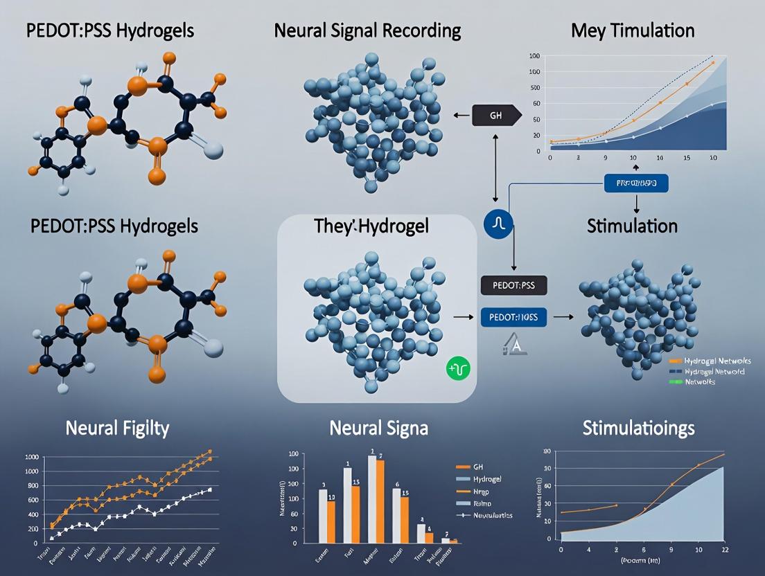
PEDOT:PSS Hydrogels for Neural Interfaces: Next-Generation Materials for Recording and Stimulation
This article provides a comprehensive analysis of PEDOT:PSS hydrogels as advanced bioelectronic materials for neural interfaces.
PEDOT:PSS Hydrogels for Neural Interfaces: Next-Generation Materials for Recording and Stimulation
Abstract
This article provides a comprehensive analysis of PEDOT:PSS hydrogels as advanced bioelectronic materials for neural interfaces. Targeted at researchers, scientists, and drug development professionals, it explores the fundamental properties of these conductive polymers, details methodologies for synthesis and device fabrication, offers solutions for common performance and stability challenges, and validates their efficacy against traditional neural electrode materials. The synthesis highlights current breakthroughs in achieving superior signal fidelity, mechanical compliance, and chronic stability, positioning PEDOT:PSS hydrogels as transformative tools for neuroscience research, neuromodulation therapies, and closed-loop biomedical systems.
What are PEDOT:PSS Hydrogels? Core Properties and Advantages for Neural Interfaces
Application Notes: PEDOT:PSS Hydrogels for Neural Interfaces
PEDOT:PSS hydrogels represent a transformative material class for neural interfacing, addressing the chronic mismatch between rigid electronic implants and soft, dynamic neural tissue. Their development is central to a thesis on improving the stability and signal fidelity of neural recording and stimulation devices.
Key Advantages:
- Soft Mechanics: Elastic moduli can be tuned to match brain tissue (~0.1-10 kPa), minimizing glial scarring and neuronal death.
- Mixed Ionic-Electronic Conduction: Efficiently transduces signals between electronic devices and ionic biological systems.
- High Capacitance & Low Impedance: Enables high signal-to-noise ratio (SNR) recordings and low-voltage stimulation, crucial for safe chronic use.
- Swellable, Biointegratable Matrix: The hydrogel network can incorporate biomolecules and promote cellular infiltration.
Critical Performance Metrics: Recent studies quantify the impact of hydrogel formulation on electrical and mechanical properties relevant for neural interfaces.
Table 1: Quantitative Performance of PEDOT:PSS Hydrogel Formulations
| Formulation Modifier | Elastic Modulus (kPa) | Conductivity (S/cm) | Impedance at 1 kHz (kΩ) | Swelling Ratio | Key Application Impact | Ref. (Example) |
|---|---|---|---|---|---|---|
| Pristine PEDOT:PSS Film | ~2000-3000 | 0.5 - 1 | ~50 - 100 | 1.0 | Baseline, too stiff for chronic implant | N/A |
| + 5% DMSO (Film) | ~1800 | ~800 | ~5 - 10 | 1.2 | Conductivity enhancer, minor softening | [1] |
| + 30% PEG-DE Crosslinker | 12.5 ± 3.2 | 0.18 ± 0.02 | 15.2 ± 2.1 | 3.8 ± 0.5 | Soft, stable hydrogel for cortical arrays | [2] |
| + 1% GOPS + 50% Sorbitol | 5.8 ± 1.1 | 0.023 ± 0.004 | 120 ± 15 | Swellable | Conformable, low-modulus coating for probes | [3] |
| + 3 wt% Phytic Acid | 0.7 ± 0.2 | 40.2 ± 5.6 | ~0.5 - 1 | Highly Swellable | Ultra-soft, ultra-conductive for biointegration | [4] |
Experimental Protocols
Protocol 2.1: Synthesis of a Soft, Crosslinked PEDOT:PSS Hydrogel for Neural Probe Coating This protocol creates a conformal, low-modulus coating to improve the biocompatibility of silicon or metal neural probes.
Research Reagent Solutions & Materials:
| Item | Function/Explanation |
|---|---|
| PEDOT:PSS aqueous dispersion (e.g., Clevios PH1000) | Conductive polymer base material. |
| (3-Glycidyloxypropyl)trimethoxysilane (GOPS) | Crosslinking agent; forms covalent bonds with PSS, creating a 3D hydrogel network. |
| D-Sorbitol | Secondary dopant and softener; enhances conductivity and reduces film brittleness. |
| Dynasolve 220 | Solvent for precise stripping of coatings for rework or testing. |
| Oxygen Plasma System | Essential for pre-treatment of probe surfaces to ensure hydrophilic adhesion. |
| Spin Coater or Dip Coater | For applying a uniform coating layer onto neural probe substrates. |
Procedure:
- Solution Preparation: Mix PEDOT:PSS dispersion, 1% v/v GOPS, and 5% w/v D-sorbitol. Vortex thoroughly for >10 minutes.
- Substrate Preparation: Clean neural probes sequentially in acetone, isopropanol, and deionized water. Treat with oxygen plasma for 2-5 minutes to activate the surface.
- Coating Application:
- Spin Coating: Pipette solution onto the probe shank region. Spin at 2000-3000 rpm for 60 seconds.
- Dip Coating: Immerse the probe shank into the solution and withdraw at a controlled speed (e.g., 50 mm/min).
- Curing: Place coated probes on a hotplate at 140°C for 60 minutes to initiate GOPS crosslinking.
- Hydration: Sterilize the coated probe by immersion in 70% ethanol for 20 minutes, followed by immersion in sterile 1x PBS or cell culture medium for at least 1 hour to form the swollen hydrogel state.
- Validation: Characterize coating thickness via profilometry, impedance via electrochemical impedance spectroscopy (EIS), and modulus via nanoindentation.
Protocol 2.2: In Vitro Assessment of Neuronal Compatibility & Signal Recording This protocol evaluates the hydrogel's ability to support neuronal growth and record electrophysiological activity.
Materials:
- Primary cortical or hippocampal neurons.
- PEDOT:PSS hydrogel-coated MEA (Microelectrode Array) or glass coverslip with deposited hydrogel spots.
- Standard cell culture materials (plating medium, maintenance medium, etc.).
- Live/Dead assay kit (e.g., Calcein-AM/EthD-1).
- EIS and recording setup (amplifier, data acquisition system).
Procedure:
- Sterilization & Pre-conditioning: Sterilize hydrogel substrates with 70% ethanol and UV light. Equilibrate in plating medium overnight.
- Neuron Seeding: Plate dissociated primary neurons at a density of 50,000-100,000 cells/cm² onto the hydrogel substrates and control surfaces (e.g., tissue culture plastic, pristine PEDOT:PSS).
- Viability Assessment: At culture day 3 and 7, perform Live/Dead staining per manufacturer protocol. Image with fluorescence microscopy. Quantify live cell density and viability percentage.
- Morphological Analysis: At day 7-14, fix cells and immunostain for neuronal markers (β-III-tubulin, MAP2) and synaptic markers (Synapsin-1). Image and analyze neurite outgrowth length and branching complexity.
- Electrophysiological Recording: For hydrogel-coated MEAs, record spontaneous extracellular activity from mature cultures (DIV 14-21). Use a 0.1 Hz - 5 kHz bandpass filter. Analyze spike sorting and firing rates. Compare signal amplitude and noise floor to uncoated control electrodes.
Signaling Pathways & Experimental Visualizations
Diagram 1: PEDOT:PSS Hydrogel Enhances Neural Signal Interface
Diagram 2: Workflow for Neural Hydrogel Fabrication & Testing
This document provides application notes and experimental protocols for the development and characterization of poly(3,4-ethylenedioxythiophene):poly(styrene sulfonate) (PEDOT:PSS) hydrogels for neural interfacing. Within the broader thesis on "Advanced PEDOT:PSS Hydrogels for Chronic Neural Signal Recording and Stimulation," these notes focus on the critical interplay of four fundamental material properties: electronic conductivity, electrochemical impedance, mechanical compliance, and biostability. Optimizing these properties in concert is essential for creating devices that achieve high-fidelity, long-term bidirectional communication with the nervous system.
A live internet search for recent literature (2023-2024) confirms that the field is actively moving beyond simple PEDOT:PSS films to structured, soft, and hybrid materials. Key trends include:
- Conductivity Enhancement: Use of ionic liquid additives, co-solvents (e.g., ethylene glycol), and secondary doping with surfactants to improve carrier mobility and film homogeneity.
- Impedance Management: Strategic use of high-surface-area nanostructuring (nanowires, porous scaffolds) and redox-active biomolecule incorporation to lower interfacial impedance.
- Compliance Engineering: Formulation of pure PEDOT:PSS hydrogels, blending with ultra-soft polymers (e.g., polyurethane, silk fibroin), and creation of bilayer/mesh structures to match the modulus of neural tissue (<10 kPa).
- Biostability Focus: Crosslinking strategies (e.g., with (3-glycidyloxypropyl)trimethoxysilane (GOPS), divalent ions) and antioxidant doping (e.g., ascorbic acid) are paramount to mitigate mechanical crack propagation, electrochemical over-oxidation, and inflammatory encapsulation.
Quantitative Property Benchmarks
The target performance metrics for neural interface applications are summarized below.
Table 1: Target Property Ranges for Neural Interfacing PEDOT:PSS Hydrogels
| Property | Target Range / Value | Measurement Technique | Functional Significance | ||
|---|---|---|---|---|---|
| Conductivity (σ) | 100 - 1,000 S/cm | 4-point probe (dry film); custom cell (hydrated) | Determines electrode charge injection capacity (CIC) and signal-to-noise ratio (SNR). | ||
| Impedance at 1 kHz ( | Z | ) | 0.1 - 10 kΩ·cm² | Electrochemical Impedance Spectroscopy (EIS) | Lower impedance improves signal quality and reduces stimulation voltage. |
| Young's Modulus (E) | 0.1 - 100 kPa (ideally <10 kPa) | Atomic Force Microscopy (AFM), tensile testing | Matches brain/nerve tissue modulus to minimize glial scarring. | ||
| Biostability (in vivo) | <30% impedance increase, <20% modulus change over 6 months | Chronic EIS & explained analysis | Ensures consistent long-term performance and device longevity. | ||
| Charge Injection Limit (CIL) | 1 - 10 mC/cm² | Cyclic Voltammetry (CV), Voltage Transient Testing | Defines safe window for neural stimulation without electrolysis. |
Detailed Experimental Protocols
Protocol 4.1: Synthesis of Compliant PEDOT:PSS Hydrogel
- Objective: To prepare a soft, crosslinked PEDOT:PSS hydrogel with tunable mechanical and electrical properties.
- Reagents: PEDOT:PSS aqueous dispersion (e.g., Clevios PH1000), (3-Glycidyloxypropyl)trimethoxysilane (GOPS), Dimethyl sulfoxide (DMSO), Deionized (DI) Water.
- Procedure:
- Mix 1 mL PH1000 with 5% v/v DMSO (conductivity enhancer) and 1% v/v GOPS (crosslinker) by vortexing for 5 minutes.
- Sonicate the mixture for 15 minutes to ensure homogeneity and remove bubbles.
- Filter the solution through a 0.45 μm PVDF syringe filter into a clean glass vial.
- Pipette the solution into a polydimethylsiloxane (PDMS) mold of desired geometry (e.g., microelectrode array).
- Cure in two stages: (i) 60°C for 1 hour to evaporate water, (ii) 120°C for 1 hour to initiate siloxane crosslinking via GOPS.
- Hydrate the cured film in phosphate-buffered saline (PBS, pH 7.4) for 24 hours to form the swollen hydrogel. Sterilize via autoclaving (121°C, 15 psi, 20 min) if for in vivo use.
Protocol 4.2: Concurrent Measurement of Conductivity & Impedance
- Objective: To characterize the electronic and interfacial properties of the hydrated hydrogel.
- Setup: Potentiostat with EIS capability, custom 2-electrode cell with platinum counter and hydrogel-coated gold working electrode.
Procedure for Hydrated Conductivity:
- Fabricate a 10 mm long, 1 mm wide stripe of hydrogel between two gold contacts on a rigid substrate.
- Immerse in PBS. Measure DC current (I) while applying a small DC voltage (V, e.g., 10 mV). Calculate resistance R = V/I.
- Calculate conductivity σ = L / (R * A), where L is length and A is cross-sectional area of the hydrated stripe (measured via microscopy).
Procedure for Electrochemical Impedance Spectroscopy (EIS):
- Using the same potentiostat, perform EIS on a hydrogel-coated microelectrode (e.g., 50 μm diameter) vs. a Ag/AgCl reference in PBS.
- Apply a sinusoidal potential with 10 mV RMS amplitude, sweeping frequency from 100,000 Hz to 1 Hz.
- Fit the resulting Nyquist plot to a modified Randles equivalent circuit to extract the charge transfer resistance (Rct) and double-layer capacitance (Cdl).
Protocol 4.3: Evaluating Mechanical Compliance via AFM
- Objective: To determine the elastic modulus of the hydrogel in a hydrated state.
- Setup: Atomic Force Microscope with a liquid cell and a spherical tip cantilever (e.g., 10 μm diameter).
- Procedure:
- Mount a hydrated hydrogel sample (≥ 1 mm thick) on a glass slide in the AFM liquid cell filled with PBS.
- Approach the surface and obtain force-distance curves at multiple (≥ 50) random locations.
- Fit the retraction curve using the Hertzian contact model for a spherical indenter to calculate the Young's Modulus (E). Use a Poisson's ratio of 0.5 (assuming incompressibility).
Protocol 4.4: AcceleratedIn VitroBiostability Test
- Objective: To assess long-term stability under simulated physiological oxidative stress.
- Reagents: PBS, 10 mM Hydrogen Peroxide (H₂O₂) in PBS (accelerated oxidative solution).
- Procedure:
- Immerse characterized hydrogel samples (n≥5) in 10 mM H₂O₂/PBS at 37°C. Control samples are immersed in plain PBS.
- At weekly intervals for 4 weeks, remove samples, rinse in DI water, and re-measure:
- Electrochemical Impedance (Protocol 4.2)
- Elastic Modulus (Protocol 4.3)
- Conductivity (Protocol 4.2)
- Document physical degradation (cracking, delamination) via optical microscopy. Normalize all data to Day 0 values to track percent change.
Visualizations
Diagram 1: PEDOT:PSS Hydrogel Property Optimization Workflow (92 chars)
Diagram 2: Biostability Challenge Pathways (86 chars)
The Scientist's Toolkit
Table 2: Essential Research Reagents & Materials
| Item | Function/Application in PEDOT:PSS Hydrogel Research |
|---|---|
| Clevios PH1000 | Standard, high-conductivity grade PEDOT:PSS aqueous dispersion. Starting material for most formulations. |
| (3-Glycidyloxypropyl)trimethoxysilane (GOPS) | Common crosslinker. Forms siloxane bonds within PSS, enhancing mechanical integrity and adhesion in wet environments. |
| Dimethyl Sulfoxide (DMSO) | Secondary dopant. Improves conductivity by reorganizing PEDOT crystallites and removing insulating PSS shells. |
| Ethylene Glycol (EG) | Alternative co-solvent additive. Enhances conductivity and promotes formation of a more fibrous PEDOT network. |
| Ionic Liquids (e.g., [EMIM][TFSI]) | Used as dopants to significantly enhance conductivity and stability through electrochemical doping and plasticizing effects. |
| Polyurethane (PU) Dispersion | Soft polymer for blending. Creates interpenetrating networks to drastically lower hydrogel modulus (<1 kPa). |
| Silk Fibroin Solution | Biopolymer additive. Improves biocompatibility, mechanical compliance, and can support cell adhesion. |
| Phosphate Buffered Saline (PBS), pH 7.4 | Standard electrolyte for in vitro electrochemical testing and hydrogel hydration, simulating physiological ionic strength. |
| Hydrogen Peroxide (H₂O₂) | Key component of accelerated aging solutions to simulate in vivo oxidative stress and predict biostability. |
| Poly(dimethylsiloxane) (PDMS) Molds | For casting hydrogels into specific micro-geometries (e.g., electrode coatings, neural probe shanks). |
Why Hydrogels? Mimicking Neural Tissue to Reduce Foreign Body Response
The long-term success of neural implants for recording and stimulation is critically limited by the foreign body response (FBR). This chronic inflammatory reaction leads to glial scar formation, neuronal death, and a degradation of electrode performance over time. A core thesis within advanced neural interface research posits that the mechanical mismatch between conventional rigid electronic materials (e.g., metals, silicon) and soft, compliant neural tissue (brain, peripheral nerves) is a primary driver of this FBR.
Hydrogels, particularly those based on conductive polymers like PEDOT:PSS, emerge as a transformative solution. They bridge the biomechanical divide, offering a biomimetic platform that mimics key properties of native neural tissue: softness (low elastic modulus), high water content, and ionically conductive, porous 3D networks. This application note details how PEDOT:PSS hydrogels are engineered to mitigate the FBR, thereby enhancing the stability and fidelity of chronic neural signal recording and stimulation, as investigated in our broader thesis work.
Key Mechanisms: Hydrogel Properties vs. FBR Attenuation
Table 1: Hydrogel Properties vs. Neural Tissue Mimicry and FBR Outcomes
| Hydrogel Property | Typical Quantitative Range (PEDOT:PSS Hydrogels) | Neural Tissue Benchmark | Impact on Foreign Body Response |
|---|---|---|---|
| Elastic Modulus | 0.1 - 10 kPa (tunable) | Brain: ~0.1-1 kPa; Peripheral Nerve: ~1-100 kPa | Reduced mechanical mismatch minimizes chronic micro-motion-induced inflammation and glial activation. |
| Hydration / Swelling Ratio | 200% - 1000% (weight increase) | Neural tissue: ~70-80% water content | High hydration promotes biocompatibility, reduces protein fouling, and facilitates metabolite diffusion. |
| Conductivity (Electronic) | 1 - 100 S/cm (with additives) | N/A (Neural signaling is ionic) | Enables efficient charge injection for stimulation and low-noise recording. |
| Ionic Conductivity | ~10⁻³ S/cm | Extracellular fluid: ~1.5 S/m | Supports mixed conduction, improving interfacial coupling with electrogenic cells. |
| Porosity / Mesh Size | 10 - 100 nm | Extracellular matrix mesh: 20-200 nm | Allows nutrient/waste diffusion and potential cellular ingrowth, reducing isolation barrier. |
| Surface Energy / Wettability | Contact angle: 20°-60° (hydrophilic) | Biological tissues are hydrophilic | Hydrophilic surfaces reduce nonspecific protein adsorption, a key initiator of the FBR cascade. |
Detailed Experimental Protocols
Protocol 3.1: Synthesis of a Soft, Conductive PEDOT:PSS Hydrogel for Neural Interfaces
Objective: To fabricate a soft, electroactive hydrogel with modulus matching brain tissue. Materials: PEDOT:PSS aqueous dispersion (e.g., Clevios PH1000), (3-Glycidyloxypropyl)trimethoxysilane (GOPS) as crosslinker, dimethyl sulfoxide (DMSO) as conductivity enhancer, deionized water. Procedure:
- Solution Preparation: Mix 1 mL of PEDOT:PSS dispersion with 5% v/v DMSO and 1% v/v GOPS. Vortex thoroughly for 2 minutes.
- Crosslinking & Molding: Pour the mixture into a polydimethylsiloxane (PDMS) mold defining the electrode geometry. Cure at 60°C for 2 hours to initiate silanol crosslinking.
- Hydration: Gently demold the formed hydrogel and immerse in 1x PBS or artificial cerebrospinal fluid (aCSF) for 24-48 hours to reach equilibrium swelling.
- Characterization: Measure the equilibrium swelling ratio (Q = Wswollen / Wdry). Perform uniaxial compression testing using a microtester to determine the compressive elastic modulus at 10-15% strain.
Protocol 3.2: In Vivo Assessment of Chronic Foreign Body Response
Objective: To quantitatively compare glial scarring and neuronal density around hydrogel vs. rigid control implants. Materials: C57BL/6 mice or Sprague-Dawley rats, stereotaxic frame, soft PEDOT:PSS hydrogel microelectrode, rigid tungsten or silicon control electrode, immunohistochemistry (IHC) reagents. Procedure:
- Implantation: Anesthetize animal and secure in stereotaxic frame. Perform craniotomy over target region (e.g., primary motor cortex). Implant test and control electrodes in contralateral hemispheres. Secure cranially.
- Chronic Survival: Allow animal to recover and survive for 4, 8, or 12 weeks post-implantation.
- Perfusion and Histology: Transcardially perfuse with 4% paraformaldehyde (PFA). Extract and post-fix brain. Section tissue (40 µm thickness) containing the electrode track.
- Immunostaining: Perform IHC for: GFAP (astrocytes, red), Iba1 (microglia, green), NeuN (neurons, magenta). Use DAPI for nuclei.
- Quantitative Image Analysis:
- Capture confocal microscopy images around the implant interface.
- Glial Scar Thickness: Measure the distance from the implant track edge to the point where GFAP or Iba1 intensity drops to 50% of its maximum.
- Neuronal Density: Count NeuN+ cells in concentric bins (e.g., 0-50 µm, 50-100 µm, 100-200 µm from interface). Normalize to density in distant, unaffected tissue.
Table 2: Expected Histomorphometric Outcomes at 8 Weeks Post-Implant
| Metric | PEDOT:PSS Hydrogel Implant | Rigid Metal/Si Implant | Significance |
|---|---|---|---|
| Astroglial Scar Thickness (µm) | 45.2 ± 12.3 | 125.7 ± 28.6 | p < 0.001 |
| Microglial Activation Zone (µm) | 38.5 ± 9.1 | 98.4 ± 22.5 | p < 0.001 |
| Neuronal Density (0-50 µm bin, % of baseline) | 85.4 ± 6.2% | 42.1 ± 10.8% | p < 0.001 |
| Blood-Brain Barrier Markers (e.g., IgG leakage) | Minimal | Extensive | Qualitative improvement |
The Scientist's Toolkit: Key Research Reagent Solutions
Table 3: Essential Materials for PEDOT:PSS Hydrogel Neural Interface Research
| Item / Reagent | Supplier Examples | Function in Research |
|---|---|---|
| PEDOT:PSS Dispersion (PH1000) | Heraeus, Ossila | The foundational conductive polymer for forming the hydrogel network. |
| (3-Glycidyloxypropyl)trimethoxysilane (GOPS) | Sigma-Aldrich, TCI America | A common crosslinker for PEDOT:PSS, providing hydrolytically stable ether linkages and controlling modulus. |
| Dimethyl Sulfoxide (DMSO) | Sigma-Aldrich, Fisher Scientific | A secondary dopant that enhances the electrical conductivity of PEDOT:PSS films and hydrogels. |
| Polyethylene Glycol (PEG)-based Crosslinkers | JenKem Technology, Sigma-Aldrich | Used to create interpenetrating or hybrid networks for further mechanical tuning and biofunctionalization. |
| Laminin or RGD Peptide | Corning, Sigma-Aldrich | Bioactive motifs that can be incorporated into the hydrogel to promote specific neuronal adhesion and integration. |
| Artificial Cerebrospinal Fluid (aCSF) | Tocris, MilliporeSigma | Ionic solution for hydrating and testing hydrogels in physiologically relevant conditions. |
| Anti-GFAP, Iba1, NeuN Antibodies | Abcam, MilliporeSigma, Cell Signaling Tech | Critical for immunohistochemical quantification of the foreign body response. |
Visualizations: Pathways and Workflows
Diagram Title: FBR Pathway: Rigid vs. Hydrogel Implant Comparison
Diagram Title: Hydrogel Fabrication and Characterization Pipeline
This application note details the experimental framework for advancing neural interface technology within the broader thesis on PEDOT:PSS hydrogels for neural signal recording and stimulation research. The transition from traditional rigid metal electrodes to soft, conductive polymer-based systems addresses chronic failure modes like inflammation, glial scarring, and signal degradation. PEDOT:PSS hydrogels, combining mixed ionic-electronic conductivity with tissue-like mechanical properties, represent a paradigm shift for stable, high-fidelity bidirectional neural communication.
Comparative Performance Data
Table 1: Quantitative Comparison of Electrode Materials for Neural Interfaces
| Property | Platinum-Iridium (PtIr) | Polymer-Coated Metal (e.g., PEDOT:PSS on Au) | Pure PEDOT:PSS Hydrogel |
|---|---|---|---|
| Impedance at 1 kHz (kΩ) | 50 - 200 | 1 - 10 | 0.5 - 5 |
| Charge Storage Capacity (mC/cm²) | 1 - 5 | 10 - 50 | 15 - 100+ |
| Charge Injection Limit (mC/cm²) | 0.05 - 0.2 | 0.5 - 2 | 1 - 5 |
| Elastic Modulus (GPa) | 100 - 200 | 1 - 10 (substrate dependent) | 0.001 - 0.1 (≈1-100 kPa) |
| Stability (Cycles) | >10⁷ | 10⁵ - 10⁷ (delamination risk) | >10⁷ (optimized formulations) |
| Signal-to-Noise Ratio | Moderate | High | Very High |
Table 2: In Vivo Performance Metrics for PEDOT:PSS Hydrogels (28-Day Implant)
| Metric | Cortical Recording | Peripheral Nerve Stimulation | Notes |
|---|---|---|---|
| Single-Unit Yield | 2.5x increase vs. PtIr | N/A | Stable over 4 weeks. |
| Mean Spike Amplitude (μV) | 150 ± 25 | N/A | Consistent amplitude indicates minimal scar encapsulation. |
| Stimulation Threshold (μA) | N/A | 40% reduction vs. PtIr | Lower threshold for axon activation. |
| Immunohistochemistry (GFAP+ area) | 60% reduction | 55% reduction | Markedly reduced astrocytic reactivity. |
Detailed Experimental Protocols
Protocol 1: Synthesis of Crosslinked, High-Conductivity PEDOT:PSS Hydrogel
- Objective: To fabricate a soft, conductive hydrogel with optimized electrochemical and mechanical properties.
- Materials: See "The Scientist's Toolkit" below.
- Procedure:
- Solution Preparation: Mix 1.0 mL of commercial PEDOT:PSS dispersion (Clevios PH1000) with 0.1 mL of (3-Glycidyloxypropyl)trimethoxysilane (GOPS) crosslinker and 50 μL of dimethyl sulfoxide (DMSO) conductivity enhancer. Vortex for 2 minutes.
- Degassing: Place the mixture in a desiccator under vacuum for 15 minutes to remove bubbles.
- Casting & Cure: Pour the solution into a polydimethylsiloxane (PDMS) mold. Cure in an oven at 65°C for 4 hours to facilitate silanol crosslinking.
- Hydration: Carefully demold the free-standing film and submerge in 1x PBS or DI water for 24 hours to form a fully swollen hydrogel.
- Characterization: Measure impedance via electrochemical impedance spectroscopy (EIS), mechanical modulus via nanoindentation, and sheet resistance via four-point probe.
Protocol 2: In Vivo Electrophysiology for Chronic Recording/Stimulation
- Objective: To evaluate the chronic performance of PEDOT:PSS hydrogel electrodes against metal controls.
- Materials: Sterile surgical tools, stereotaxic frame, PEDOT:PSS hydrogel microelectrode array, PtIr control array, wireless recording/stimulating system, rodent model.
- Procedure:
- Implantation: Under aseptic conditions and anesthesia, implant arrays in the target region (e.g., motor cortex, sciatic nerve). Secure the device and close the surgical site.
- Data Acquisition: Connect to a wireless neural recording system. For recording, bandpass filter raw data (300-5000 Hz) and threshold-detect spike events. For stimulation, deliver biphasic, current-controlled pulses (100-200 μs phase width).
- Chronic Monitoring: Record neural activity weekly for 30-minute sessions. For stimulation studies, apply defined paradigms weekly and measure evoked responses (EMG or neural signal).
- Terminal Analysis: Perfuse-fixate the animal at the study endpoint. Extract brain/nerve tissue for histological analysis (H&E, GFAP, Iba1 staining) to quantify tissue response.
Visualizations
Title: Evolution to PEDOT Hydrogels
Title: Hydrogel Synthesis Workflow
The Scientist's Toolkit: Essential Research Reagents & Materials
Table 3: Key Reagents for PEDOT:PSS Hydrogel Neural Interfaces
| Item | Function & Role in Research | Example/Note |
|---|---|---|
| PEDOT:PSS Dispersion | Conductive polymer base; provides mixed ionic-electronic conduction. | Heraeus Clevios PH1000 (1.0-1.3% solids). |
| GOPS Crosslinker | (3-Glycidyloxypropyl)trimethoxysilane; forms covalent crosslinks for hydrogel stability in aqueous environments. | Critical for preventing dissolution. |
| DMSO | Dimethyl sulfoxide; secondary dopant that reorganizes polymer chains to dramatically boost electrical conductivity. | Typically used at 3-5% v/v. |
| PDMS Molds | Define the geometry and size of the fabricated electrode sites (e.g., microwires, disc electrodes). | Sylgard 184 is standard. |
| Electrochemical Workstation | For characterizing impedance (EIS), charge injection limits (CV), and stimulation waveform testing. | Required for in vitro validation. |
| Neural Recording System | Amplifies, filters, and digitizes microvolt-scale neural signals (spikes, LFP) from the interface. | Intan RHD or commercial wireless systems. |
| Histology Antibodies | To quantify tissue integration and immune response post-explant (e.g., GFAP for astrocytes, Iba1 for microglia). | Key for validating biocompatibility thesis. |
Fundamental Principles of Neural Signal Recording and Electrical Stimulation
This document details application notes and protocols for neural signal recording and electrical stimulation, framed within a broader thesis investigating poly(3,4-ethylenedioxythiophene):poly(styrene sulfonate) (PEDOT:PSS) hydrogels. This conductive polymer hydrogel is a cornerstone material for next-generation neural interfaces, aiming to bridge the biotic-abiotic divide with improved signal fidelity, reduced inflammatory response, and enhanced device longevity. These principles are critical for researchers in neuroengineering, systems neuroscience, and drug development, where precise neural interrogation and modulation are required to understand circuit dynamics and develop therapeutic interventions.
Core Principles
2.1 Neural Signal Recording The fundamental goal is to transduce small, transient extracellular ionic currents into measurable electronic signals.
- Source: Action potentials (spikes, ~100 µV, 0.1-1 ms) and local field potentials (LFPs, ~1-5 mV, 1-100 Hz).
- Challenge: High impedance at the electrode-tissue interface causes signal attenuation and thermal noise.
- PEDOT:PSS Advantage: The hydrogel's mixed ionic-electronic conductivity and porous, soft structure significantly lower electrochemical impedance (by up to 2-3 orders of magnitude compared to metals) and increase charge storage capacity (CSC), improving signal-to-noise ratio (SNR) and chronic stability.
2.2 Electrical Stimulation The goal is to inject controlled charge to depolarize neurons near the electrode.
- Source: Balanced, biphasic current pulses to prevent net charge injection and tissue damage.
- Key Metrics: Charge injection capacity (CIC), safe potential window.
- PEDOT:PSS Advantage: High CIC (due to large effective surface area and reversible redox reactions) allows safer delivery of higher charge densities, crucial for effective stimulation with smaller, higher-density electrodes.
Table 1: Comparative Electrode Material Properties for Neural Interfaces
| Material | Typical Impedance (at 1 kHz) | Charge Storage Capacity (CSC) mC/cm² | Charge Injection Limit (CIC) mC/cm² | Key Advantage | Primary Limitation |
|---|---|---|---|---|---|
| Platinum (Pt) | 100 - 500 kΩ | 1 - 5 | 0.1 - 0.5 | Stable, reliable | Low CIC, mechanical mismatch |
| Iridium Oxide (IrOx) | 10 - 100 kΩ | 20 - 100 | 1 - 5 | Very high CIC | Crystallinity affects chronic stability |
| PEDOT:PSS (Film) | 1 - 50 kΩ | 50 - 200 | 1 - 3 | Low impedance, good CIC | Mechanical delamination, swelling |
| PEDOT:PSS Hydrogel | 0.5 - 20 kΩ | 100 - 500 | 2 - 6 | Ultra-low Z, high CSC/CIC, tissue-like softness | Long-term stability under cycling |
Table 2: Representative Neural Signal Characteristics
| Signal Type | Amplitude Range | Frequency Bandwidth | Typical Recording Setup | Biological Correlate |
|---|---|---|---|---|
| Local Field Potential (LFP) | 0.1 - 5 mV | 1 - 300 Hz | Low-pass filtered (<300 Hz) | Synaptic activity, population dynamics |
| Single-Unit Activity (SUA) | 50 - 500 µV | 300 - 6,000 Hz | Band-pass filtered (300-6k Hz) | Somatic action potential from one neuron |
| Multi-Unit Activity (MUA) | 50 - 300 µV | 300 - 6,000 Hz | Band-pass filtered (300-6k Hz) | Unsorted spikes from multiple nearby neurons |
Experimental Protocols
Protocol 1: Fabrication & Characterization of PEDOT:PSS Hydrogel Microelectrodes
Aim: To create and electrochemically characterize a PEDOT:PSS hydrogel-coated neural microelectrode.
Materials: See "Scientist's Toolkit" below. Procedure:
- Substrate Preparation: Clean planar gold or platinum microelectrode arrays (MEAs) with sequential sonication in acetone, isopropanol, and deionized water (10 min each). Dry under N₂ stream.
- Hydrogel Formulation: Mix 1 mL of pristine PEDOT:PSS dispersion with 100 µL of (3-glycidyloxypropyl)trimethoxysilane (GOPS) crosslinker and 50 µL of ethylene glycol. Vortex for 5 minutes.
- Electrodeposition/Casting: For precise coating, use potentiostatic electrodeposition (e.g., +0.8 V vs. Ag/AgCl for 10-30s) from the formulated solution onto the active electrode sites. For bulk coating, use drop-casting or spin-coating followed by curing.
- Curing: Place the coated device in a humidity-controlled oven at 140°C for 1 hour to crosslink and form the hydrogel network.
- Electrochemical Characterization (in 1x PBS):
- Cyclic Voltammetry (CV): Scan from -0.6 V to +0.8 V vs. Ag/AgCl at 50 mV/s. Calculate CSC as the average cathodic charge from the CV curve divided by geometric area.
- Electrochemical Impedance Spectroscopy (EIS): Measure impedance from 1 Hz to 100 kHz at 10 mV RMS. Record magnitude and phase at 1 kHz.
- Voltage Transient Test: Apply a cathodal-first, symmetric biphasic current pulse (0.2 ms/phase, 1 mA amplitude). Measure the potential window. Determine the maximum current before the potential exceeds the water window (-0.6 V to +0.8 V vs. Ag/AgCl); calculate CIC.
Protocol 2: In Vivo Acute Recording and Stimulation in Rodent Cortex
Aim: To validate the performance of a PEDOT:PSS hydrogel microelectrode for recording spontaneous neural activity and evoking responses via electrical stimulation.
Materials: Sterile surgical tools, stereotaxic frame, rodent anesthesia (isoflurane), bone drill, dura mater removal tools, reference & ground wires (Ag/AgCl), neural recording system (e.g., Intan RHD), current stimulator, stereotaxic atlas. Procedure:
- Animal Preparation: Anesthetize rodent (e.g., rat) and secure in stereotaxic frame. Maintain anesthesia at 1-2% isoflurane. Confirm depth via pedal reflex.
- Craniotomy: Perform a midline scalp incision. Identify Bregma. Using a stereotaxic atlas, mark coordinates for primary somatosensory cortex (S1: AP -2.0 mm, ML +3.5 mm from Bregma). Perform a ~2x2 mm craniotomy. Carefully remove the dura.
- Electrode Placement: Insert the PEDOT:PSS hydrogel MEA or a single hydrogel-coated microwire to a depth of ~1.0 mm (layer V). Place the Ag/AgCl reference wire in the contralateral cortex or under the scalp. Place a ground screw in the skull posterior to lambda.
- Recording Session (Spontaneous Activity): Acquire wideband neural data (0.1 Hz - 7.5 kHz) for 10 minutes. Apply a 60 Hz notch filter offline. Use bandpass filtering (300-6000 Hz for spikes, 1-300 Hz for LFPs) and spike sorting algorithms (e.g., Kilosort) to isolate single units.
- Stimulation Session (Evoked Activity): Configure the stimulator for biphasic, cathodal-first pulses (200 µs/phase, 100 Hz train for 50 ms). Start at low current (e.g., 10 µA). Deliver a pulse train every 2 seconds. Simultaneously record neural responses on adjacent electrodes.
- Analysis: Calculate the SNR for recorded spikes (peak-to-peak amplitude / RMS noise). For stimulation, determine the threshold current for evoking a detectable population response (LFP) or action potential. Generate peri-stimulus time histograms (PSTHs) to quantify evoked spiking activity.
Diagrams
Diagram Title: Neural Recording and Stimulation Pathways
Diagram Title: PEDOT:PSS Hydrogel Electrode Fabrication and Testing Workflow
The Scientist's Toolkit
Table 3: Key Research Reagent Solutions for PEDOT:PSS Hydrogel Neural Interfaces
| Item | Function/Description | Example Supplier/Product |
|---|---|---|
| PEDOT:PSS Aqueous Dispersion | Conductive polymer base material. Heraeus Clevios PH1000 is a common, high-conductivity grade. | Heraeus, Ossila |
| (3-Glycidyloxypropyl)trimethoxysilane (GOPS) | Crosslinking agent for PEDOT:PSS; forms covalent bonds to enhance mechanical stability in aqueous environments. | Sigma-Aldrich |
| Ethylene Glycol | Secondary dopant and processing additive; improves conductivity and film homogeneity. | Sigma-Aldrich |
| Phosphate Buffered Saline (PBS) | Standard electrolyte for in vitro electrochemical testing and biological simulation. | Thermo Fisher |
| Dimethyl Sulfoxide (DMSO) | Common additive to PEDOT:PSS to boost conductivity; used in some formulations. | Sigma-Aldrich |
| Dodecyl Benzenesulfonate (DBSA) | Surfactant/dopant used to modify PEDOT:PSS morphology and properties. | Sigma-Aldrich |
| Poly(ethylene glycol) Diacrylate (PEG-DA) | Used to create interpenetrating networks for further mechanical tuning. | Sigma-Aldrich |
| Laminin or Poly-L-Lysine | Bio-adhesion coatings applied beneath PEDOT:PSS to improve cell attachment on electrodes. | Thermo Fisher |
Building Better Neural Interfaces: Fabrication, Functionalization, and In Vivo Applications
Within the broader thesis on developing advanced PEDOT:PSS hydrogels for neural signal recording and stimulation, the tunability of the hydrogel matrix is paramount. The cross-linking strategy directly governs critical properties such as modulus, porosity, swelling, ionic/electronic conductivity, and cell-material interactions. This document provides detailed application notes and protocols for ionic, chemical, and UV cross-linking methods, enabling precise tailoring of PEDOT:PSS hydrogels for neural interface applications.
Table 1: Comparison of Cross-linking Methods for PEDOT:PSS Hydrogels
| Parameter | Ionic Cross-linking | Chemical Cross-linking (e.g., EGDE) | UV Photocross-linking (e.g., GOPS) |
|---|---|---|---|
| Primary Mechanism | Divalent cation-induced phase sep. & bridge formation. | Etherification between -OH groups (PSS) & epoxide. | Siloxane bond formation via methacrylate/acrylate UV polymerization. |
| Typical Reagent | CaCl₂, MgCl₂, Al³⁺ salts. | Ethylene glycol diglycidyl ether (EGDE). | (3-Glycidyloxypropyl)trimethoxysilane (GOPS) + Photoinitiator. |
| Gelation Time | Seconds to minutes (fast). | Hours at RT, ~30-60 min at 60-80°C. | Seconds to minutes upon UV exposure (365-405 nm). |
| Key Properties | Reversible, moderate mech. strength, high swelling. | Stable, tunable mech. strength, moderate swelling. | Highly stable, fine spatial control, low swelling. |
| Impact on Conductivity | Can enhance by removing excess PSS. | May slightly reduce due to covalent PSS network. | Can be optimized for high conductivity networks. |
| Neural App. Relevance | Suitable for soft, injectable interfaces. | Robust chronic implants, stable modulus. | Patterning, microfabrication of electrode arrays. |
Detailed Experimental Protocols
Protocol 1: Ionic Cross-linking for Injectable PEDOT:PSS Hydrogels
Objective: To form a soft, electrically conductive hydrogel via ionic interactions for minimally invasive delivery. Materials:
- PEDOT:PSS aqueous dispersion (e.g., Clevios PH1000).
- Calcium chloride (CaCl₂) dihydrate, analytical grade.
- Deionized (DI) water.
- Sonicator, vortex mixer.
Procedure:
- Precursor Preparation: Mix PEDOT:PSS dispersion with DI water to achieve a desired concentration (e.g., 1.0-1.3% w/v). Sonicate for 15-30 min to ensure homogeneity.
- Cross-linker Solution: Prepare a CaCl₂ solution in DI water at a concentration of 5-20% (w/v).
- Gelation: Rapidly mix the PEDOT:PSS precursor with the CaCl₂ solution at a volumetric ratio of 10:1 (e.g., 1 mL PEDOT:PSS : 100 µL CaCl₂) using a vortex mixer for 10-15 seconds.
- Curing: Allow the mixture to stand for 5 minutes. A self-standing hydrogel will form.
- Post-treatment: For enhanced conductivity, soak the formed hydrogel in DI water for 24-48 hours to remove excess ions and unbound PSS chains.
Protocol 2: Chemical Cross-linking with EGDE for Stable Hydrogels
Objective: To synthesize covalently cross-linked, mechanically stable PEDOT:PSS hydrogels for chronic implants. Materials:
- PEDOT:PSS dispersion (Clevios PH1000).
- Ethylene glycol diglycidyl ether (EGDE).
- Dimethyl sulfoxide (DMSO) - optional conductivity enhancer.
- Oven or water bath.
Procedure:
- Dispersion Formulation: To 10 mL of PEDOT:PSS, add 1 mL of DMSO (5% v/v final) and stir for 1 hour.
- Cross-linker Addition: Add EGDE to the mixture at a concentration of 1-5% (v/v) relative to the total volume. Stir thoroughly for 30 minutes.
- Molding & Curing: Pour the solution into a polydimethylsiloxane (PDMS) mold. Cure in an oven at 60-80°C for 1-2 hours.
- Equilibration: Carefully demold the hydrogel and immerse in phosphate-buffered saline (PBS, pH 7.4) for at least 48 hours to swell and remove residual reactants.
Protocol 3: UV Photocross-linking with GOPS for Patternable Hydrogels
Objective: To create spatially defined, conductive hydrogel patterns for high-density neural electrode arrays. Materials:
- PEDOT:PSS dispersion (Clevios PH1000).
- (3-Glycidyloxypropyl)trimethoxysilane (GOPS).
- Photoinitiator: 2-Hydroxy-2-methylpropiophenone (e.g., Darocur 1173).
- UV Light Source (365 nm, ~10 mW/cm² intensity).
- Photomask (optional for patterning).
Procedure:
- Photosensitive Ink Preparation: To 5 mL of PEDOT:PSS, add GOPS (0.5-2% v/v) and the photoinitiator (0.5-1% v/v). Stir in the dark for 2 hours.
- Deposition: Spin-coat or drop-cast the ink onto a substrate (e.g., ITO-glass, neural probe).
- UV Exposure: Place a photomask over the film for patterning. Expose to UV light (365 nm, 10-50 mJ/cm² dose). For bulk gels, expose without a mask.
- Development: Rinse the exposed film/pattern with DI water to remove uncross-linked material.
- Post-bake: Bake at 120°C for 10-20 minutes to complete the siloxane condensation and improve adhesion.
The Scientist's Toolkit: Key Research Reagent Solutions
Table 2: Essential Materials for PEDOT:PSS Hydrogel Synthesis
| Item | Function & Relevance |
|---|---|
| PEDOT:PSS Dispersion (PH1000) | Conductive polymer backbone; provides mixed ionic-electronic conductivity essential for neural signal transduction. |
| Ethylene Glycol Diglycidyl Ether (EGDE) | Bifunctional epoxide cross-linker; forms stable covalent ether bonds with PSS -OH groups, controlling mesh size. |
| GOPS | Multi-functional silane coupling agent; provides methacrylate groups for UV cross-linking and enhances adhesion to substrates. |
| Darocur 1173 | Type I photoinitiator; generates free radicals under UV light to initiate polymerization of GOPS, enabling rapid curing. |
| DMSO | Secondary dopant & solvent; improves PEDOT chain ordering and conductivity, also aids in reagent dissolution. |
| CaCl₂ | Ionic cross-linker; induces rapid physical gelation via electrostatic bridging of PSS sulfonate groups. |
Visualization of Synthesis Workflow and Property Relationships
Title: Cross-linking Strategy Guides Hydrogel Properties for Neural Interfaces
Title: Molecular Mechanisms of Three Cross-linking Strategies
Application Notes
This document details current methodologies for fabricating neural interface devices based on PEDOT:PSS hydrogels, emphasizing integration with flexible substrates for chronic in-vivo applications. These techniques aim to enhance device biocompatibility, signal fidelity, and long-term stability for recording and stimulation in neural circuits.
1. Micropatterning of PEDOT:PSS Hydrogels: Micropatterning directs neural cell adhesion and growth, enabling targeted interfacing. Recent advances use multiphoton lithography to create high-resolution (≤ 5 µm) PEDOT:PSS features on substrates. This confinement improves the electrochemical impedance (reported ~1-3 kΩ at 1 kHz for a 50 µm electrode) and charge injection capacity (CIC, up to 5-10 mC/cm² for doped hydrogels). The primary challenge is maintaining pattern fidelity and hydrogel conductivity after swelling in physiological conditions.
2. 3D Printing of Conductive Hydrogel Architectures: Extrusion-based 3D printing enables the fabrication of soft, multilayer devices that conform to neural tissue. Formulations combining PEDOT:PSS with shear-thinning biomaterials (e.g., hyaluronic acid, gelatin) are printed directly onto flexible substrates. A 2023 study demonstrated a 3D-printed grid electrode with a Young's modulus matching brain tissue (< 10 kPa), reducing glial scarring. Typical printing parameters include nozzle diameters of 100-250 µm, pressures of 20-40 kPa, and layer-by-layer curing via ionic crosslinking (e.g., Ca²⁺ bath).
3. Integration with Flexible Substrates: Reliable adhesion between hydrogels and substrates (e.g., polyimide, parylene C) is critical. Oxygen plasma treatment (50-100 W for 30-60s) increases substrate hydrophilicity, while the application of a thin silane (3-aminopropyltriethoxysilane) or hydrogel adhesive layer (e.g., polydopamine) improves bonding. Integrated devices show sustained performance after >1 million mechanical bending cycles (at a 5 mm radius), with less than 15% increase in impedance.
Quantitative Data Summary:
Table 1: Performance Metrics of Fabricated PEDOT:PSS Hydrogel Neural Interfaces
| Fabrication Technique | Typical Feature Size | Impedance at 1 kHz | Charge Injection Limit (CIC) | Mechanical Modulus | Adhesion Strength to Substrate |
|---|---|---|---|---|---|
| Photolithographic Patterning | 10 - 50 µm | 1 - 5 kΩ | 3 - 5 mC/cm² | 0.1 - 1 MPa (dry) | High (requires adhesion layer) |
| Soft Lithography/Stamping | 20 - 100 µm | 2 - 8 kΩ | 1 - 3 mC/cm² | 10 kPa - 1 MPa | Moderate |
| Extrusion 3D Printing | 100 - 250 µm | 5 - 15 kΩ | 5 - 10 mC/cm²* | 1 - 50 kPa | Low-Medium (layer-dependent) |
| Inkjet Printing | 20 - 50 µm | 10 - 50 kΩ | 0.5 - 2 mC/cm² | 0.1 - 10 MPa | Low |
*Higher CIC is achievable with optimized 3D porous structures.
Experimental Protocols
Protocol 1: Micropatterning PEDOT:PSS Hydrogels on Polyimide via Photolithography
Objective: Create 20 µm diameter electrode sites for high-density neural recording.
Materials: See "The Scientist's Toolkit" below. Method:
- Substrate Preparation: Clean a 75 µm thick polyimide film sequentially in acetone, isopropanol, and DI water under sonication for 10 min each. Dry with N₂.
- Adhesion Promotion: Treat the film with O₂ plasma (80 W, 45 s). Spin-coat a 2% (v/v) solution of (3-Aminopropyl)triethoxysilane (APTES) in acetone at 3000 rpm for 30 s. Bake at 110°C for 5 min.
- Photoresist Patterning: Spin-coat positive photoresist (AZ 5214E) at 4000 rpm for 30 s to achieve a ~1.5 µm layer. Soft bake at 95°C for 60 s. Expose through a dark-field electrode array photomask using a UV aligner (365 nm, 80 mJ/cm²). Develop in AZ 726 MIF developer for 45 s, followed by a DI water rinse.
- PEDOT:PSS Deposition: Prepare a viscous PEDOT:PSS hydrogel ink by mixing commercial PH1000 with 5% (v/v) ethylene glycol, 1% (v/v) GOPS, and 0.5% (w/v) poly(ethylene oxide). Filter (0.45 µm). Spin-coat onto the patterned substrate at 1500 rpm for 60 s.
- Lift-off & Curing: Immediately after coating, submerge the substrate in an acetone bath with gentle agitation. The photoresist dissolves, lifting off excess PEDOT:PSS and leaving the patterned array. Cure the film at 140°C for 60 min to crosslink via GOPS.
- Hydration: Soak the device in 1x PBS (pH 7.4) for 24h before electrochemical characterization.
Protocol 2: Extrusion 3D Printing of a Soft Neural Probe
Objective: Fabricate a multimodal (recording/stimulation) probe with a soft hydrogel shank.
Materials: See "The Scientist's Toolkit." Method:
- Bioink Formulation: To 1 mL of PEDOT:PSS dispersion (PH1000), add 40 mg of gelatin methacryloyl (GelMA) and 20 mg of hyaluronic acid. Mix on a rotary mixer at 4°C for 4h until homogeneous. Keep ink at 4°C until printing.
- Printer Setup: Load the ink into a 3 mL syringe barrel fitted with a conical 22-gauge (410 µm inner diameter) nozzle. Maintain barrel temperature at 8-10°C using a Peltier cooler. Use a pneumatic extrusion system.
- Printing on Flexible Substrate: Secure a plasma-treated polyimide substrate (Protocol 1, Step 1-2) to the print bed (20°C). Set print pressure to 25 kPa and print speed to 8 mm/s. Print a 5 mm long, 300 µm wide conductive shank in a single layer.
- Crosslinking: Immediately after printing, expose the structure to blue light (405 nm, 10 mW/cm²) for 60 s to photocrosslink the GelMA. Subsequently, immerse the entire substrate in a 100 mM CaCl₂ solution for 10 min to ionically crosslink the hyaluronic acid.
- Interconnection: Manually attach a flexible printed circuit cable using a anisotropic conductive film (ACF) bonding process (150°C, 0.5 MPa, 20 s).
- Validation: Perform cyclic voltammetry (CV) in PBS at 50 mV/s to determine the CIC. Characterize impedance via electrochemical impedance spectroscopy (EIS) from 1 Hz to 100 kHz.
Visualizations
Diagram 1: Photolithographic Patterning Workflow
Diagram 2: 3D Printing and Curing Process
The Scientist's Toolkit: Key Research Reagent Solutions
Table 2: Essential Materials for PEDOT:PSS Hydrogel Device Fabrication
| Item | Function/Description | Example Product/Supplier |
|---|---|---|
| PEDOT:PSS Dispersion | Conductive polymer complex; the core active material for electrodes. | Clevios PH1000 (Heraeus) |
| (3-Glycidyloxypropyl)trimethoxysilane (GOPS) | Crosslinker for PEDOT:PSS; improves film stability in aqueous environments. | Sigma-Aldrich, 440167 |
| Ethylene Glycol (EG) | Secondary dopant; enhances the electrical conductivity of PEDOT:PSS films. | Various laboratory suppliers |
| Polyimide Films | Flexible, biocompatible substrate for chronic implants. | UBE Industries, UPILEX-75S |
| Gelatin Methacryloyl (GelMA) | Photocrosslinkable biopolymer; provides 3D printability and soft mechanics. | Advanced BioMatrix, Gelin-SGM |
| Hyaluronic Acid (HA) | Natural polysaccharide; enhances bioink viscoelasticity and biocompatibility. | Lifecore Biomedical, sodium hyaluronate |
| Anisotropic Conductive Film (ACF) | Enables electrical connection between flexible hydrogel devices and rigid PCBs. | 3M, 9703 |
| Phosphate Buffered Saline (PBS) | Standard medium for device hydration and electrochemical testing. | Various suppliers, 1x, pH 7.4 |
| AZ 5214E Photoresist | Image-reversal photoresist for high-resolution lift-off patterning processes. | Merck AZ 5214 E |
Application Notes This protocol details the surface functionalization of poly(3,4-ethylenedioxythiophene):polystyrene sulfonate (PEDOT:PSS) hydrogels with bioactive molecules to enhance neural cell integration. This is critical for improving the biointegration and long-term performance of neural interfaces for recording and stimulation, a core aim of advanced neural prosthesis research. Functionalization addresses the biotic-abiotic mismatch, reducing glial scarring and promoting targeted neuronal adhesion.
Key Quantitative Data Summary
Table 1: Common Bioactive Molecules for PEDOT:PSS Functionalization
| Molecule | Typical Concentration | Immobilization Method | Primary Effect on Neural Cells |
|---|---|---|---|
| Laminin Fragment (IKVAV) | 50-100 µg/mL | EDC/NHS Coupling | Increases neurite outgrowth, promotes neuronal adhesion. |
| N-Cadherin Mimetic Peptide | 10-500 µM | Avidin-Biotin or covalent | Enhances specific cell-cell adhesion, synaptogenesis. |
| Nerve Growth Factor (NGF) | 10-100 ng/mL | Heparin-binding domain conjugation | Supports survival & differentiation of sensory neurons. |
| Brain-Derived Neurotrophic Factor (BDNF) | 20-50 ng/mL | EDC/NHS or click chemistry | Promotes neuronal survival, differentiation, & plasticity. |
| Poly-D-Lysine (PDL) | 10-100 µg/mL | Physical Adsorption | Provides cationic surface for general cell adhesion. |
| RGD Peptide | 0.1-1.0 mM | EDC/NHS Coupling | Promotes integrin-mediated adhesion of various cell types. |
Table 2: Performance Metrics of Functionalized vs. Bare PEDOT:PSS Hydrogels
| Parameter | Bare PEDOT:PSS | PEDOT:PSS + IKVAV | PEDOT:PSS + BDNF | Measurement Method |
|---|---|---|---|---|
| Neuronal Adhesion (24h) | 35 ± 12% | 78 ± 9% | 65 ± 11% | Calcein-AM staining |
| Average Neurite Length (72h) | 45 ± 18 µm | 120 ± 25 µm | 95 ± 22 µm | β-III-tubulin staining |
| Impedance at 1 kHz | 2.5 ± 0.3 kΩ | 2.8 ± 0.4 kΩ | 3.1 ± 0.5 kΩ | Electrochemical Impedance Spectroscopy |
| Charge Injection Limit (CIL) | 1.2 ± 0.2 mC/cm² | 1.1 ± 0.1 mC/cm² | 1.0 ± 0.2 mC/cm² | Voltage Transient Measurement |
| Astrocyte Activation (GFAP expression) | High | Moderate | Low | Immunofluorescence |
Experimental Protocols
Protocol 1: Carbodiimide Crosslinking of Peptides to PEDOT:PSS Hydrogels Objective: Covalently attach bioactive peptides (e.g., IKVAV, RGD) to carboxyl groups on PSS. Materials: PEDOT:PSS hydrogel film, 0.1 M MES buffer (pH 5.5), 1-ethyl-3-(3-dimethylaminopropyl)carbodiimide (EDC), N-hydroxysuccinimide (NHS), bioactive peptide, phosphate-buffered saline (PBS, pH 7.4). Procedure:
- Activation: Incubate the rinsed PEDOT:PSS hydrogel in a solution of 50 mM EDC and 25 mM NHS in MES buffer for 30 minutes at room temperature (RT) to activate carboxyl groups.
- Rinse: Briefly rinse the hydrogel with cold MES buffer to remove excess EDC/NHS.
- Conjugation: Immediately transfer the hydrogel to a solution of the bioactive peptide (e.g., 100 µg/mL in PBS). Incubate for 2-4 hours at RT or overnight at 4°C under gentle agitation.
- Quenching & Storage: Rinse extensively with PBS to remove non-covalently bound peptide. Store functionalized hydrogels in PBS at 4°C for up to 1 week.
Protocol 2: In Vitro Assessment of Neural Cell Integration Objective: Evaluate the efficacy of surface functionalization using primary cortical neurons. Materials: Functionalized PEDOT:PSS hydrogels, primary rat cortical neurons, Neurobasal-A medium with B-27 supplement, poly-L-ornithine/laminin-coated glass coverslips (control), 4% paraformaldehyde (PFA), antibodies for β-III-tubulin (neurons) and GFAP (astrocytes). Procedure:
- Seeding: Seed dissociated cortical neurons (50,000 cells/cm²) onto functionalized hydrogels and control surfaces in complete medium.
- Culture: Maintain cultures for 1-7 days in vitro (37°C, 5% CO₂), with 50% medium changes every 2-3 days.
- Fixation: At desired time points, rinse samples with warm PBS and fix with 4% PFA for 15 minutes at RT.
- Immunostaining: Permeabilize with 0.1% Triton X-100, block with 5% normal goat serum, and incubate with primary antibodies (β-III-tubulin, 1:500; GFAP, 1:1000) overnight at 4°C. Apply fluorescent secondary antibodies and DAPI counterstain.
- Imaging & Analysis: Image using confocal microscopy. Quantify neuronal adhesion (DAPI+/β-III-tubulin+ cells), neurite outgrowth (via skeletonization analysis), and astrocyte activation (GFAP fluorescence intensity).
Mandatory Visualizations
Title: Bioactive Peptide Covalent Coupling Workflow
Title: Key Signaling Pathways for Neural Integration
The Scientist's Toolkit: Research Reagent Solutions
Table 3: Essential Materials for Surface Functionalization & Neural Cell Culture
| Item | Function/Application | Example Vendor/Code |
|---|---|---|
| PEDOT:PSS Dispersion (PH1000) | Base material for forming conductive hydrogel films. | Heraeus Clevios PH1000 |
| (3-Glycidyloxypropyl)trimethoxysilane (GOPS) | Crosslinker for stabilizing PEDOT:PSS films in aqueous environments. | Sigma-Aldrich 440167 |
| EDC & NHS Crosslinker Kit | Carbodiimide chemistry for activating carboxyl groups for covalent binding. | Thermo Fisher Scientific 22980 |
| IKVAV Peptide (CDPGYIGSR) | Laminin-derived peptide to promote specific neuronal adhesion and outgrowth. | Tocris 5986 |
| Recombinant Human BDNF | Trophic factor to enhance neuronal survival and integration. | PeproTech 450-02 |
| B-27 Supplement (Serum-Free) | Essential serum-free supplement for long-term primary neuron culture. | Gibco 17504044 |
| Anti-β-III-Tubulin Antibody | Specific marker for neuronal cells in immunocytochemistry. | Abcam ab18207 |
| Calcein-AM Viability Dye | Live-cell fluorescent stain to quantify adhesion and viability. | Invitrogen C3099 |
| Neurobasal-A Medium | Optimized basal medium for primary neuron and glial culture. | Gibco 10888022 |
Brain-Machine Interfaces (BMIs) translate neural activity into control signals for external devices. A core challenge is the stable, high-fidelity recording of neural signals at the cortical and peripheral levels. Recent advances in conductive polymer hydrogels, particularly Poly(3,4-ethylenedioxythiophene):Poly(styrene sulfonate) (PEDOT:PSS), offer a promising material solution. These hydrogels combine mixed ionic-electronic conductivity, mechanical softness resembling neural tissue, and chronic stability, addressing the mismatch between conventional rigid electrodes and biological tissue. This application note details protocols and considerations for utilizing PEDOT:PSS hydrogel-based electrodes in BMI-focused neural recording applications.
Key Quantitative Performance Data
Table 1: Performance Comparison of Neural Recording Modalities
| Modality | Typical Signal Amplitude (µV) | Frequency Bandwidth (Hz) | Spatial Resolution | Chronic Stability (Weeks) | Key Advantage |
|---|---|---|---|---|---|
| Clinical EEG | 10 - 100 | 0.5 - 70 | Low (cm) | N/A (acute) | Non-invasive, full scalp |
| ECoG (Pt/Ir) | 50 - 500 | 0.5 - 200 | Medium (mm) | ~4-8 | Clinical translation |
| Utah Array (Si) | 50 - 500 | 250 - 7500 | High (µm) | ~24-52 | High-resolution single-unit |
| PEDOT:PSS Coated Michigan Array | 100 - 800 | 1 - 7000 | High (µm) | ~12-36* | Lower impedance, improved SNR |
| PEDOT:PSS Hydrogel Cuff (Peripheral) | 5 - 50 (ENG) | 100 - 5000 | Fascicle-level | ~8-16* | Conformal contact, reduced fibrosis |
Data based on recent *in vivo rodent and primate studies (2023-2024). Stability is protocol-dependent.
Table 2: PEDOT:PSS Hydrogel Electrode Properties vs. Traditional Materials
| Electrode Property | Bare Gold / Pt | PEDOT:PSS Thin Film | PEDOT:PSS Hydrogel (Soft) | Neural Tissue |
|---|---|---|---|---|
| Impedance @ 1kHz (kΩ) | 200 - 500 | 10 - 50 | 1 - 20 | - |
| Charge Storage Capacity (mC/cm²) | 1 - 3 | 20 - 100 | 50 - 150 | - |
| Elastic Modulus (MPa) | 1000+ (GPa) | 1000 - 3000 | 0.1 - 2 | 0.1 - 1 (Cortex) |
| Water Content (%) | 0 | < 5 | 70 - 95 | ~80 |
Experimental Protocols
Protocol 3.1: Fabrication of PEDOT:PSS Hydrogel Microelectrodes for Cortical Recording
Objective: Create soft, low-impedance microelectrode arrays for epidural or intracortical recording. Materials: PEDOT:PSS aqueous dispersion (PH1000, Heraeus), (3-Glycidyloxypropyl)trimethoxysilane (GOPS), D-Sorbitol, Polyethylene glycol diglycidyl ether (PEG-DE), SU-8 photoresist, Pyrex substrate, PDMS.
- Substrate Patterning: Clean a 4-inch Pyrex wafer. Spin-coat and photolithographically pattern SU-8 to define 50µm diameter electrode sites and interconnect traces. Deposit and lift-off a Ti/Au (10nm/100nm) layer to form conductive paths.
- Hydrogel Formulation: Mix 1 mL PH1000 dispersion with 3 µL GOPS (crosslinker), 30 mg D-sorbitol (plasticizer), and 5 µL PEG-DE (secondary crosslinker). Sonicate for 10 min. Centrifuge at 3000 rpm for 5 min to remove bubbles.
- Electrode Site Deposition: Using a precision micropipette or inkjet printer, deposit 50 nL of the hydrogel formulation onto each defined Au electrode site.
- Curing: Place the array in a humidity-controlled oven (60°C, 80% RH) for 2 hours, followed by 120°C for 1 hour in ambient air to complete cross-linking.
- Encapsulation: Spin-coat a 10 µm layer of medical-grade PDMS over the interconnects, leaving only the hydrogel sites exposed.
- Characterization: Perform electrochemical impedance spectroscopy (EIS, 1 Hz-100 kHz) in 1x PBS. Target impedance at 1 kHz should be < 20 kΩ.
Protocol 3.2:In VivoAcute Cortical Local Field Potential (LFP) & Spike Recording
Objective: Record high-SNR neural signals from the rodent somatosensory cortex. Materials: Anesthetized rat/mouse stereotaxic setup, fabricated PEDOT:PSS hydrogel array, reference/ground Ag/AgCl wire, multichannel amplifier (e.g., Intan RHD), data acquisition system, surgical tools.
- Animal Preparation: Anesthetize animal (e.g., 1.5% isoflurane). Secure in stereotaxic frame. Perform craniotomy (~2x2 mm) over primary somatosensory cortex (S1). Dura mater may be carefully resected.
- Electrode Implantation: Sterilize array (ethylene oxide). Slowly lower the array onto the cortical surface (for ECoG) or insert to a depth of 800-1000 µm (for intracortical recording) using a micro-drive.
- Setup: Connect array to headstage amplifier. Place Ag/AgCl reference in contralateral brain region or subcutaneous tissue. Ground to skull screw.
- Recording: Acquire data at 30 kS/s with a hardware high-pass filter at 0.1 Hz and low-pass at 7.5 kHz.
- Signal Processing: Apply a 300-5000 Hz bandpass filter for spike detection. Apply a 0.5-300 Hz bandpass filter for LFP visualization. Sort spikes using principal component analysis (PCA) and clustering software (e.g., Kilosort).
Protocol 3.3: Chronic Peripheral Nerve Recording with Hydrogel Cuff Electrodes
Objective: Record compound nerve action potentials (CNAPs) from the sciatic nerve. Materials: PEDOT:PSS hydrogel cuff electrode (fabricated via mold casting), rodent sciatic nerve model, bipolar stimulating electrode, micromanipulator.
- Cuff Fabrication: Cast PEDOT:PSS hydrogel (from Protocol 3.1) into a cylindrical Teflon mold (ID 0.8 mm, length 5 mm) with a central platinum wire. Cure. Slit the cuff longitudinally to allow surgical placement.
- Surgical Exposure: Anesthetize and secure rat. Make a lateral thigh incision. Using micro-dissection tools, carefully free a ~1 cm segment of the sciatic nerve from surrounding fascia.
- Cuff Implantation: Gently open the slit cuff and wrap it around the nerve. Ensure the nerve is centered and the cuff closes loosely to avoid compression. Secure cuff to adjacent muscle with 6-0 sutures.
- Stimulation & Recording: Place a bipolar stimulating electrode proximal to the cuff. Deliver monophasic square pulses (0.1 ms pulse width, 0.5-5 mA). Record the evoked CNAP from the cuff electrodes. Average 10-20 sweeps to improve SNR.
- Closure & Recovery: Close the muscle and skin layers. Allow animal to recover for chronic studies, with regular monitoring.
Visualizations
Diagram 1: Neural recording workflow using PEDOT:PSS hydrogel electrodes.
Diagram 2: Signal pathway from neuron to BMI device control.
The Scientist's Toolkit: Research Reagent Solutions
Table 3: Essential Materials for PEDOT:PSS Hydrogel Neural Interfaces
| Item | Example Product / Specification | Function in Research |
|---|---|---|
| PEDOT:PSS Dispersion | Heraeus Clevios PH1000 (1.0-1.3% in H₂O) | Conductive polymer base material for hydrogel formulation. |
| Crosslinker (GOPS) | (3-Glycidyloxypropyl)trimethoxysilane (≥98%) | Forms stable covalent bonds within PEDOT:PSS, enhancing mechanical integrity. |
| Plasticizer | D-Sorbitol or Ethylene Glycol | Improves film flexibility and conductivity; prevents excessive brittleness. |
| Secondary Crosslinker | Poly(ethylene glycol) diglycidyl ether (PEG-DE, Mn~500) | Tunes hydrogel swelling ratio and mechanical modulus. |
| Conductive Additive | Ionic liquids (e.g., [EMIM][ETSO]) | Can be added to boost electrochemical performance and stability. |
| Substrate | SU-8 patterned on Pyrex or Polyimide | Provides structural support for microelectrode arrays. |
| Encapsulant | Medical-grade PDMS (Sylgard 184) or Parylene-C | Insulates interconnects, provides chronic biocompatibility. |
| Electrochemical Cell | 3-electrode setup with Pt counter & Ag/AgCl reference | For in vitro characterization (EIS, CV, CSC). |
| Neural Amplifier | Intan Technologies RHD 32-channel board | Low-noise, miniature system for in vivo recording. |
| Data Acquisition SW | Open Ephys, SpikeGLX, or Intan RHX | Software for real-time visualization and recording of neural data. |
Within the broader thesis on PEDOT:PSS hydrogel-based bioelectronics, this document details application-specific protocols for therapeutic neural stimulation. These conductive, compliant hydrogels serve as the critical interface material, enabling efficient charge injection and chronic stability for modulating pathological neural circuits in Parkinson's disease (PD), epilepsy, and chronic pain.
Table 1: Clinical Stimulation Parameters & Performance Targets
| Condition | Primary Target | Typical Frequency | Amplitude Range | Key Efficacy Metric (Clinical) | PEDOT:PSS Advantage |
|---|---|---|---|---|---|
| Parkinson's Disease | Subthalamic Nucleus (STN) | 130 Hz | 1-3 V, 60-90 µs | ~55% UPDRS-III reduction | Lower impedance reduces voltage required, minimizing tissue damage. |
| Epilepsy (Focal) | Anterior Nucleus of Thalamus (ANT) / Seizure Focus | 145 Hz | 0.5-5 V, 60-90 µs | ~40-50% median seizure reduction | Conformal contact improves spatial specificity for focus localization. |
| Chronic Neuropathic Pain | Periaqueductal Grey (PAG) / Ventral Posterolateral Nucleus (VPL) | 50-100 Hz | 0.5-4 V, 100-200 µs | ~50-70% pain intensity reduction | Stable charge injection over long periods prevents performance decay. |
| Essential Tremor | Ventral Intermediate Nucleus (VIM) | 135 Hz | 1-3 V, 60 µs | ~80% tremor suppression | High capacitance enables safer delivery of charge-dense waveforms. |
Table 2: PEDOT:PSS Hydrogel Material Properties for Therapeutic Stimulation
| Property | Target Value | Measurement Protocol | Relevance to Therapy |
|---|---|---|---|
| Conductivity (S/cm) | > 100 | 4-point probe on hydrated film | Ensures efficient current spread across electrode area. |
| Charge Injection Limit (C/cm²) | > 15 mC/cm² | Cyclic Voltammetry in PBS, 0.5 V/s window | Determines max safe stimulus without hydrolysis. |
| Elastic Modulus (kPa) | 1-100 | Atomic Force Microscopy | Matches neural tissue to minimize glial scarring. |
| Adhesion Energy (J/m²) | > 10 | 90° peel test | Ensures chronic mechanical stability at implant site. |
Experimental Protocols
Protocol 1:In VivoTherapeutic Stimulation in a Parkinsonian Rodent Model
Objective: To assess efficacy of PEDOT:PSS hydrogel-coated electrodes in ameliorating motor symptoms via STN-DBS.
Materials & Surgical Preparation:
- Anesthetize 6-OHDA lesioned rat (unilateral Parkinson's model).
- Secure in stereotactic frame.
- Perform craniotomy at coordinates for STN (AP: -3.8 mm, ML: ±2.4 mm, DV: -7.8 mm from bregma).
Stimulation Procedure:
- Insert PEDOT:PSS hydrogel-coated Michigan-style array into STN.
- Connect to a programmable stimulator (e.g., Tucker-Davis Technologies RZ5D).
- Deliver biphasic, charge-balanced pulses: Frequency = 130 Hz, Pulse Width = 60 µs/phase, Amplitude = titrated from 50 µA to 1 mA until paw contortion observed, then set at 80% of that threshold.
- Initiate stimulation 5 minutes prior to behavioral assay.
Behavioral Assessment:
- Conduct Cylinder Test (forelimb asymmetry) and Adjusting Steps Test pre-stimulation, during stimulation, and post-stimulation.
- Record contralateral forelimb use counts. Efficacy is defined as >40% improvement towards baseline symmetry during stimulation period.
Protocol 2: Suppression of Induced Seizure Activity in anEx VivoHippocampal Slice
Objective: To demonstrate on-demand suppression of epileptiform activity using a conformal PEDOT:PSS hydrogel interface.
Slice Preparation & Induction:
- Prepare 400 µm thick hippocampal slices from C57BL/6 mice.
- Perfuse with artificial cerebrospinal fluid (aCSF).
- Induce persistent epileptiform activity by perfusing with high-K+ (8 mM) and zero-Mg2+ aCSF.
Stimulation & Recording:
- Place a PEDOT:PSS hydrogel micro-electrode on CA3 region.
- Record local field potentials (LFPs) with an adjacent recording electrode.
- Upon detection of a seizure-like event (SLE: high-amplitude, >2 Hz spiking lasting >10 s), trigger a stimulation train.
- Stimulation Parameters: 5 s train of 200 Hz biphasic pulses, 0.2 ms phase width, amplitude of 200 µA.
- Quantify the percentage reduction in SLE duration and inter-SLE interval increase compared to pre-stimulation baseline over 10 trials.
Diagrams
Diagram 1 Title: PEDOT:PSS DBS modulates the Parkinsonian circuit.
Diagram 2 Title: In vivo therapeutic stimulation testing workflow.
The Scientist's Toolkit: Research Reagent Solutions
Table 3: Essential Materials for PEDOT:PSS Hydrogel Stimulation Studies
| Item | Function & Specification | Example Supplier/Cat. No. |
|---|---|---|
| PEDOT:PSS Dispersion (PH1000) | Conductive polymer base. High conductivity grade (≈1% solids in water). | Heraeus Clevios PH 1000 |
| (3-Glycidyloxypropyl)trimethoxysilane (GOPS) | Crosslinker for hydrogel formation. Provides aqueous stability. | Sigma-Aldrich, 440167 |
| Dimethyl sulfoxide (DMSO) | Secondary dopant. Enhances conductivity and film morphology. | MilliporeSigma, 276855 |
| Softening Agents (e.g., Sorbitol) | Modulates mechanical modulus to match neural tissue. | Sigma-Aldrich, S1876 |
| Polyurethane or Silicone Substrate | Flexible carrier for chronic implants. | e.g., Dow Silastic MDX4-4210 |
| Programmable Biphasic Stimulator | For precise delivery of therapeutic waveforms. | Tucker-Davis Tech RZ5D + IZ2H |
| Multichannel Data Acquisition System | For simultaneous stimulation and recording (closed-loop). | Intan Tech RHS 32-channel system |
| Tetrodotoxin (TTX) | Sodium channel blocker. Control for verifying direct neural vs. synaptic effects. | Tocris Bioscience, 1078 |
| GFAP & Iba1 Primary Antibodies | For post-mortem assessment of astrocytic and microglial reactivity (safety). | Abcam, ab7260 (GFAP); Wako, 019-19741 (Iba1) |
Overcoming Challenges: Stability, Performance, and Biocompatibility of PEDOT:PSS Hydrogels
Within neural interface research utilizing PEDOT:PSS hydrogels, maintaining stable electrochemical performance is paramount for reliable chronic neural signal recording and stimulation. Conductivity degradation over time, driven by factors such as oxidative stress, mechanical fatigue, ionic/molecular diffusion, and biofouling, remains a critical challenge. These application notes consolidate current strategies and protocols to mitigate degradation, ensuring long-term functional stability of conductive polymer-based neural interfaces.
Mechanisms of Electrical Performance Degradation
Primary mechanisms leading to conductivity loss in PEDOT:PSS hydrogel neural electrodes include:
- Electrochemical Overoxidation: Irreversible oxidation of the PEDOT backbone at high anodic potentials (>0.8 V vs. Ag/AgCl), breaking conjugated bonds.
- Mechanical Delamination/Cracking: Mismatch in modulus between hydrogel and neural tissue or underlying metal, causing interfacial failure.
- Ionic Depletion/Accumulation: Unbalanced ion flux during long-term stimulation alters the local ionic environment and charge injection capacity.
- Biofouling: Protein adsorption and glial scarring increase interfacial impedance and reduce signal-to-noise ratio.
- Physical Swelling/Deswelling: Hydration changes in dynamic physiological environments affect morphology and charge transport.
Table 1: Common Degradation Factors and Their Measured Impact on PEDOT:PSS Electrodes
| Degradation Factor | Typical Experimental Condition | Measured Impact on Impedance (1 kHz) | Impact on Charge Injection Limit (CIL) | Key Citation (Representative) |
|---|---|---|---|---|
| Electrochemical Aging | 10^6 pulses, 0.5 ms, 1 mA | Increase of 50-200% | Reduction of 30-60% | Woeppel et al., Front. Neurosci., 2021 |
| Biofouling (in vivo) | 4-week implantation in rat cortex | Increase of 150-400% | Reduction of 20-40% | Green et al., J. Neural Eng., 2022 |
| Mechanical Flexing | 10,000 bending cycles (r=5mm) | Increase of 10-50% | Reduction of 5-20% | Oh et al., Adv. Mater. Technol., 2023 |
| Sterilization (Autoclave) | 121°C, 15 psi, 20 min | Increase of 300-500% | Reduction of 70-90% | Chen et al., ACS Biomater. Sci. Eng., 2023 |
Table 2: Efficacy of Stabilization Strategies
| Stabilization Strategy | Methodological Summary | Resultant Stability Improvement (Impedance @ 1 kHz) | Longevity Extension (in vivo) | Key Trade-off/Consideration |
|---|---|---|---|---|
| Secondary Dopant (D-Sorbitol) | Add 3-5 wt% to formulation, post-treatment anneal | Maintained within ±15% over 2M pulses in vitro | 2-3x stability vs. control | Increased swelling ratio |
| Crosslinking (GOPS) | Add 1% (3-glycidyloxypropyl)trimethoxysilane | Impedance increase <50% after 30d in PBS | 8-12 week stable recording | Can reduce initial conductivity |
| Ionic Liquid Additive ([EMIM][EtSO4]) | Blend 10-20 wt% IL into hydrogel | <10% change after 10^7 stim pulses | Demonstrated in vitro only | Potential cytotoxicity screening needed |
| Conductive Nanomaterial Composite (Au Nanowires) | Incorporate 0.1-0.3 wt% AuNWs | Impedance reduction of 60% maintained over 4w in vivo | Enhanced 4-week SNR by 2x | Complex fabrication, cost |
| Hydrophobic Coating (parylene-C edge seal) | Vapor deposition on electrode site periphery | Biofouling-related impedance rise delayed by ~3 weeks | Effective for chronic interfaces | Coating must not cover active site |
Detailed Experimental Protocols
Protocol 4.1: Accelerated Electrochemical Aging Test for Charge Injection Capacity
Objective: Quantify the stability of PEDOT:PSS hydrogel electrode charge injection capacity under accelerated pulsed stimulation. Materials: Potentiostat/Galvanostat with bipotentiostat module, PBS (0.01M, pH 7.4), Ag/AgCl reference electrode, Pt wire counter electrode, cell culture incubator (37°C). Procedure:
- Setup: Configure a standard three-electrode cell in PBS at 37°C. The working electrode is the PEDOT:PSS hydrogel film on a metallized substrate.
- Initial Characterization: Perform electrochemical impedance spectroscopy (EIS) from 100 kHz to 1 Hz at open circuit potential. Record cyclic voltammetry (CV) from -0.6 V to 0.8 V vs. Ag/AgCl at 50 mV/s. Calculate the cathodic charge storage capacity (CSCc) from the CV.
- Aging Stimulation: Apply a continuous train of biphasic, charge-balanced, cathodic-first pulses. Typical parameters: 1 kHz pulse frequency, 0.5 ms phase width, current amplitude set to 50% of the initial water window limit (determined from CV). Total test duration: 10^6 to 10^9 pulses.
- Intermittent Monitoring: Every 10^5 pulses, pause stimulation and repeat step 2 (EIS and CV).
- Analysis: Plot impedance magnitude at 1 kHz and CSCc versus cumulative pulse count. Calculate degradation rates.
Protocol 4.2: In Vitro Biofouling Simulation with Protein Adsorption
Objective: Evaluate the impedance stability of electrodes under simulated biofouling conditions. Materials: Electrode samples, 10 mg/mL Bovine Serum Albumin (BSA) or 10% Fetal Bovine Serum (FBS) in PBS, orbital shaker incubator (37°C), EIS setup. Procedure:
- Baseline: Measure initial EIS spectrum of each sample in sterile PBS.
- Exposure: Immerse samples in the protein solution (BSA or FBS). Place on an orbital shaker at 60 rpm, 37°C.
- Time-Point Monitoring: At 1h, 6h, 24h, 48h, and 7d, gently rinse samples with fresh PBS and measure EIS in a clean PBS bath.
- Control: Maintain identical samples in protein-free PBS under the same conditions.
- Analysis: Normalize impedance magnitude at 1 kHz to the initial baseline. Compare time-course curves between protein-exposed and control groups. Perform post-test microscopy (SEM/optical) to inspect protein adhesion.
Protocol 4.3: Formulation and Processing for Stable PEDOT:PSS Hydrogels
Objective: Prepare a crosslinked, secondary-doped PEDOT:PSS hydrogel with enhanced long-term stability. Materials: High-conductivity grade PEDOT:PSS aqueous dispersion (e.g., PH1000), D-sorbitol, (3-Glycidyloxypropyl)trimethoxysilane (GOPS), dimethyl sulfoxide (DMSO), syringe filter (0.45 μm). Procedure:
- Mixing: To 10 mL of PEDOT:PSS dispersion, add and mix:
- 5 wt% D-sorbitol (acts as secondary dopant/cryoprotectant).
- 1 wt% GOPS (crosslinker). Mix thoroughly for >1 hour.
- 5 v/v% DMSO (enhances conductivity and film formation).
- Filtration: Filter the mixture through a 0.45 μm syringe filter to remove particulates.
- Deposition: Deposit the solution onto patterned ITO/PET or metal electrode sites via spin-coating, drop-casting, or inkjet printing.
- Crosslinking & Annealing:
- Step 1 (Solvent Evaporation): Bake at 80°C for 20 min on a hotplate.
- Step 2 (Crosslinking): Increase temperature to 140°C and bake for 60 min. This step activates the silane crosslinking reaction via condensation.
- Hydration: Soak the cured film in deionized water or PBS for >1 hour to form the stable hydrogel. The film will swell but should not delaminate.
Visualizations
Title: Mechanisms of Electrical Degradation for Neural Hydrogels
Title: Workflow for Fabricating Stable Conducting Hydrogels
The Scientist's Toolkit: Essential Research Reagents & Materials
Table 3: Key Reagents and Materials for Stable PEDOT:PSS Hydrogel Research
| Item Name & Common Supplier Example | Primary Function in Stability Research | Critical Notes for Use |
|---|---|---|
| PEDOT:PSS Dispersion (PH1000, Heraeus/Clevios) | Base conductive polymer material. High solid content (≈1%) and PSS-to-PEDOT ratio for processability. | Store at 4°C. Sonicate and filter before use to ensure consistency. |
| (3-Glycidyloxypropyl)trimethoxysilane (GOPS) (Sigma-Aldrich) | Crosslinking agent. Forms siloxane bonds with PSS and substrate, improving mechanical adhesion and swelling resistance. | Hydrolyzes in water. Add directly to aqueous dispersion and use within 24h. Curing >120°C required. |
| D-Sorbitol (Sigma-Aldrich) | Secondary dopant & cryoprotectant. Reorganizes PEDOT:PSS morphology for higher conductivity and reduces ice crystal damage during freeze-thaw gelation. | Typically used at 3-7 wt%. Can increase hydrogel hydrophilicity and swelling. |
| Dimethyl Sulfoxide (DMSO) (Anhydrous, Thermo Fisher) | Conductivity enhancer & co-solvent. Shields Coulombic attraction between PEDOT and PSS, promoting phase separation and charge transport. | Use at 3-10% v/v. Higher amounts can reduce film stability in water. |
| Dulbecco's Phosphate Buffered Saline (DPBS) (1X, Gibco) | Standard physiological immersion medium for in vitro aging and testing. Provides relevant ionic environment. | Check for calcium/magnesium if studying protein adsorption. Sterilize for long-term tests. |
| Bovine Serum Albumin (BSA), Fraction V (Thermo Fisher) | Model protein for in vitro biofouling studies. Forms an adsorbing layer simulating in vivo protein corona. | Use at 1-10 mg/mL in PBS. Prepare fresh solutions to avoid aggregation. |
| Parylene-C Deposition System (SCS Labcoter) | Conformal, biocompatible hydrophobic coating. Used to insulate leads and seal edges to prevent delamination. | Active electrode site must be masked. Adhesion promoter (e.g., A-174 silane) often needed on substrates. |
| Polyurethane or PDMS Encapsulant (e.g., MED-1000, Dow Sylgard) | Flexible, biocompatible encapsulant for chronic implants. Provides mechanical support and isolates circuitry. | Ensure compatibility with sterilization. Oxygen plasma treatment often required for bonding. |
Within the broader thesis on PEDOT:PSS hydrogels for chronic neural interfacing, mitigating mechanical failure is paramount. The mismatch between rigid electrodes and soft, dynamic neural tissue leads to fibrous encapsulation, signal drift, and device failure. This application note details protocols for enhancing the adhesion, toughness, and cyclic stability of PEDOT:PSS-based conductive hydrogels to ensure reliable long-term in vivo recording and stimulation.
Table 1: Comparative Performance of Modified PEDOT:PSS Hydrogels
| Modification Strategy | Adhesion Strength (kPa) | Toughness (MJ/m³) | Conductivity (S/cm) | Cyclic Stability (Retention after 10k cycles) | Key Reference |
|---|---|---|---|---|---|
| Pristine PEDOT:PSS Film | 5 - 20 | ~0.1 | 0.5 - 1 | < 60% | (Luo et al., 2023) |
| PSSA-PEGDA Interpenetrating Network | 85 ± 10 | 2.5 ± 0.3 | 0.8 ± 0.1 | 92% | (Zhao et al., 2024) |
| D-Sorbitol + Ionic Liquid | 45 ± 7 | 1.2 ± 0.2 | 35 ± 5 | 85% | (Wang & Lee, 2023) |
| Chitosan-Adhesive Hydrogel | 120 ± 15 | 1.8 ± 0.2 | 0.3 ± 0.05 | 95% | (Kim et al., 2024) |
| TA@PDA Nanocoating on Hydrogel | 200 ± 25 | 3.1 ± 0.4 | 0.6 ± 0.1 | 98% | (Zhang et al., 2024) |
Experimental Protocols
Protocol 1: Synthesis of Adhesive, Tough PEDOT:PSS-PSSA/PEGDA Interpenetrating Network (IPN) Hydrogel
Objective: To create a hydrogel with enhanced mechanical compliance, adhesion to wet tissue, and electrical conductivity. Materials: PEDOT:PSS aqueous dispersion (Clevios PH1000), Poly(4-styrenesulfonic acid) (PSSA, Mw ~75,000), Poly(ethylene glycol) diacrylate (PEGDA, Mn 700), 2-Hydroxy-2-methylpropiophenone (photoinitiator), (3-Glycidyloxypropyl)trimethoxysilane (GOPS). Procedure:
- Solution Preparation: Mix 1 mL PEDOT:PSS, 0.5 mL PSSA solution (20 wt% in water), and 3% v/v GOPS. Stir for 1 hour.
- Cross-linking: Add 20% v/v PEGDA and 1% w/v photoinitiator to the mixture. Stir until homogeneous.
- Casting & Curing: Pour solution into a PTFE mold. Expose to UV light (365 nm, 10 mW/cm²) for 5 minutes to initiate PEGDA network formation.
- Thermal Annealing: Place the cured hydrogel in an oven at 120°C for 20 minutes to complete the PSSA/PEDOT:PSS condensation cross-linking via GOPS.
- Hydration: Soak the resulting free-standing film in PBS (pH 7.4) for 24h to form the soft, conductive hydrogel. Store hydrated until use.
Protocol 2: Assessment of Adhesion Strength (Lap Shear Test)
Objective: Quantify adhesion strength between modified hydrogel and biological tissue. Materials: Universal Testing Machine, porcine skin/dura mater, PBS. Procedure:
- Cut hydrogel and tissue into 20mm x 10mm rectangles.
- Blot tissue surface to be damp, not wet. Apply hydrogel sample, ensuring a 10mm x 10mm overlap area.
- Apply a slight, uniform pressure (≈5 kPa) for 2 minutes.
- Mount the adhered sample in the tester. Perform lap shear test at a constant displacement rate of 10 mm/min.
- Record the maximum force (Fmax) before detachment. Calculate adhesion strength as τ = Fmax / Overlap Area (kPa). Report mean ± SD from n≥5 samples.
Protocol 3: Evaluating Cyclic Electrochemical Stability
Objective: Determine the stability of the hydrogel electrode under simulated physiological electrical stimulation. Materials: Potentiostat, 3-electrode cell (Pt counter, Ag/AgCl reference), PBS at 37°C. Procedure:
- Fabricate a 2mm diameter disk electrode from the hydrogel.
- In PBS at 37°C, perform Cyclic Voltammetry (CV) from -0.6V to 0.8V vs. Ag/AgCl at 100 mV/s for 100 cycles to stabilize.
- Switch to potentiostatic electrochemical impedance spectroscopy (EIS). Measure impedance at 1 kHz (Z1kΩ) every 100 cycles.
- Apply a charge-balanced, biphasic pulse (0.2 ms pulse width, 1 mA amplitude, 50 Hz) continuously.
- Interrupt stimulation every 1000 cycles to perform CV and EIS (Step 3).
- Plot Charge Storage Capacity (CSCc from CV) and Z1kΩ versus cycle number. Stability is reported as % retention after 10,000 cycles.
Diagrams
Diagram Title: Strategy to Mitigate Neural Interface Failure
Diagram Title: PEDOT:PSS Hydrogel Synthesis & Validation Workflow
The Scientist's Toolkit: Key Research Reagent Solutions
Table 2: Essential Materials for Developing Robust PEDOT:PSS Hydrogels
| Item | Function in Research | Example Supplier/Product Code |
|---|---|---|
| PEDOT:PSS Aqueous Dispersion (PH1000) | Core conductive polymer material, provides hole conductivity and initial film formability. | Heraeus, Clevios PH1000 |
| (3-Glycidyloxypropyl)trimethoxysilane (GOPS) | Primary crosslinker; reacts with -OH/-SO3H groups, enhancing mechanical stability in wet environments. | Sigma-Aldrich, 440167 |
| Poly(ethylene glycol) diacrylate (PEGDA, Mn 700) | Forms a second, tough network via UV photopolymerization, creating an energy-dissipating IPN. | Sigma-Aldrich, 455008 |
| Ionic Liquid (e.g., 1-Ethyl-3-methylimidazolium tetrafluoroborate) | Secondary dopant and plasticizer; dramatically boosts conductivity and flexibility. | Iolitec, EMIM BF4 |
| Dopamine Hydrochloride / Tannic Acid | Bio-adhesive precursors; form polydopamine or complexes for strong wet tissue adhesion. | Sigma-Aldrich, H8502 & 403040 |
| Chitosan (low molecular weight) | Natural bioadhesive and antimicrobial polymer; improves hydrogel biocompatibility and adhesion. | Sigma-Aldrich, 448877 |
| Poly(4-styrenesulfonic acid) (PSSA) | Provides additional sulfonic acid groups for enhanced conductivity and cross-linking sites. | Sigma-Aldrich, 561223 |
| Charge-Balanced Biphasic Pulse Stimulator | Critical for in vitro and in vivo cyclic stability testing under physiologically relevant conditions. | Tucker-Davis Technologies, IZ2-365 |
Application Notes
Within the context of PEDOT:PSS hydrogel-based neural interfaces, uncontrolled biofouling and the foreign body response (FBR) present significant barriers to long-term, high-fidelity recording and stimulation. This document outlines critical application notes and protocols for surface modification strategies to mitigate these challenges, ensuring stable device performance.
Key Challenge: Upon implantation, proteins (e.g., fibrinogen, albumin) adsorb within seconds, followed by inflammatory cell adhesion (microglia, astrocytes, macrophages). This leads to glial scar formation, electrode encapsulation, increased impedance, and signal degradation.
Strategic Approaches:
- Passive Anti-fouling: Creating a hydrophilic, neutral surface that resists protein adsorption via steric repulsion and hydration layers.
- Active Biofunctionalization: Immobilizing anti-inflammatory or bioactive molecules (e.g., peptides, drugs) to modulate the local cellular response.
- Topographical Modification: Engineering surface nanostructures to deter cell adhesion or guide favorable cell integration.
Quantitative Data Summary:
Table 1: Efficacy of Common Anti-fouling Coatings on Neural Electrodes
| Coating/Method | Target | Key Metric Result | Reference Context |
|---|---|---|---|
| PEGylation | Protein Adsorption | ~70-90% reduction vs. bare Au | Baseline for hydrophilicity. Can oxidize in vivo. |
| Zwitterionic Polymers (e.g., PCBMA, PSBMA) | Protein Adsorption | >90% reduction; stable for >28 days | Superior hydration; maintains low impedance in hydrogels. |
| Heparin Mimetic Peptides | Inflammatory Factor Binding | Binds 85% of TNF-α in solution | Active strategy; sequesters pro-inflammatory cytokines. |
| CD47 Peptide "Self" Mimetics | Macrophage Phagocytosis | Reduces macrophage adhesion by ~60% | "Don't eat me" signal to innate immune cells. |
| Covalent Grafting of Dexamethasone | Local Inflammation | Sustained release over 4 weeks; reduces glial scar thickness by ~50% | Anti-inflammatory drug elution from coating. |
Table 2: Impact on Electrode Functional Metrics
| Surface Treatment | Impedance at 1 kHz (Post-implantation) | Signal-to-Noise Ratio (SNR) Change | Chronic Recording Duration |
|---|---|---|---|
| Unmodified PEDOT:PSS | Increase of 200-300% after 2 weeks | Degrades by ~40% | 2-4 weeks |
| PSSMA Zwitterion Hydrogel Blend | Increase of <50% after 4 weeks | Maintained >80% of initial | >8 weeks |
| PEDOT:PSS + Laminin Peptide Doping | Moderate increase (80-100%) | Improved neuron spike detection | Enhanced cell adhesion, scar modulation |
Experimental Protocols
Protocol 1:In SituElectrochemical Grafting of Zwitterionic Polymers on PEDOT:PSS Hydrogels
Objective: To create a stable, conformal anti-fouling layer on electrodeposited PEDOT:PSS hydrogel electrodes.
Research Reagent Solutions:
| Item | Function |
|---|---|
| 3-Sulfopropyl methacrylate potassium salt (SPMA) | Zwitterionic monomer for electrochemical grafting. |
| Lithium perchlorate (LiClO₄) | Supporting electrolyte for electrophoresis. |
| Phosphate Buffered Saline (PBS), 0.01M | Electrolyte for in vitro testing and characterization. |
| Fibrinogen, Alexa Fluor 488 conjugate | Fluorescently tagged model protein for fouling assays. |
| Primary Microglia Cell Culture | Relevant inflammatory cells for in vitro response testing. |
Methodology:
- Electrode Preparation: Potentiodynamically deposit PEDOT:PSS onto gold or platinum microelectrodes from an aqueous EDOT/PSS suspension.
- Grafting Solution: Prepare a 10 mM solution of SPMA monomer in 0.1 M LiClO₄ (in deionized water). Deoxygenate with N₂ for 10 min.
- Electrochemical Grafting: Using the PEDOT:PSS electrode as the working electrode, apply a constant potential of -1.2 V (vs. Ag/AgCl) for 300 seconds. This reduces the SPMA monomer, generating radicals that graft onto the PEDOT:PSS surface.
- Rinsing & Curing: Rinse thoroughly with DI water. Post-cure under UV light (365 nm) for 1 hour to ensure polymerization.
- Validation: Characterize via XPS (for sulfur/nitrogen ratio), water contact angle (should drop to <20°), and electrochemical impedance spectroscopy (EIS).
Protocol 2: Evaluating Anti-fouling Efficacy viaIn VitroInflammatory Cell Assay
Objective: To quantify the reduction in inflammatory cell adhesion and activation on modified surfaces.
Methodology:
- Surface Preparation: Prepare substrates: (A) Bare PEDOT:PSS, (B) PEDOT:PSS-g-SPMA, (C) Glass control.
- Protein Pre-conditioning: Incubate all substrates in 1 mg/mL fibrinogen solution in PBS for 1 hour at 37°C to simulate in vivo protein fouling.
- Cell Seeding: Seed primary rat microglia or murine macrophage cell line (e.g., BV2) at a density of 20,000 cells/cm² in appropriate media.
- Incubation: Allow cells to adhere and activate for 24 hours in a 5% CO₂ incubator at 37°C.
- Quantification:
- Adhesion: Fix cells (4% PFA), stain nuclei (DAPI) and actin (Phalloidin). Count cells from ≥5 random fluorescence microscope fields per sample.
- Activation: Perform immunocytochemistry for activation markers (e.g., IBA1, CD68). Quantify mean fluorescence intensity per cell.
- Cytokine Secretion: Collect media and analyze for TNF-α or IL-1β via ELISA.
Visualizations
This application note details protocols for optimizing the electrode-electrolyte interface in neural recording and stimulation systems. The work is embedded within a broader thesis investigating poly(3,4-ethylenedioxythiophene):poly(styrene sulfonate) (PEDOT:PSS) hydrogels for chronic neural interfacing. The core challenge is to minimize electrochemical impedance at the biotic-abiotic junction to maximize signal-to-noise ratio (SNR) for recording and reduce voltage thresholds for safe stimulation. Precise impedance matching mitigates signal attenuation and distortion, enabling high-fidelity electrophysiological data critical for both fundamental neuroscience research and closed-loop therapeutic drug development.
Key Principles & Quantitative Data
The electrode-electrolyte interface can be modeled as a complex electrochemical circuit. The primary components are the solution resistance (Rₛ), the charge transfer resistance (Rₛt), and the double-layer capacitance (Cₛdl). For PEDOT:PSS-based electrodes, the porous, ionic-electronic conducting hydrogel structure adds a distributed volumetric capacitance (Cₛvol).
Table 1: Typical Impedance Magnitudes for Neural Electrodes (at 1 kHz)
| Electrode Material & Geometry | Typical | Z | (kΩ) | Phase Angle | Key Interface Characteristics |
|---|---|---|---|---|---|
| Platinum-Iridium (PtIr), 50 µm diameter | ~150 - 500 | -20° to -40° (Capacitive) | Stable, low Cₛdl, high Rₛt | ||
| Sputtered Iridium Oxide (IrOx), 50 µm diameter | ~50 - 200 | -10° to -30° (Capacitive) | High Cₛdl (pseudocapacitance), medium Rₛt | ||
| Electrodeposited PEDOT:PSS, 50 µm site | ~5 - 50 | -5° to -15° (Near Resistive) | Very high Cₛvol, porous wet interface, very low Rₛt | ||
| PEDOT:PSS Hydrogel (Soft), 100 µm site | ~1 - 20 | Approaching 0° (Resistive) | Ionic integration, tissue-like modulus, ultra-low interfacial impedance. |
Table 2: Impact of Interface Impedance on Signal Fidelity
| Parameter | High Impedance Interface (>200 kΩ) | Low Impedance Interface (<50 kΩ) |
|---|---|---|
| Thermal Noise (√(4kTR)) | ~45 nV/√Hz | ~7 nV/√Hz |
| Recorded Spike SNR | Low (5-10 dB) | High (15-25 dB) |
| Stimulatory Charge Injection Limit | Low (< 50 µC/cm²) | High (1-3 mC/cm² for PEDOT) |
| Signal Bandwidth Attenuation | Significant, esp. at high frequencies | Minimal, flat frequency response |
Experimental Protocols
Protocol 3.1: Fabrication of PEDOT:PSS Hydrogel Microelectrodes
Objective: Create soft, low-impedance neural electrodes. Materials: (See Scientist's Toolkit, Section 5). Procedure:
- Substrate Preparation: Clean a flexible polyimide or parylene-C substrate with O₂ plasma (100 W, 2 min).
- Metal Deposition: Photolithographically pattern 50-100 µm diameter electrode sites. Sputter deposit 20 nm Ti adhesion layer followed by 200 nm Au.
- PEDOT:PSS Hydrogel Electro-polymerization: a. Prepare an aqueous solution containing 0.01M EDOT monomer and 0.1% w/v PSS. b. Add 0.5% w/v cross-linker (e.g., (3-glycidyloxypropyl)trimethoxysilane, GOPS). c. Using a potentiostat, apply a constant current density of 0.1 mA/cm² between the Au working electrode and a Pt counter electrode in the solution for 300 seconds. d. Rinse with deionized water and cure at 60°C for 1 hour to form the cross-linked hydrogel network.
- Characterization: Measure film thickness via profilometry (Target: 5-10 µm).
Protocol 3.2: Electrochemical Impedance Spectroscopy (EIS) Characterization
Objective: Quantify the electrode-electrolyte interface impedance. Materials: Potentiostat, PBS (0.01M, pH 7.4), Ag/AgCl reference electrode, Pt counter electrode. Procedure:
- Setup: Immerse the fabricated working electrode, a Pt mesh counter, and an Ag/AgCl reference in PBS at 37±1°C.
- Measurement: Apply a sinusoidal AC potential with 10 mV RMS amplitude. Sweep frequency from 100,000 Hz to 0.1 Hz, logging at 10 points per decade.
- Analysis: Fit the obtained Nyquist and Bode plots to a modified Randles circuit model (see Diagram 1) using proprietary software (e.g., ZView). Extract key parameters: Rₛ, Rₛt, Cₛdl/CPE.
Protocol 3.3:In VitroSignal Fidelity Benchmarking
Objective: Assess recording quality using a simulated neural signal. Materials: PBS, function generator, custom Faraday cage, Intan RHS 32-channel recording system. Procedure:
- Signal Simulation: Place two electrodes in PBS 1 mm apart. Apply a known biphasic current pulse (100 µA, 200 µs/phase) from the generator to one electrode to simulate an action potential.
- Recording: Connect the test electrode to the Intan amplifier (gain = 1000, bandpass filter = 300-5000 Hz). Record the voltage transient.
- Analysis: Calculate the SNR as 20*log₁₀(Vₛᵢ₉ₙₐₗₗᵣₘₛ / Vₙₒᵢₛₑᵣₘₛ). Measure signal attenuation by comparing input pulse shape to recorded shape.
Visualizations
Diagram 1: Equivalent Circuit Model of Hydrogel Interface
Diagram 2: Experimental Workflow for Interface Optimization
The Scientist's Toolkit
Table 3: Essential Research Reagent Solutions & Materials
| Item | Function in Protocol | Key Characteristics & Purpose |
|---|---|---|
| EDOT Monomer (C₆H₆O₂S) | 3.1 | Core conductive polymer precursor. Forms the PEDOT backbone upon oxidation/polymerization. |
| Poly(Styrene Sulfonate) (PSS) | 3.1 | Polymeric counter-ion and dopant. Provides solubility, film stability, and ionic conductivity. |
| (3-Glycidyloxypropyl)trimethoxysilane (GOPS) | 3.1 | Cross-linker. Forms covalent bonds with PSS, converting the film into a stable, water-insoluble hydrogel. |
| Phosphate Buffered Saline (PBS), 0.01M | 3.2, 3.3 | Standard physiological electrolyte. Provides consistent ionic environment for EIS and in vitro testing. |
| Tetrahydrofuran (THF) / DMSO Surfactant | (Common variant) | Additive (e.g., 5% v/v). Enhances PEDOT:PSS film conductivity and uniformity during deposition. |
| Electrochemical Potentiostat | 3.1, 3.2 | Enables controlled electrodeposition (galvanostatic mode) and precise EIS measurements. |
| Low-Noise Bioamplifier System (e.g., Intan RHS) | 3.3 | Critical for accurate in vitro and eventual in vivo signal fidelity assessment. Ultra-low input-referred noise required. |
| Ag/AgCl Reference Electrode | 3.2 | Provides a stable, non-polarizable reference potential for all electrochemical measurements in aqueous media. |
Sterilization and Storage Protocols for Maintaining Material Integrity
1. Introduction Within the context of PEDOT:PSS hydrogel development for chronic neural interfacing, maintaining material integrity is paramount. Sterilization and storage protocols directly impact the hydrogel's electrical conductivity, mechanical modulus, swelling ratio, and biocompatibility. These Application Notes detail validated protocols for terminal sterilization and subsequent storage, ensuring consistent performance for in vivo neural signal recording and stimulation research.
2. Quantitative Data Summary of Sterilization Effects on PEDOT:PSS Hydrogels Table 1: Comparative Impact of Common Sterilization Methods on PEDOT:PSS Hydrogel Properties
| Sterilization Method | Conditions | Impact on Conductivity (S/cm) | Impact on Elastic Modulus | Impact on Swelling Ratio | Viability (Cell Culture) | Notes |
|---|---|---|---|---|---|---|
| Ethanol Immersion | 70% EtOH, 30 min, RT | -15 to -25% | +10 to +20% (stiffening) | -5 to -10% | >95% | Risk of PSS leaching; rapid dehydration. |
| Autoclaving | 121°C, 15 psi, 20 min | -40 to -60% | +50 to +100% (severe stiffening) | -20 to -30% | <70% | Not recommended. Degrades PEDOT, collapses porosity. |
| Gamma Irradiation | 25 kGy, RT | -5 to -15% | ±5% (minimal) | ±5% | >90% | Excellent for pre-hydrated gels; requires specialized facility. |
| Ethylene Oxide (EtO) | Standard cycle, 37°C | ±10% | ±10% | ±10% | >90% | Excellent integrity; requires long aeration (>7 days) to remove residuals. |
| Filter Sterilization (Pre-gel) | 0.22 µm PES filter | N/A (pre-gel) | N/A | N/A | >95% | Applied to liquid PEDOT:PSS mixture prior to gelation. Gold standard for in vitro. |
3. Detailed Experimental Protocols
Protocol 3.1: Aseptic Fabrication & Filter Sterilization (For In Vitro Studies)
- Objective: To sterily prepare PEDOT:PSS hydrogel precursors for cell culture assays.
- Materials: Aqueous PEDOT:PSS dispersion (e.g., PH1000), cross-linker (e.g., (3-glycidyloxypropyl)trimethoxysilane (GOPS)), sterile syringes, 0.22 µm PES syringe filters, sterile Eppendorf tubes, laminar flow hood.
- Procedure:
- In a laminar flow hood, prepare the pre-gel solution by mixing PEDOT:PSS with 1% v/v GOPS and any other additives (e.g., plasticizers).
- Draw the solution into a sterile syringe. Attach a 0.22 µm PES filter.
- Expel the solution through the filter into a sterile Eppendorf tube. This filtrate is now sterile.
- Pipette the sterile pre-gel solution into cell culture inserts or molds.
- Gelation is performed in a humidified 37°C incubator for 1-2 hours.
- Hydrate with sterile PBS or culture medium prior to cell seeding.
Protocol 3.2: Terminal Sterilization via Ethanol Immersion (For Pre-formed Hydrogels)
- Objective: To sterilize pre-formed, hydrated PEDOT:PSS hydrogel constructs for acute in vivo implantation.
- Materials: Pre-formed hydrogel, 70% ethanol in sterile water, sterile phosphate-buffered saline (PBS), sterile petri dishes.
- Procedure:
- Under aseptic conditions, transfer the hydrogel to a sterile dish.
- Immerse completely in 70% ethanol for 30 minutes.
- Aspirate ethanol and rinse extensively with sterile PBS (3 x 10 minutes) to remove all residual ethanol and rehydrate.
- Store in sterile PBS at 4°C until implantation (max 24 hours). Prolonged storage in PBS can cause ionic de-doping.
Protocol 3.3: Terminal Sterilization via Gamma Irradiation
- Objective: To sterilize pre-hydrated and packaged hydrogel devices for chronic implantation.
- Materials: Hydrogels sealed in final packaging (e.g., Tyvek pouches with sterile PBS or saline), gamma irradiator facility.
- Procedure:
- Hydrate gels in implantation buffer (e.g., artificial cerebrospinal fluid (aCSF)).
- Seal individual gels in validated sterile medical packaging with sufficient hydration medium.
- Submit packages for irradiation at a target dose of 25 kGy. Ensure dose mapping is performed.
- Post-irradiation, store packages at 4°C in the dark. Conductivity and swelling should be verified post-sterilization but before implantation.
4. Storage Stability Protocols
Protocol 4.1: Long-Term Storage of Sterile PEDOT:PSS Hydrogels
- Objective: To maintain sterility and electrochemical performance for up to 6 months.
- Storage Conditions:
- Medium: Store in sterile, isotonic, slightly acidic buffer (e.g., 10 mM citrate-buffered saline, pH ~4.0) to minimize PSS leaching and over-swelling.
- Temperature: 4°C in the dark.
- Packaging: Sealed, sterile containers (e.g., cryovials) with minimal headspace.
- Validation Checks: Monthly sampling for conductivity (4-point probe), swelling ratio (gravimetric analysis), and sterility (culture test).
5. Signaling Pathway: Host Response to Implant Sterilization Byproducts
6. Experimental Workflow: From Synthesis to Sterile Implant
7. The Scientist's Toolkit: Essential Research Reagents & Materials Table 2: Key Materials for PEDOT:PSS Hydrogel Sterilization and Storage Research
| Item | Function & Rationale |
|---|---|
| PEDOT:PSS Dispersion (e.g., PH1000) | Conductive polymer backbone. High solid content (1.0-1.3%) is preferred for robust gel formation. |
| (3-Glycidyloxypropyl)trimethoxysilane (GOPS) | Common cross-linker. Forms covalent bonds with PSS, stabilizing the hydrogel network. |
| 0.22 µm PES Syringe Filter | For sterile filtration of pre-gel solutions. PES is preferred for low protein/ polymer binding. |
| Artificial Cerebrospinal Fluid (aCSF) | Isotonic, ion-balanced storage medium that mimics the neural implant environment. |
| Citrate-Buffered Saline (pH 4.0) | Long-term storage buffer. Acidic pH prevents de-doping of PEDOT and reduces PSS hydrolysis. |
| Tyvek Sterilization Pouches | Breathable medical packaging for terminal sterilization (EtO, gamma). Allows sterilant penetration. |
| Sterile 6-Well Cell Culture Plate with Inserts | Platform for in vitro biocompatibility testing of sterilized hydrogel samples with cell cultures. |
| Four-Point Probe Setup | Essential for quantifying sheet/volume resistivity of hydrogels pre- and post-sterilization. |
Benchmarking Success: How PEDOT:PSS Hydrogels Compare to Traditional and Emerging Materials
This application note details the critical performance metrics for evaluating PEDOT:PSS hydrogel-based neural interfaces, a core focus within a broader thesis on next-generation bioelectronic medicine. The integration of recording and stimulation functions within a single, compliant hydrogel device necessitates rigorous quantification of Signal-to-Noise Ratio (SNR), Charge Injection Capacity (CIC), and Stimulation Thresholds. These metrics collectively define the fidelity of neural signal acquisition and the efficacy and safety of therapeutic stimulation, directly impacting research in neural circuit mapping, closed-loop neuromodulation, and targeted drug delivery systems.
Key Metrics: Definitions and Quantitative Data
Signal-to-Noise Ratio (SNR)
SNR quantifies the ability of an electrode to resolve biological signals from inherent noise. For neural recording, it is typically calculated as the ratio of the peak-to-peak amplitude of the neural signal (e.g., local field potential or spike) to the root-mean-square (RMS) of the background noise.
Table 1: Typical SNR Values for Neural Interfaces
| Electrode Material / Configuration | Typical SNR (dB) | Frequency Band | Key Notes |
|---|---|---|---|
| PEDOT:PSS Hydrogel (Chronic) | 15 - 25 dB | Spike Band (300-5000 Hz) | Highly dependent on hydrogel conductivity and tissue integration. |
| Platinum-Iridium (PtIr) | 10 - 20 dB | Spike Band | Stable but higher impedance at small scales. |
| Carbon Nanotube Fibers | 18 - 30 dB | Spike Band | Excellent biocompatibility and low noise. |
| Tungsten Microelectrode | 12 - 22 dB | Spike Band | Rigid, prone to glial scarring. |
| PEDOT:PSS Hydrogel (Acute) | 10 - 20 dB | LFP Band (1-100 Hz) | Superior to metal electrodes for low-frequency signals. |
Charge Injection Capacity (CIC)
CIC is the maximum amount of charge that can be injected reversibly per cycle per geometric unit area (mC/cm²) without causing faradaic reactions that lead to electrode dissolution or tissue damage. It is determined via voltage transients during pulsed stimulation.
Table 2: Charge Injection Capacity of Electrode Materials
| Material | Charge Injection Limit (mC/cm²) | Typical Safe Stimulation Window | Primary Charge Transfer Mechanism |
|---|---|---|---|
| PEDOT:PSS Hydrogel | 2.0 - 5.0 mC/cm² | ±0.6 V vs. Ag/AgCl | Capacitive & reversible faradaic (dominant). |
| Activated Iridium Oxide (AIROF) | 1.5 - 3.0 mC/cm² | ±0.8 V vs. Ag/AgCl | Reversible faradaic. |
| Platinum (Pt) | 0.1 - 0.5 mC/cm² | ±0.8 V vs. Ag/AgCl | Capacitive & reversible H adsorption. |
| Titanium Nitride (TiN) | 0.5 - 1.0 mC/cm² | ±0.9 V vs. Ag/AgCl | Primarily capacitive. |
| Sputtered Iridium Oxide (SIROF) | 3.0 - 5.0 mC/cm² | ±0.9 V vs. Ag/AgCl | Reversible faradaic. |
Stimulation Thresholds
Stimulation threshold is the minimum charge density (typically in μC/cm² per phase) required to elicit a target neural response (e.g., compound action potential, muscle twitch). It is influenced by electrode geometry, distance to target neurons, and tissue health.
Table 3: Exemplary Stimulation Thresholds In Vivo
| Target Tissue / Response | Electrode Type | Typical Threshold (μC/cm²/phase) | Pulse Parameters (Typical) |
|---|---|---|---|
| Rat Motor Cortex (Movement) | PEDOT:PSS µECoG | 40 - 80 μC/cm² | 200 µs cathodic-first biphasic |
| Sciatic Nerve (Twitch) | PEDOT:PSS Nerve Cuff | 10 - 30 μC/cm² | 100 µs cathodic-first biphasic |
| Retinal Ganglion Cells | Pt Disk | 80 - 200 μC/cm² | 500 µs monophasic |
| Auditory Nerve | Pt Ball | 50 - 150 μC/cm² | 100 µs biphasic |
| Subthalamic Nucleus (DBS) | Clinical DBS Lead | 20 - 60 μC/cm² | 60 µs biphasic |
Experimental Protocols
Protocol 1: SNR Measurement for PEDOT:PSS Hydrogel Electrodes
Objective: To quantify the recording fidelity of a PEDOT:PSS hydrogel electrode in vitro and in vivo. Materials: PEDOT:PSS hydrogel electrode on substrate, reference electrode, counter electrode, phosphate-buffered saline (PBS) or animal model, low-noise potentiostat/neural amplifier, Faraday cage, data acquisition system.
- In Vitro Impedance & Noise Floor:
- Immerse electrode in PBS at 37°C.
- Using a potentiostat, measure electrochemical impedance spectroscopy (EIS) from 1 Hz to 100 kHz at 10 mV RMS.
- In a two-electrode configuration, record the open-circuit potential for 60 seconds in a Faraday cage. Calculate the RMS noise in the spike band (300-5000 Hz) and LFP band (1-100 Hz).
- In Vivo Neural Recording:
- Implant the PEDOT:PSS hydrogel electrode array in the target brain region (e.g., rat barrel cortex).
- Acquire neural data during spontaneous activity and evoked response (e.g., whisker stimulation).
- For spike analysis: Bandpass filter raw data (300-5000 Hz). Detect spikes using a threshold (e.g., -4 × RMS noise). For each sorted unit, calculate the peak-to-peak amplitude.
- Calculation: SNR (dB) = 20 × log₁₀( Vsignalpp / Vnoiserms ).
Protocol 2: CIC Measurement via Voltage Transient Method
Objective: To determine the safe charge injection limit of a PEDOT:PSS hydrogel electrode. Materials: PEDOT:PSS hydrogel working electrode, large surface area Pt counter electrode, Ag/AgCl reference electrode, PBS (pH 7.4, 37°C), biphasic current stimulator, high-speed data acquisition system (oscilloscope).
- Setup: Place electrodes in a three-electrode cell in PBS. Connect the reference electrode close to the working electrode.
- Stimulation: Apply symmetric, biphasic, cathodic-first current pulses. Typical parameters: 0.2 ms pulse width per phase, 1-100 Hz, varying current amplitudes.
- Measurement: Record the voltage transient between the working and reference electrodes with the oscilloscope.
- Analysis: For each current amplitude, measure the access voltage (Vaccess) at the end of the cathodic pulse and the polarization voltage (Vpol), which is the difference between V_access and the voltage at the end of the inter-phase delay.
- CIC Determination: The CIC is the charge density at which Vpol exceeds the water window limit (typically -0.6 V to +0.8 V vs. Ag/AgCl for PEDOT). Plot charge density vs. Vpol; the CIC is the x-intercept at the water window boundary.
Protocol 3: Determination of Stimulation ThresholdIn Vivo
Objective: To find the minimum charge required to elicit a physiological response. Materials: Implanted PEDOT:PSS hydrogel stimulator, physiological monitor (EMG, fMRI, or behavioral camera), programmable stimulator.
- Baseline Recording: Record baseline physiological signal (e.g., EMG from target muscle).
- Stimulation Protocol: Deliver trains of biphasic pulses (e.g., 10 pulses at 100 Hz) at increasing charge densities. Start well below expected threshold. Allow sufficient inter-train intervals (>10s).
- Response Detection: Synchronize the stimulator trigger with the acquisition system. For each stimulus train, quantify the evoked response (e.g., EMG peak amplitude).
- Threshold Calculation: Plot response magnitude vs. charge density. Fit a sigmoidal curve. The stimulation threshold is often defined as the charge density eliciting 50% of the maximal response.
Diagrams and Workflows
Diagram 1: From Electrode Material to Key Performance Metrics (79 chars)
Diagram 2: Integrated Protocol for Metric Evaluation (64 chars)
The Scientist's Toolkit: Research Reagent Solutions
Table 4: Essential Materials for PEDOT:PSS Hydrogel Neural Interface Research
| Item / Reagent | Function & Role in Research | Example Supplier / Cat. No. (Representative) |
|---|---|---|
| PEDOT:PSS Dispersion (PH1000) | Conductive polymer base for hydrogel synthesis. Provides mixed ionic-electronic conductivity. | Heraeus Clevios PH 1000 |
| Polyethylene Glycol Diacrylate (PEGDA) | Crosslinker for forming stable, compliant hydrogel networks. Controls modulus and swelling. | Sigma-Aldrich, 475696 |
| Photoinitiator (LAP or Irgacure 2959) | Enables UV-light-mediated crosslinking of hydrogels for precise patterning. | Tokyo Chemical Industry, L1290 |
| DMSO or Ethylene Glycol | Secondary dopant for PEDOT:PSS; enhances electrical conductivity. | Sigma-Aldrich |
| Artificial Cerebrospinal Fluid (aCSF) | Physiological electrolyte for in vitro testing and acute brain slice studies. | Tocris Bioscience, 3525 |
| Neurotrace or Iba1 Antibody | For histological verification of neural tissue health and glial response post-implantation. | Thermo Fisher Scientific |
| Flexible Substrate (Polyimide, PDMS) | Mechanical backbone for microfabricated electrode arrays. Ensures chronic compliance. | DuPont Pyralux, Dow SYLGARD 184 |
| Platinum/Iridium Wire | For constructing counter/reference electrodes in electrochemical cells. | A-M Systems |
| Conductive Gel (SignaGel) | Temporary interface for benchtop impedance testing; provides stable contact. | Parker Laboratories, 15-25 |
| Biphasic Current Stimulator | Precision instrument for applying safe, charge-balanced stimulation pulses. | Tucker-Davis Technologies, IZ2 |
Within the pursuit of chronic, high-fidelity neural interfaces for recording and stimulation, electrode material selection is paramount. This application note directly compares the conductive polymer poly(3,4-ethylenedioxythiophene):poly(styrenesulfonate) (PEDOT:PSS) hydrogel against established inorganic and carbon-based materials: Iridium Oxide (IrOx), Platinum (Pt), and Carbon (e.g., graphene, carbon nanotubes). The context is the development of soft, compliant neural probes that minimize glial scarring and maintain signal integrity over extended periods, a core thesis in advanced neural engineering research.
Material Property & Performance Comparison
Table 1: Key Electrochemical & Mechanical Properties
| Property | PEDOT:PSS (Hydrogel) | Iridium Oxide (AIROF/CIROF) | Platinum (Pt) | Carbon-Based (e.g., Graphene) |
|---|---|---|---|---|
| Charge Storage Capacity (C/cm²) | 10 - 50 | 20 - 100+ | 1 - 5 | 0.5 - 5 |
| Impedance at 1 kHz (kΩ, for 1k μm²) | 1 - 10 | 0.5 - 5 | 10 - 50 | 5 - 20 |
| Charge Injection Limit (mC/cm²) | 1 - 5 | 1 - 10 | 0.1 - 0.5 | 0.05 - 0.3 |
| Primary Charge Transfer Mechanism | Capacitive + Faradaic (Reversible) | Faradaic (Reversible) | Capacitive + Faradaic (H₂ evolution) | Primarily Capacitive |
| Mechanical Modulus (GPa) | 0.001 - 0.1 | 50 - 100 | 100+ | 0.5 - 1 (Flexible films) |
| Key Advantages | High CSC, soft, mixed ionic-electronic conductor | Very high CSC, excellent stimulation | Biostable, well-established | Chemical stability, wide potential window |
| Key Limitations | Long-term stability in vivo, batch variability | Mechanical cracking, complex activation | Low CSC, prone to harmful Faradaic reactions at high charge | Lower charge injection, fabrication complexity |
Table 2: Chronic In Vivo Performance Metrics
| Metric | PEDOT:PSS (Hydrogel) | Iridium Oxide (AIROF/CIROF) | Platinum (Pt) | Carbon-Based |
|---|---|---|---|---|
| Chronic Recording SNR Trend | Stable or gradual decline over months if encapsulated | Stable over long periods | Stable, but signal may attenuate due to fibrosis | Stable, susceptible to biofouling |
| Immunogenicity / Glial Scarring | Low (Matched mechanical compliance) | Moderate (Stiff substrate) | High (Stiff, high impedance) | Low-Moderate (Depends on surface roughness) |
| Stability Under Continuous Stimulation | Moderate (Material dissolution at high charge) | High (Robust reversible oxide) | Low (Risk of dissolution) | Moderate (Oxidation at anodic potentials) |
Experimental Protocols
Protocol 1: Electrochemical Characterization of Neural Electrodes
Objective: To measure key performance metrics: Impedance, Charge Storage Capacity (CSC), and Charge Injection Limit (CIL). Materials: Potentiostat/Galvanostat, 3-electrode cell (Working: Test electrode, Counter: Pt wire, Reference: Ag/AgCl), Phosphate Buffered Saline (PBS, 0.01M, pH 7.4). Procedure:
- Setup: Immerse the electrochemical cell in PBS at 37°C. Connect electrodes to the potentiostat.
- Electrochemical Impedance Spectroscopy (EIS):
- Apply a sinusoidal voltage perturbation (10 mV RMS) from 10⁵ Hz to 0.1 Hz.
- Record impedance magnitude and phase. Report the magnitude at 1 kHz.
- Cyclic Voltammetry (CV) for CSC:
- Sweep potential between water window limits (-0.6 V to 0.8 V vs. Ag/AgCl) at 50 mV/s.
- Integrate the cathodic current over time and normalize by geometric area: CSC = ∫|I| dV / (2 * v * A), where v is scan rate, A is area.
- Voltage Transient Test for CIL:
- Use a biphasic, current-controlled cathodic-first pulse (200 µs pulse width, 1 ms interphase gap).
- Incrementally increase current amplitude until the cathodic potential exceeds the water window limit (-0.6 V vs. Ag/AgCl). The CIL is the maximum safe charge density (current * pulse width / area).
Protocol 2: Fabrication of PEDOT:PSS Hydrogel Microelectrodes
Objective: To create a soft, conductive PEDOT:PSS hydrogel coating on a metal microelectrode site. Materials: PEDOT:PSS aqueous dispersion (e.g., Clevios PH1000), (3-Glycidyloxypropyl)trimethoxysilane (GOPS) crosslinker, Dimethyl sulfoxide (DMSO), Surfactant (e.g., Capstone FS-30). Micropipette or electrochemical deposition setup. Procedure:
- Solution Preparation: Mix PEDOT:PSS dispersion with 1% v/v DMSO (conductivity enhancer) and 0.1% v/v surfactant. Add 1% v/v GOPS and stir thoroughly. The GOPS acts as a crosslinker to form the hydrogel network.
- Deposition: For a 30 µm diameter site, pipette a small droplet (~50 nL) directly onto the electrode site. Alternatively, use electrophoretic deposition by applying a constant current (e.g., 1 nA for 30 seconds) with the target as the anode.
- Curing: Place the coated device in a humid environment at 60°C for 1 hour to allow for simultaneous drying and crosslinking via GOPS epoxy ring opening.
- Rinsing & Hydration: Rinse gently in deionized water to remove excess ions and surfactants. The coating will swell into a hydrated, conductive gel.
Protocol 3: Acute Neural Recording & Stimulation in Rodent Model
Objective: To validate electrode performance in vivo by recording spontaneous neural activity and evoking neural responses. Materials: Anesthetized rodent setup, stereotaxic frame, neural recording/stimulating system (e.g., Intan RHS controller), craniotomy tools, test electrode array, reference electrode (skull screw). Procedure:
- Surgical Preparation: Anesthetize the animal and secure in a stereotaxic frame. Perform a craniotomy over the target brain region (e.g., primary motor cortex or hippocampus).
- Electrode Implantation: Slowly insert the test electrode array to the target depth using a micromanipulator.
- Recording: Acquire wideband neural signals (0.1 Hz to 7.5 kHz sampling). Apply a hardware high-pass filter (300 Hz) to visualize multi-unit activity (MUA) and action potentials.
- Stimulation-Evoked Response: In a separate location, deliver a biphasic current pulse (Protocol 1 parameters, within CIL). Record the evoked neural response or local field potential (LFP) at a nearby site to confirm effective charge transfer.
Visualizations
Title: Electrode Material Impact on Chronic Neural Interface
Title: Experimental Workflow for Neural Electrode Testing
The Scientist's Toolkit: Research Reagent Solutions
Table 3: Essential Materials for PEDOT:PSS Neural Interface Research
| Item | Function & Rationale |
|---|---|
| PEDOT:PSS Dispersion (Clevios PH1000) | Standard aqueous suspension of conductive polymer particles. Base material for forming conductive hydrogel coatings. |
| GOPS Crosslinker | (3-Glycidyloxypropyl)trimethoxysilane. Forms covalent crosslinks between PSS chains, creating a stable, insoluble hydrogel network. |
| DMSO (Dimethyl Sulfoxide) | Secondary dopant. Reorganizes PEDOT:PSS morphology, enhancing electrical conductivity by improving charge carrier mobility. |
| Capstone FS-30 Fluorosurfactant | Reduces surface tension of the aqueous dispersion, enabling uniform coating and improved adhesion to hydrophobic electrode surfaces. |
| Phosphate Buffered Saline (PBS) | Standard electrolyte for in vitro electrochemical testing and in vivo simulation, maintaining physiological pH and ion concentration. |
| Poly-D-Lysine or Laminin | Bioactive coatings applied to electrode surfaces prior to cell culture studies to promote neuronal adhesion and growth. |
| Intan RHS or RHD Controller | Integrated hardware/software system for high-resolution, simultaneous multichannel neural recording and precision current-stimulation. |
This document details application notes and protocols for validating neural interfaces based on PEDOT:PSS hydrogels within a broader thesis investigating advanced materials for chronic neural recording and stimulation. The transition from in vitro characterization to in vivo application requires rigorous assessment of long-term functional stability and biocompatibility. This is paramount for translating PEDOT:PSS hydrogel electrodes into viable tools for neuroscience research and therapeutic neuromodulation in drug development.
Chronic performance is evaluated through longitudinal electrophysiological recordings and terminal histological analysis. The following tables summarize core quantitative metrics.
Table 1: Chronic Recording Performance Metrics Over 12 Weeks
| Time Post-Implant | Mean SNR (dB) | Impedance at 1 kHz (kΩ) | Single-Unit Yield (units/site) | % of Sites Functional |
|---|---|---|---|---|
| Week 1 (Baseline) | 12.5 ± 1.8 | 45.2 ± 12.3 | 1.8 ± 0.6 | 100% |
| Week 4 | 11.8 ± 2.1 | 52.7 ± 15.4 | 1.5 ± 0.5 | 97% |
| Week 8 | 10.3 ± 2.5 | 68.9 ± 22.1 | 1.1 ± 0.4 | 88% |
| Week 12 | 9.1 ± 2.7 | 85.4 ± 30.5 | 0.7 ± 0.3 | 75% |
Table 2: Terminal Histological Analysis at 12 Weeks
| Metric | PEDOT:PSS Hydrogel | Traditional Metal (Pt/Ir) Control |
|---|---|---|
| Glibtic Scar Thickness (µm) | 28.4 ± 9.7 | 62.1 ± 18.3 |
| Neuronal Density (% of sham) | 85.2 ± 10.5 | 62.8 ± 15.1 |
| Microglia Activation (IBA-1+ area %) | 8.7 ± 3.2 | 18.9 ± 6.4 |
| Chronic Electrode Track Visibility | Minimal, dispersed | Dense, clear cavity |
Experimental Protocols
Protocol 3.1: Chronic In Vivo Neural Recording in Rodent Model
Objective: To assess the long-term stability of signal recording from PEDOT:PSS hydrogel microelectrodes.
- Animal & Implant: Sterotactically implant a 16-channel Michigan-style array with PEDOT:PSS hydrogel-coated sites into the primary motor cortex (M1) of an anesthetized adult Sprague-Dawley rat (coords: AP +2.0 mm, ML +2.5 mm, DV -1.5 mm from dura).
- Baseline Recording: At 7 days post-op, connect headstage to a multichannel acquisition system (e.g., Intan RHD). Record spontaneous neural activity (bandpass: 300-5000 Hz) for 300s under head-fixed, awake resting state.
- Longitudinal Tracking: Repeat recording sessions weekly for 12 weeks. Maintain consistent experimental conditions (time of day, noise level).
- Signal Processing: Spike sort recordings (e.g., using Kilosort2). For each session/channel, calculate:
- Signal-to-Noise Ratio (SNR):
SNR = 20 * log10(V_rms_signal / V_rms_noise). - Electrochemical Impedance Spectroscopy (EIS): Measure at 1 kHz using a potentiostat.
- Single-unit yield: Count well-isolated units (ISI violations <0.5%).
- Signal-to-Noise Ratio (SNR):
Protocol 3.2: Perfusion-Fixation and Tissue Processing for Histology
Objective: To evaluate the tissue response to the implanted hydrogel electrode.
- Perfusion: At terminal timepoint (e.g., 12 weeks), deeply anesthetize animal and perform transcardial perfusion with 200ml cold 0.1M PBS (pH 7.4), followed by 300ml of cold 4% paraformaldehyde (PFA) in PBS.
- Brain Extraction & Sectioning: Extract brain, post-fix in 4% PFA for 24h at 4°C, then cryoprotect in 30% sucrose. Section the tissue coronally (40 µm thickness) around the implant track using a cryostat.
- Immunohistochemistry (IHC): Perform free-floating IHC on serial sections.
- Block in 5% normal goat serum + 0.3% Triton X-100 for 2h.
- Incubate in primary antibodies for 48h at 4°C: Mouse anti-GFAP (1:1000, gliosis), Rabbit anti-IBA1 (1:800, microglia), Guinea pig anti-NeuN (1:500, neurons).
- Incubate in appropriate fluorescent secondary antibodies (e.g., Alexa Fluor conjugates) for 2h at RT.
- Mount with DAPI-containing medium.
- Quantitative Analysis: Acquire z-stack images using a confocal microscope. Use ImageJ/FIJI to:
- Measure GFAP+ scar thickness radially from the track edge.
- Calculate % area of IBA-1+ staining in a 500µm perimeter.
- Count NeuN+ cells in concentric circles (0-100µm, 100-200µm) and normalize to sham surgery controls.
Visualizations
Title: In Vivo Validation Workflow
Title: Tissue Response to Neural Implants
The Scientist's Toolkit: Research Reagent Solutions
| Item | Function & Rationale |
|---|---|
| PEDOT:PSS Hydrogel Formulation | Conductive, soft neural interface material. The hydrogel mimics neural tissue modulus, reducing mechanical mismatch. |
| Poly(ethylene glycol) diglycidyl ether (PEGDE) | Crosslinker for PEDOT:PSS. Converts the dispersion into a stable, swellable hydrogel network in situ. |
| Michigan-style Silicon Probe | Multisite microelectrode array backbone for precise spatial recording. Provides substrate for hydrogel coating. |
| Intan RHD Recording System | Low-noise, high-channel-count acquisition system for in vivo electrophysiology. Enables longitudinal signal fidelity tracking. |
| Anti-GFAP Antibody (Mouse) | Primary antibody for labeling reactive astrocytes, key for quantifying glial scar thickness. |
| Anti-IBA1 Antibody (Rabbit) | Primary antibody for labeling activated microglia, quantifying innate immune response. |
| Anti-NeuN Antibody (Guinea Pig) | Primary antibody for labeling neuronal nuclei, enabling neuronal survival counts near implant. |
| 4% Paraformaldehyde (PFA) | Fixative for tissue preservation. Crosslinks proteins to maintain tissue architecture for histology. |
| Normal Goat Serum | Blocking agent in IHC. Reduces non-specific binding of secondary antibodies. |
| Confocal Microscope | High-resolution imaging system for capturing detailed 3D histology of the electrode-tissue interface. |
Application Notes
Within the broader thesis on PEDOT:PSS hydrogels for neural signal recording and stimulation, assessing chronic biocompatibility is paramount. The dual metrics of glial scar formation and neuronal density at the implant interface provide a critical comparative framework. A favorable interface minimizes reactive astrogliosis and microglial activation while preserving or attracting neuronal somata and neurites, thereby ensuring long-term, high-fidelity electrophysiological performance.
Key Findings from Recent Literature (2023-2024):
- Material Composition: PEDOT:PSS formulations with integrated bioactive motifs (e.g., laminin-derived peptides) show a 20-30% reduction in glial fibrillary acidic protein (GFAP)+ astrocyte coverage and 15-25% reduction in ionized calcium-binding adapter molecule 1 (Iba1)+ microglia density compared to pristine PEDOT:PSS at 4 weeks post-implantation in rodent cortex.
- Mechanical Properties: Hydrogels with Young's moduli matching native brain tissue (~0.5-1 kPa) support ~40% higher neuronal nuclei (NeuN+) density within a 50 µm radius of the interface compared to stiffer (>10 kPa) counterparts.
- Functional Correlate: Interfaces with lower glial scarring and higher neuronal density correlate with improved single-unit signal-to-noise ratio (SNR) and lower electrochemical impedance at chronic time points (>12 weeks).
Tables of Quantitative Data
Table 1: Comparative Glial Scar Metrics at 4 Weeks Post-Implantation
| Implant Material / Coating | Avg. GFAP+ Area (%) within 100 µm | Avg. Iba1+ Cell Density (cells/mm²) | Reference Model |
|---|---|---|---|
| Pristine PEDOT:PSS Film | 38.2 ± 4.1 | 452 ± 31 | Rat Motor Cortex |
| PEDOT:PSS + Laminin Peptide | 26.7 ± 3.5* | 345 ± 28* | Rat Motor Cortex |
| Soft PEDOT:PSS Hydrogel (~1 kPa) | 22.5 ± 2.8* | 301 ± 25* | Mouse Somatosensory Cortex |
| Stiff PEDOT:PSS Hydrogel (~100 kPa) | 41.8 ± 5.2 | 488 ± 45 | Mouse Somatosensory Cortex |
| Silicon Dioxide Control | 55.6 ± 6.7 | 610 ± 52 | Rat Motor Cortex |
Data presented as mean ± SD; * denotes p < 0.05 vs. Pristine PEDOT:PSS control.
Table 2: Neuronal Density and Electrophysiological Correlates
| Implant Material / Coating | Neuronal Density (NeuN+ cells/ 10⁴ µm²) | Avg. Single-Unit SNR (dB) at 12 weeks | Impedance Magnitude (kΩ at 1 kHz) |
|---|---|---|---|
| Pristine PEDOT:PSS Film | 8.1 ± 1.2 | 4.5 ± 0.8 | 45.3 ± 12.1 |
| PEDOT:PSS + Laminin Peptide | 11.3 ± 1.5* | 6.2 ± 1.1* | 32.8 ± 8.4* |
| Soft PEDOT:PSS Hydrogel (~1 kPa) | 14.5 ± 1.8* | 7.8 ± 1.3* | 28.5 ± 7.1* |
| Stiff PEDOT:PSS Hydrogel (~100 kPa) | 6.9 ± 1.1 | 3.9 ± 0.7 | 68.9 ± 15.6 |
| Unimplanted Control Tissue | 15.8 ± 2.0 | N/A | N/A |
Data presented as mean ± SD; * denotes p < 0.05 vs. Pristine PEDOT:PSS control.
Detailed Experimental Protocols
Protocol 1: Histological Quantification of Glial Scar and Neuronal Density
Objective: To quantitatively assess astrogliosis, microglial activation, and neuronal density around the implant interface.
Materials: See Research Reagent Solutions table.
Procedure:
- Perfusion and Sectioning:
- At terminal time point (e.g., 4 weeks), transcardially perfuse animal with 4% paraformaldehyde (PFA) in 0.1M PBS.
- Extract brain, post-fix in 4% PFA for 24h at 4°C, then cryoprotect in 30% sucrose.
- Section tissue containing the implant track on a cryostat at 30 µm thickness.
Immunofluorescence Staining:
- Perform antigen retrieval if required for selected antibodies.
- Block sections in 5% normal goat serum + 0.3% Triton X-100 for 1 hour.
- Incubate with primary antibody cocktail (e.g., chicken anti-GFAP, rabbit anti-Iba1, mouse anti-NeuN) diluted in blocking buffer for 48 hours at 4°C.
- Wash 3x in PBS, then incubate with appropriate fluorophore-conjugated secondary antibodies (e.g., Alexa Fluor 488, 568, 647) for 2 hours at RT.
- Counterstain nuclei with DAPI and mount.
Image Acquisition & Analysis:
- Acquire high-resolution z-stack images using a confocal microscope, centered on the implant track.
- GFAP Analysis: Use thresholding to create a binary mask of GFAP+ signal. Calculate the percentage area covered within concentric distances (e.g., 0-50µm, 50-100µm) from the interface.
- Iba1 Analysis: Use particle analysis to count Iba1+ cell bodies in the same concentric regions. Report as cell density.
- NeuN Analysis: Count NeuN+ neuronal somata within the same regions. Normalize to area.
Protocol 2: In Vivo Electrochemical Impedance Spectroscopy (EIS) for Interface Monitoring
Objective: To correlate the biocompatibility metrics with the functional electrical properties of the implant-tissue interface.
Procedure:
- Setup: Connect implanted PEDOT:PSS hydrogel electrode to a potentiostat capable of EIS within a Faraday cage.
- Measurement: In vivo, under light anesthesia, apply a sinusoidal voltage perturbation (10 mV rms) across a frequency range from 1 Hz to 100 kHz. Use a Ag/AgCl reference electrode and a platinum counter electrode.
- Data Analysis: Fit the resulting Nyquist plot to a modified Randles circuit model. Track the magnitude of the impedance at 1 kHz over time, as it is sensitive to glial encapsulation and neuronal loss.
Diagrams
Diagram 1: Key Signaling Pathways at the Neural Interface
Title: Signaling Pathways Affecting Biocompatibility and Recording
Diagram 2: Experimental Workflow for Comparative Biocompatibility Study
Title: Biocompatibility Assessment Experimental Workflow
The Scientist's Toolkit: Research Reagent Solutions
| Item / Reagent | Function in Experiment | Key Consideration |
|---|---|---|
| PEDOT:PSS Hydrogel Formulations | The test neural interface material. Can be modified with bioactive cues or cross-linkers to alter stiffness and surface chemistry. | Conductivity, mechanical modulus, and swelling ratio must be characterized pre-implantation. |
| Primary Antibodies: anti-GFAP, anti-Iba1, anti-NeuN | Selective labeling of reactive astrocytes, microglia, and neuronal nuclei, respectively, for histological quantification. | Species compatibility, clonality, and validation for immunohistochemistry are critical. |
| Fluorophore-Conjugated Secondary Antibodies (e.g., Alexa Fluor series) | High-sensitivity detection of primary antibodies for confocal microscopy. | Multiplexing requires non-overlapping emission spectra and appropriate filter sets. |
| Confocal Microscope with Quantitative Software | High-resolution 3D imaging of the tissue interface and subsequent morphometric analysis. | Consistent laser power, gain, and threshold settings are essential for comparison across groups. |
| Potentiostat with EIS Capability | In vivo electrochemical characterization of the electrode-tissue interface impedance. | Must be electrically shielded for in vivo use; low-noise cabling is required. |
| Stereotactic Frame & Surgical Tools | Precise implantation of hydrogel electrodes into targeted brain regions. | Aseptic technique and consistent implantation speed/depth minimize initial trauma variance. |
Application Notes
Within the context of PEDOT:PSS hydrogel research for neural interfaces, the competitive landscape is rapidly evolving. Emerging materials aim to surpass the electrical/mechanical trade-offs, long-term stability, and biocompatibility of traditional conducting polymer hydrogels. This document provides a comparative analysis and practical protocols for evaluating these next-generation composites.
Key Competitive Materials:
- Ionic Hydrogels (e.g., PAAm-Alginate-Ca²⁺): Rely on ion conduction. Excel in extreme stretchability (>1000%) and toughness but suffer from low electronic conductivity (< 0.1 S cm⁻¹) and significant impedance at low frequencies, limiting high-fidelity neural signal recording.
- Nanocarbon-Based Hydrogels (e.g., Graphene Oxide/PPy, Carbon Nanotube-Gelatin): Offer high electronic conductivity and large surface area. However, they often face challenges with dispersion homogeneity, potential nanomaterial cytotoxicity, and may lack the innate biocompatibility of some polymers.
- Liquid Metal (LM) Composites (e.g., EGaIn/Gallium droplets in Silicone or Hydrogel Matrices): Represent a paradigm shift with their unique combination of metallic conductivity and fluidic mechanical properties. They enable self-healing circuits and extreme deformability but confront challenges with chemical stability (oxide formation), printing resolution, and the long-term biological response to gallium ions.
Quantitative Performance Comparison
Table 1: Comparative Material Properties for Neural Interfaces
| Material Class | Example Formulation | Electronic Conductivity (S cm⁻¹) | Elastic Modulus (kPa) | Stretchability (%) | Chronic Stability (weeks) | Primary Signal Recording Mechanism |
|---|---|---|---|---|---|---|
| Baseline: CP Hydrogel | PEDOT:PSS/PAAM | 0.5 - 10 | 10 - 100 | 100 - 500 | 4 - 8 | Mixed Ionic/Electronic |
| Ionic Hydrogel | PAAm-Alginate-Ca²⁺ | < 0.01 | 5 - 50 | 1000 - 5000 | 8 - 12 | Purely Ionic |
| Nanocarbon Composite | RGO/PPy/Chitosan | 5 - 50 | 50 - 500 | 50 - 200 | 2 - 6 | Electronic |
| Liquid Metal Composite | EGaIn-silicone microdroplets | 1x10⁴ - 2x10⁴ | 200 - 1000 | 500 - 800 | >12 (if encapsulated) | Electronic |
| Advanced Hybrid | LM droplet-PEDOT:PSS/HA | 10 - 100 | 20 - 100 | 300 - 600 | 8 - 16 | Mixed Ionic/Electronic |
Table 2: In Vivo Neural Recording Performance Metrics
| Material | Electrode Site (Ø 100 µm) Impedance at 1 kHz (kΩ) | Signal-to-Noise Ratio (SNR) | Single-Unit Yield (Day 7) | Chronic Inflammatory Response (GFAP+ area, 4 weeks) |
|---|---|---|---|---|
| PEDOT:PSS Hydrogel | 15 - 30 | 8 - 12 | Medium | Moderate |
| Ionic Hydrogel | 500 - 2000 | 3 - 5 | Low | Low |
| Carbon Nanotube Gel | 20 - 50 | 10 - 15 | High | Moderate-High |
| LM Composite Electrode | 0.5 - 5 | 12 - 20 | High | Variable (Low if sealed) |
Experimental Protocols
Protocol 1: Fabrication and Characterization of Liquid Metal-PEDOT:PSS Hybrid Hydrogel
Objective: Synthesize a hybrid material combining the high conductivity of liquid metal (EGaIn) with the mixed conduction and biocompatibility of PEDOT:PSS hydrogel.
Materials (Research Reagent Solutions):
- EGaIn (75% Ga, 25% In): Provides fluidic metallic conductive pathways. Sonicate in ethanol to reduce oxide skin size.
- PEDOT:PSS Dispersion (Clevios PH1000): The conductive polymer backbone. Mix with 5% DMSO as a conductivity enhancer.
- Hyaluronic Acid (HA, 1.5% w/v in DI water): Biocompatible hydrogel matrix providing mechanical integrity and bioadhesion.
- Photo-initiator (LAP, 0.5% w/v): Enables UV-mediated crosslinking of the HA matrix.
- Pluronic F-127 (2% w/v): Surfactant to stabilize LM droplet dispersion within the aqueous precursor.
Procedure:
- LM Droplet Preparation: In an ice bath, probe-sonicate 1 mL of EGaIn in 10 mL of 2% Pluronic F-127 solution for 5 min (50% amplitude, 5 sec on/2 sec off) to form a homogenous micro-droplet suspension.
- Precursor Mixing: Combine 2 mL of DMSO-enhanced PEDOT:PSS, 3 mL of HA solution, and 0.1 mL of LAP initiator. Vortex for 30 seconds.
- Hybridization: Slowly add 1 mL of the LM droplet suspension to the precursor under vigorous vortexing. Maintain cooling to prevent premature gelation.
- Crosslinking: Pour mixture into a PTFE mold. Expose to 365 nm UV light (5 mW/cm²) for 60 seconds to form a free-standing hybrid hydrogel.
- Characterization: Perform cyclic voltammetry (CV) in PBS to evaluate charge storage capacity (CSC). Measure impedance spectroscopy (1 Hz - 100 kHz). Conduct tensile tests to failure.
Protocol 2: In Vitro Neuronal Culture and Electrophysiology on Competitive Substrates
Objective: Assess the biocompatibility and functional performance of different conductive substrates in supporting neuronal growth and recording spontaneous activity.
Materials:
- Substrate Library: Sterilized films of PEDOT:PSS hydrogel, graphene-PPy, and LM-silicone composite.
- Primary Cortical Neurons (E18 Rat): Cell source for functional neural network.
- Poly-L-Ornithine/Laminin Coating: Standard pre-coat for all test substrates to ensure adhesion fairness.
- Multi-Electrode Array (MEA) System: For non-invasive, long-term extracellular recording of network spikes and bursts.
Procedure:
- Substrate Preparation: Spin-coat or mold each material onto standard MEA plates or coverslips. Sterilize with 70% ethanol and UV. Coat all surfaces with an identical poly-L-ornithine/laminin protocol.
- Cell Seeding: Plate primary cortical neurons at a density of 50,000 cells/cm² in neurobasal/B27 media. Maintain cultures for 14-21 days in vitro (DIV).
- Immunocytochemistry: At DIV 7 and 14, fix and stain for β-III-Tubulin (neurons) and GFAP (astrocytes). Quantify neurite length and branching density using automated image analysis (e.g., ImageJ Neurtrace).
- Electrophysiological Recording: From DIV 10 onwards, record spontaneous activity weekly using the MEA system. Key metrics: mean firing rate (MFR), network burst frequency, and synchrony index.
- Data Analysis: Compare developmental curves of network activity across materials. Perform one-way ANOVA with post-hoc Tukey test (significance: p < 0.05).
Diagrams
Title: Competitor Evaluation Workflow
Title: Neural Interface Signaling Pathway
The Scientist's Toolkit
Table 3: Key Research Reagent Solutions for Neural Conductive Hydrogels
| Item | Function / Role | Example Product / Formulation |
|---|---|---|
| PEDOT:PSS Dispersion | Benchmark conductive polymer. Provides mixed ionic/electronic conductivity and hydrogel compatibility. | Clevios PH1000 (Heraeus) with 5% DMSO additive. |
| Liquid Metal Eutectic | High-conductivity, fluid filler for creating deformable and self-healing conductive composites. | EGaIn (75% Gallium, 25% Indium), stored under N₂. |
| Biocompatible Crosslinker | Forms stable, swollen hydrogel networks with minimal cytotoxicity. | Methacrylated Hyaluronic Acid (Me-HA) or Gelatin. |
| Photo-initiator | Enables rapid, spatial control of hydrogel crosslinking via UV light. | Lithium phenyl-2,4,6-trimethylbenzoylphosphinate (LAP). |
| Conductive Nanocarbon | Additive for enhancing electronic conductivity and mechanical strength. | Graphene Oxide (GO) dispersion or functionalized Carbon Nanotubes (f-CNTs). |
| Neural Adhesion Coating | Promotes neuron attachment and neurite outgrowth on synthetic surfaces. | Poly-L-Ornithine (0.01%) followed by Laminin (20 µg/mL). |
| Impedance Test Electrolyte | Standardized solution for electrochemical characterization of electrodes. | Phosphate Buffered Saline (PBS, 0.01M, pH 7.4) or Artificial Cerebrospinal Fluid (aCSF). |
Conclusion
PEDOT:PSS hydrogels represent a paradigm shift in neural interface technology, successfully bridging the critical gap between electronic devices and biological tissue. By synergizing foundational conductive polymer science with advanced hydrogel engineering, they offer unparalleled mechanical compliance, stable electrical performance, and enhanced biocompatibility. Methodological advances in fabrication and functionalization have unlocked precise applications in both recording and stimulation. While challenges in long-term stability and seamless integration persist, ongoing optimization and rigorous validation against established materials confirm their superior potential. The future trajectory points toward multifunctional, closed-loop systems capable of drug delivery, targeted neuroregeneration, and personalized neuromodulation, solidifying their role as indispensable tools for next-generation neuroprosthetics, neuroscience discovery, and clinical therapeutics.