OECT vs OFET Biosensors: A Comparative Guide for Biomedical Researchers and Developers
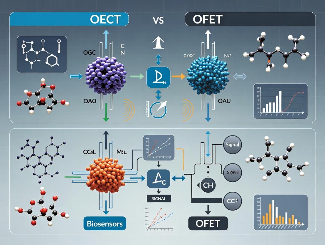
This article provides a comprehensive, up-to-date comparison of Organic Electrochemical Transistors (OECTs) and Organic Field-Effect Transistors (OFETs) as biosensing platforms.
OECT vs OFET Biosensors: A Comparative Guide for Biomedical Researchers and Developers
Abstract
This article provides a comprehensive, up-to-date comparison of Organic Electrochemical Transistors (OECTs) and Organic Field-Effect Transistors (OFETs) as biosensing platforms. Tailored for researchers, scientists, and drug development professionals, it explores the foundational device physics and materials, details fabrication methods and real-world applications, addresses common challenges and optimization strategies, and delivers a critical, side-by-side analysis of performance metrics and validation protocols. The synthesis offers actionable insights for selecting and advancing the optimal technology for specific biomedical sensing needs.
Unpacking the Core: The Physics and Materials Behind OECT and OFET Biosensors
The evolution of organic bioelectronics has yielded two principal transistor architectures for biosensing: Organic Electrochemical Transistors (OECTs) and Organic Field-Effect Transistors (OFETs). A fundamental distinction underpinning their operational differences lies in their gating mechanism: OECTs primarily utilize volumetric (bulk) gating, while OFETs operate via surface (interfacial) gating. This whitepaper dissects these core principles, framing them within the critical research thesis of selecting the optimal transducer for specific biosensing applications, particularly in pharmaceutical development.
Core Operational Principles
Volumetric Gating (OECT Paradigm)
In OECTs, the organic semiconductor channel (e.g., PEDOT:PSS) is ionically permeable and hydrated. Upon application of a gate voltage, electrolyte ions (e.g., Na+, Cl-) migrate into the bulk of the organic semiconductor film, dedoping/doping it throughout its entire volume. This reversible electrochemical process modulates the channel's electronic conductivity by changing the density of charge carriers (holes for PEDOT:PSS) across the film thickness.
- Key Characteristic: The capacitance is an ionic charge double-layer (EDL) capacitance in series with a volumetric capacitance, leading to very high total capacitance (~mF cm⁻²). The transconductance (gm) scales with channel volume.
Surface Gating (OFET Paradigm)
In OFET-based biosensors, the organic semiconductor (e.g., pentacene, DPPT-TT) is typically ion-impermeable. The gating effect occurs at the interface between the semiconductor and a dielectric layer (or electrolyte). Charge carriers are induced or depleted within the first few molecular layers (~1-3 nm) of the semiconductor, forming a conducting channel. In electrolyte-gated OFETs (EGOFETs), ions in the electrolyte form an EDL at the semiconductor surface, but do not penetrate the bulk.
- Key Characteristic: The capacitance is limited to the interfacial EDL capacitance (~µF cm⁻²). The transconductance depends on the semiconductor's mobility and the quality of the interface.
Quantitative Comparison
Table 1: Core Characteristics of Volumetric vs. Surface Gating
| Parameter | Volumetric Gating (OECT) | Surface Gating (OFET/EGOFET) |
|---|---|---|
| Gating Region | Entire bulk of the channel (3D) | Interface/Surface only (2D) |
| Ion Penetration | Deep, reversible penetration | No penetration (planar EDL) |
| Typical Capacitance | 1 - 10 mF cm⁻² | 1 - 10 µF cm⁻² |
| Transconductance (gm) | Very high (mS range) | Moderate (µS range) |
| Operating Voltage | Low (< 1 V) | Low to Moderate (< 3 V) |
| Channel Material | Mixed ionic-electronic conductor (MIEC) | Primarily electronic conductor |
| Biosensing Relevance | Sensitive to ionic flux & bulk property changes | Sensitive to surface potential & binding events |
| Response to pH/ Ionic Strength | Strong | Weak (unless interface is functionalized) |
Table 2: Implications for Biosensing in Drug Development Research
| Aspect | OECT (Volumetric) | OFET (Surface) |
|---|---|---|
| Target Size | Excellent for cells, large biomolecules, and metabolites | Optimal for small molecules, proteins, DNA (surface binding) |
| Signal Amplification | Exceptional due to high gm | Good, but typically lower |
| Integration with Aqueous Media | Inherently excellent | Requires careful dielectric/interface engineering |
| Spatial Resolution (e.g., for cell mapping) | Lower (bulk effect blurs localized signals) | Higher (localized surface effect) |
| Long-term Stability in Buffer | Can suffer from gradual volumetric swelling/degradation | Generally more stable, dependent on encapsulation |
Experimental Protocols for Characterization
Protocol 1: Measuring Volumetric Gating in OECTs
Objective: To characterize the bulk doping/dedoping process and extract relevant figures of merit. Materials: See "Scientist's Toolkit" (Table 3). Methodology:
- Device Fabrication: Pattern source/drain electrodes (Au) on a substrate. Spin-coat or screen-print the MIEC (e.g., PEDOT:PSS) channel. Define a well for the electrolyte and integrate a gate electrode (Ag/AgCl or Pt).
- Electrical Setup: Connect to a source measure unit (SMU) in a 3-terminal configuration (Source, Drain, Gate). Place the device in phosphate-buffered saline (PBS).
- Transfer Curve Measurement: Sweep the gate voltage (VG) from positive to negative (e.g., +0.6 V to -0.8 V) while maintaining a constant small drain voltage (VDS, e.g., -0.1 V). Record the drain current (I_D).
- Output Curve Measurement: At fixed VG steps, sweep VDS and record I_D.
- Key Analysis:
- Calculate peak transconductance: gm = δID / δVG.
- Determine the response time by applying a gate voltage step and measuring the temporal I_D response.
- Perform electrochemical impedance spectroscopy (EIS) to quantify the channel capacitance.
Protocol 2: Measuring Surface Gating in EGOFETs
Objective: To characterize interfacial gating and extract mobility and threshold voltage. Materials: See "Scientist's Toolkit" (Table 3). Methodology:
- Device Fabrication: Pattern source/drain electrodes (Au) on a substrate. Deposit a thin film of organic semiconductor (e.g., via thermal evaporation or solution-shearing). Define a well for the electrolyte and integrate a gate electrode.
- Electrical Setup: Similar to OECT setup but with careful shielding to minimize noise.
- Transfer Curve Measurement: Sweep VG while measuring ID at constant V_DS (in the linear regime, e.g., -0.1 V). Use a slower sweep rate to allow EDL formation.
- Key Analysis:
- Plot √|ID| vs. VG in the saturation regime to extract the charge carrier mobility (µ) and threshold voltage (V_Th) using the standard FET equation.
- Calculate interfacial capacitance via C-V measurements or EIS.
Visualization of Operational Principles
Diagram 1: OECT Volumetric Gating Mechanism (77 chars)
Diagram 2: OFET Surface Gating Mechanism (73 chars)
The Scientist's Toolkit: Key Research Reagents & Materials
Table 3: Essential Materials for OECT/OFET Biosensor Research
| Item | Function & Relevance | Example/Notes |
|---|---|---|
| PEDOT:PSS Dispersion | The canonical MIEC for OECTs. Provides high volumetric capacitance and ionic permeability. | Clevios PH1000, often mixed with ethylene glycol and cross-linkers for stability. |
| High-Mobility p/n-type OSC | For OFET channels. Determines baseline electronic performance (mobility, on/off ratio). | DPPT-TT (p-type), N2200 (n-type), evaporated pentacene. |
| Ion-Selective/Functionalized Membranes | To impart specificity in biosensors. Converts biological event into ionic or potentiometric signal. | Nafion (cation selector), lipid bilayers, immobilized enzymes or antibodies. |
| Stable Gate Electrodes | Provides stable potential in electrolyte. Critical for reproducible gating. | Ag/AgCl wire or patterned electrode, Platinum wire. |
| Physiological Buffer Salts | Electrolyte for gating and biomolecule environment. Ionic strength directly affects OECT response. | Phosphate Buffered Saline (PBS), Artificial Interstitial Fluid. |
| Cross-linkers & Additives | Stabilize MIEC films in aqueous media (for OECTs) or improve OSC morphology (for OFETs). | (3-glycidyloxypropyl)trimethoxysilane (GOPS), divinyl sulfone (DVS), surfactants. |
| Microfluidic Encapsulation | Defines electrolyte area, enables fluidic handling, and protects sensitive components. | PDMS gaskets, epoxy-based photoresists (e.g., SU-8). |
This whitepaper provides an in-depth technical analysis of the core architectural components—channel, electrolyte, and gate electrode—in Organic Electrochemical Transistors (OECTs), framed within a broader research thesis contrasting OECT and Organic Field-Effect Transistor (OFET) biosensors. The unique operational paradigm of OECTs, based on volumetric ion-to-electron transduction within an organic mixed conductor, fundamentally distinguishes them from the surface-dominated electrostatics of OFETs. This guide details material considerations, operational mechanisms, quantitative performance parameters, and experimental protocols for characterizing these core elements, serving as a resource for researchers and drug development professionals advancing bioelectronic sensing platforms.
The selection between OECT and OFET architectures for biosensing applications hinges on fundamental transduction mechanisms. OFETs, operating via field-effect modulation of charge carriers in a thin conduction channel, are exquisitely sensitive to surface potentials and binding events at the dielectric/semiconductor interface. In contrast, OECTs operate via the reversible, volumetric electrochemical doping/dedoping of an organic semiconductor channel by ions from an electrolyte. This bulk penetration of ions (typically from a biologically relevant aqueous medium) renders OECT transconductance several orders of magnitude higher than OFETs at low operating voltages (<1 V), making them exceptionally sensitive to ionic and biochemical fluctuations. This document deconstructs the three pillars enabling this performance: the mixed ionic-electronic conducting channel, the ionically conductive electrolyte, and the gate electrode governing ion injection.
The Mixed Ionic-Electronic Conducting (MIEC) Channel
The channel material is the cornerstone of OECT performance. It must facilitate both electronic (hole/electron) transport and ion penetration/transport. Semiconducting polymers like poly(3,4-ethylenedioxythiophene):poly(styrene sulfonate) (PEDOT:PSS) are quintessential, where PSS provides ionic conduction pathways and PEDOT provides electronic conduction.
Key Material Parameters:
- Ionic Conductivity (σᵢ): Dictates ion penetration speed and device transient response.
- Electronic Conductivity (σₑ): Determines the ON-current and electronic mobility.
- Volumetric Capacitance (C): The charge stored per unit volume per volt, a defining figure of merit. Higher C enables greater modulation for a given voltage.
- Swelling Ratio: The degree of volumetric expansion upon electrolyte uptake, affecting device stability and kinetics.
Table 1: Common OECT Channel Materials and Key Properties
| Material System | Ionic Conductivity (σᵢ) | Electronic Conductivity (σₑ) | Volumetric Capacitance (C*) | Primary Application |
|---|---|---|---|---|
| PEDOT:PSS (high-cond.) | ~0.1 S/cm | ~100-1000 S/cm | ~40-100 F/cm³ | General biosensing, amplifiers |
| PEDOT:PSS (glycol-treated) | ~0.2 S/cm | ~1000 S/cm | ~100-200 F/cm³ | High-sensitivity metabolite sensing |
| p(g2T-TT) / p(g2T-TT)-T | ~10⁻³ S/cm | ~10⁻² S/cm | ~200-300 F/cm³ | N-type OECTs, complementary logic |
| PEDOT:PSS / Ion Gel | ~1 S/cm | ~500 S/cm | >300 F/cm³ | High-frequency operation |
Experimental Protocol: Channel Characterization via Electrochemical Impedance Spectroscopy (EIS)
Objective: To determine the volumetric capacitance (C*) and ionic resistance of a channel material. Materials: Channel film on patterned Au electrodes, reference electrode (e.g., Ag/AgCl), counter electrode (Pt wire), electrolyte (e.g., 0.1 M NaCl). Procedure:
- Configure a 3-electrode cell with the channel film as the working electrode.
- Apply a small AC voltage perturbation (e.g., 10 mV RMS) across a frequency range (e.g., 1 MHz to 0.1 Hz) at a set DC bias (e.g., 0 V vs. Ag/AgCl).
- Measure impedance (Z) and phase (θ).
- Fit the Nyquist plot to an equivalent circuit model (e.g., a resistor in series with a constant phase element (CPE) parallel to another resistor).
- Extract the effective capacitance from the CPE parameters. Normalize by the channel volume to obtain C*.
Diagram 1: EIS protocol for channel characterization
The Electrolyte
The electrolyte is the ionic charge transport medium and the biorecognition element host. Its composition directly influences OECT operation, sensitivity, and biocompatibility.
Critical Factors:
- Ionic Strength: Affects Debye length and the sensing window for charged analytes.
- pH and Buffer Capacity: Critical for maintaining biological activity and interpreting sensor response.
- Specific Ion Effects: Ion size and hydration shell affect penetration into the channel.
Table 2: Electrolyte Compositions for OECT Biosensing
| Electrolyte Type | Primary Composition | Typical Concentration | Key Role/Consideration |
|---|---|---|---|
| Physiological Buffer | Phosphate Buffered Saline (PBS) | 0.01M - 0.1M | Baseline for in vitro biosensing, controls ionic strength & pH. |
| Cell Culture Medium | DMEM, RPMI with supplements | Variable (ionic ~0.15M) | For real-time cell monitoring; complex, may contain interferents. |
| Specific Ion Solution | NaCl, KCl | 1 mM - 1 M | For characterizing fundamental ion sensitivity (cation vs. anion). |
| Functionalized Electrolyte | PBS with enzymes/aptamers | Varies | Contains biorecognition element for specific analyte detection. |
The Gate Electrode
The gate electrode controls ion injection into the channel. Its potential governs the electrochemical window and can be functionalized to become the primary sensing interface.
Types & Characteristics:
- Non-Functionalized (e.g., Pt, Au): Used for characterizing channel or electrolyte properties. Provides a stable potential via reversible reactions (e.g., H⁺ reduction, oxide formation).
- Functionalized Gates: The gate surface is modified with receptors (enzymes, antibodies, aptamers). Binding events alter the gate's effective potential, modulating the channel current—a highly sensitive configuration.
- Quasi-Reference Electrodes (Ag/AgCl): Common in integrated setups, providing a stable, low-polarizability reference potential.
Table 3: Gate Electrode Configurations in OECT Biosensors
| Gate Type | Material/Modification | Stability | Typical Use Case | Transduction Mechanism |
|---|---|---|---|---|
| Simple Metal | Pt, Au wire | High | Fundamental studies, ion sensing | Capacitive charging / faradaic reactions |
| Integrated QRE | Patterned Ag/AgCl | Medium-High | Miniaturized, multiplexed devices | Stable reference potential |
| Biofunctionalized | Au with SAM + Antibody | Medium (depends on bio-layer) | Specific antigen detection (e.g., cortisol) | Binding-induced potential shift |
| Enzymatic | Carbon paste with GOx | Low-Medium (enzyme lifetime) | Metabolite sensing (e.g., glucose) | Catalytic reaction product (H₂O₂) |
Experimental Protocol: Gate Functionalization for Aptamer-Based Sensing
Objective: To immobilize thrombin-binding aptamer on a gold gate for specific protein detection. Materials: Au gate electrode, 5'-Thiol-modified aptamer, 6-mercapto-1-hexanol (MCH), Tris-EDTA buffer, thrombin protein solution.
Procedure:
- Clean Au Gate: Sonicate in acetone, ethanol, and DI water. Treat with UV-Ozone for 15 min.
- Aptamer Immobilization: Incubate gate in 1 µM thiol-aptamer solution in TE buffer for 2 hours at room temperature. This forms a self-assembled monolayer (SAM) via Au-S bonds.
- Backfilling: Rinse and incubate in 1 mM MCH solution for 1 hour to displace non-specifically adsorbed aptamers and create a well-ordered, bio-inert monolayer.
- Rinsing & Storage: Rinse thoroughly with buffer and store in clean buffer at 4°C until use.
- Measurement: Record OECT transfer characteristics (Iₛₜ vs. V₉) before and after exposure to thrombin solutions of varying concentration.
Diagram 2: Aptamer gate functionalization workflow
The Scientist's Toolkit: Research Reagent Solutions
Table 4: Essential Materials for OECT Fabrication & Characterization
| Item | Function | Example Product/Specification |
|---|---|---|
| Conductive Polymer | Forms the OECT channel. | Heraeus Clevios PH1000 (PEDOT:PSS), 1.3% in H₂O. |
| Ionic Additive | Enhances film conductivity & stability. | Ethylene Glycol (99.8%), DMSO, or surfactant Zonyl FS-300. |
| Crosslinker | Reduces film dissolution/swelling in electrolyte. | (3-Glycidyloxypropyl)trimethoxysilane (GOPS). |
| High-Resolution Photoresist | For patterning micro-scale channels/gates. | MicroChem SU-8 2000 series or LOR series. |
| Gate Electrode Material | Provides stable potential/functionalization site. | Pt/Ir wire (0.127 mm dia.), Ag/AgCl pellet, patterned Au on glass. |
| Biological Buffer | Provides stable ionic background for biosensing. | 1X PBS, pH 7.4, sterile-filtered. |
| Surface Modifier | Enables bioreceptor immobilization on Au gates. | 11-mercaptoundecanoic acid (11-MUA) for EDC/NHS coupling. |
| Blocking Agent | Reduces non-specific binding on sensor surfaces. | Bovine Serum Albumin (BSA), fraction V. |
| Electrochemical Cell | Holds electrolyte for device testing. | Custom 3D-printed well or commercial electrochemical cell. |
| Source/Measure Unit | Applies Vds/Vgs and measures Ids. | Keithley 2400/2600 Series SMU or Palmsens4 potentiostat. |
This whitepaper details the core architecture of Organic Field-Effect Transistors (OFETs), framed within a broader thesis investigating the fundamental differences between OFET and Organic Electrochemical Transistor (OECT)-based biosensors. For biosensing applications, the solid-state, three-terminal OFET structure contrasts sharply with the volumetric ionic-electronic coupling in OECTs, defining distinct operational mechanisms and sensor design philosophies.
Core Components and Their Functions
The performance of an OFET biosensor is governed by the synergistic interaction of its three core layers: the semiconductor, the dielectric, and the electrodes.
Organic Semiconductor (OSC)
The OSC layer is the charge-transporting heart of the OFET. In a biosensor context, it also often serves as the primary site for biorecognition event transduction. The two primary architectures are:
- Bottom-gate: OSC deposited on top of the dielectric.
- Top-gate: OSC deposited on the substrate, followed by dielectric and gate electrode.
Commonly used OSCs include polymers like P3HT, PEDOT:PSS (for hole transport), and small molecules like pentacene or C60 derivatives (for electron transport). Recent trends focus on donor-acceptor copolymers (e.g., DPP-based polymers) with tailored energy levels for ambient stability and specific interactions with analytes.
Dielectric Layer
The dielectric electrically insulates the semiconductor from the gate electrode while capacitively coupling them. Its properties critically affect the operating voltage and interfacial trap states.
- Inorganic Dielectrics: SiO₂, Al₂O₃, HfO₂. Offer high capacitance but rigid, high-temperature processing.
- Polymer Dielectrics: PMMA, PVP, CYTOP, parylene C. Enable low-voltage operation, flexibility, and solution processability. Parylene C is particularly favored in biosensing for its excellent biocompatibility and conformal coating ability.
- Electrolyte Dielectrics: Ionic liquids/gels. Enable ultra-high capacitance, leading to very low operating voltages (<1 V) and creating a hybrid OFET/OECT operational mode.
Electrodes (Source, Drain, Gate)
Electrodes inject and extract charge carriers from the OSC. The work function of the source/drain electrodes must align with the HOMO (p-type) or LUMO (n-type) levels of the OSC for efficient charge injection.
- Common Materials: Au (high work function, for p-type), Ca/Al (low work function, for n-type), ITO (transparent). In biosensors, Au is often functionalized with biorecognition elements (e.g., antibodies, aptamers).
- Gate Electrode: Can be a metal (Al, Au) or, in liquid-gated configurations, a reference electrode like Ag/AgCl.
Quantitative Performance Parameters & Comparison
Key performance metrics for OFETs in biosensing are summarized below. These parameters are directly modulated by biorecognition events (e.g., binding of a target biomolecule to the functionalized gate/ semiconductor surface), producing the sensor signal.
Table 1: Key OFET Performance Metrics and Typical Ranges
| Parameter | Symbol | Definition | Typical Range (OFET Biosensors) | Impact of Biomolecular Binding |
|---|---|---|---|---|
| Field-Effect Mobility | μ (cm²/V·s) | Charge carrier drift velocity per unit electric field. | 10⁻³ to >10 cm²/V·s | Decrease due to introduced scattering or trap sites. |
| Threshold Voltage | V_T (V) | Gate voltage required to turn on the channel. | -5 V to +5 V | Shift due to change in interfacial charge or capacitance. |
| On/Off Current Ratio | ION/IOFF | Ratio of maximum to minimum channel current. | 10³ to 10⁸ | Decrease if binding increases off-current or decreases on-current. |
| Subthreshold Swing | SS (mV/dec) | Gate voltage needed to increase current by one decade. | 100 - 2000 mV/dec | Increase if binding introduces additional interface traps. |
| Operational Voltage | VDS, VGS (V) | Voltages applied during operation. | < 5 V (Polymer dielectric) < 1 V (Electrolyte gate) | N/A |
Table 2: Core Material Choices for OFET Biosensors
| Component | Material Options | Key Properties for Biosensing | Common Deposition Methods |
|---|---|---|---|
| Semiconductor | P3HT, DPP-DTT, Pentacene, N2200 | Energy level alignment, environmental stability, surface functionality for bioreceptor attachment. | Spin-coating, Inkjet printing, Vacuum evaporation. |
| Dielectric | Parylene C, PMMA, CYTOP, SiO₂, Ionic Gel | Biocompatibility, low leakage, high capacitance, stability in aqueous media. | CVD (parylene), Spin-coating, Thermal evaporation. |
| Electrodes | Au, Pt, ITO, PEDOT:PSS | Work function, chemical stability, ease of functionalization (e.g., Au-thiol chemistry). | Thermal evaporation, Sputtering, Electroplating. |
Experimental Protocol: Fabrication and Characterization of a Basic OFET Biosensor
A. Substrate Preparation & Gate Electrode Deposition:
- Clean a heavily doped silicon wafer (serves as global gate) or glass/plastic substrate.
- If using a separate gate, deposit and pattern a gate electrode (e.g., 50 nm Au) via photolithography and evaporation.
B. Dielectric Deposition:
- For SiO₂: Thermally oxidize the Si wafer to grow a 100-300 nm oxide layer.
- For polymer dielectric (e.g., PMMA): Spin-coat a 5-10% solution in anisole at 2000-4000 rpm for 60 s. Anneal at 80-100°C for 1 hour to remove solvent.
C. Organic Semiconductor Deposition:
- For polymer OSC (e.g., P3HT): Spin-coat a 1-2% solution in chlorobenzene in a nitrogen glovebox. Anneal at 80-120°C for 30 min.
- For small molecule OSC (e.g., pentacene): Deposit via thermal evaporation at a rate of 0.1-0.3 Å/s under high vacuum to a thickness of 30-50 nm.
D. Source/Drain Electrode Deposition (Top-Contact Geometry):
- Use a shadow mask to define channel length (L) and width (W). Typical L = 20-100 µm, W = 100-5000 µm.
- Thermally evaporate Au (50 nm) through the mask to form electrodes.
E. Biosensor Functionalization (Example: Streptavidin-Biotin Model):
- Treat the Au S/D electrodes or the OSC surface with a thiolated or silane-based linker molecule (e.g., 11-mercaptoundecanoic acid) for 12 hours.
- Activate carboxyl groups with a mixture of EDC and NHS for 1 hour.
- Immerse the OFET in a solution of streptavidin (10 µg/mL in PBS) for 2 hours. Rinse thoroughly.
- The device is now ready for exposure to biotinylated target analytes.
F. Electrical Characterization:
- Use a semiconductor parameter analyzer (e.g., Keysight B1500A) in a shielded probe station.
- Sweep gate voltage (VGS) at constant drain voltage (VDS) to obtain transfer characteristics (IDS vs VGS).
- Extract μ, VT, ION/IOFF, and SS using standard FET equations:
- μ = (∂√|IDS|/∂VGS)² * (2L)/(W*Ci) (in saturation regime)
- where C_i is the dielectric capacitance per unit area.
The Scientist's Toolkit: Essential Research Reagents & Materials
Table 3: Key Reagents for OFET Biosensor Development
| Item | Function/Description |
|---|---|
| P3HT (Regioregular) | Benchmark p-type organic semiconductor polymer. |
| Parylene C | USP Class VI biocompatible, vapor-deposited dielectric barrier. |
| Cytop | Low-k, hydrophobic fluoropolymer dielectric, minimizes ion diffusion. |
| (3-Aminopropyl)triethoxysilane (APTES) | Silane coupling agent to introduce amine groups on oxide surfaces for biomolecule conjugation. |
| 1-Pyrenebutanoic acid succinimidyl ester | Non-covalent linker for functionalizing graphene or carbon nanotube-based OSCs via π-π stacking. |
| Phosphate Buffered Saline (PBS), pH 7.4 | Standard aqueous medium for biological functionalization and measurements. |
| N,N-Dimethylformamide (DMF) / Chlorobenzene | High-purity solvents for dissolving and processing organic semiconductors. |
| Ethylene Diamine Tetraacetic Acid (EDTA) | Chelating agent added to measurement buffers to sequester metal ions that could dope the OSC. |
Schematic Diagrams
OFET Biosensor Structure & Signal Path
OFET vs OECT Transduction Mechanism
Within the comparative research paradigm of Organic Electrochemical Transistors (OECTs) versus Organic Field-Effect Transistors (OFETs) for biosensing applications, the performance, sensitivity, and operational stability are fundamentally governed by the material selection. This whitepaper provides an in-depth technical guide to three core material classes: conducting polymers (the active channel material), ion gels (the gate dielectric/electrolyte), and biocompatible substrates (the foundational support). The synergistic integration of these materials dictates critical differences in OECT and OFET biosensor operation, including transduction mechanism, interface with biological analytes, and device architecture.
Conducting Polymers: The Active Semiconductor
Conducting polymers (CPs) are π-conjugated organic materials that can conduct electronic charge while maintaining mechanical flexibility. Their mixed ionic-electronic conduction properties are central to biosensor function.
Core Properties & Relevance:
- OECTs: Require CPs with high volumetric capacitance and efficient mixed ionic-electronic conduction (MIEC). The benchmark material is poly(3,4-ethylenedioxythiophene) doped with poly(styrene sulfonate) (PEDOT:PSS). Upon application of a gate bias, hydrated ions from the electrolyte penetrate the bulk of the CP film, dedoping it and modulating its electronic conductivity.
- OFETs: Require CPs with high charge carrier mobility and ordered microstructures for efficient lateral charge transport in a thin channel. Materials like poly(3-hexylthiophene) (P3HT) and diketopyrrolopyrrole (DPP)-based polymers are common. Sensing occurs primarily through electrostatic interactions or doping events at the surface of the semiconductor.
Quantitative Comparison of Key Conducting Polymers:
Table 1: Key Properties of Conducting Polymers in OECT vs. OFET Biosensors
| Polymer | Typical OECT µC* (F cm⁻¹ V⁻¹ s⁻¹) | Typical OFET µ (cm² V⁻¹ s⁻¹) | Key Advantages | Primary Biosensor Role |
|---|---|---|---|---|
| PEDOT:PSS | 200 - 400 | 0.01 - 1 | High MIEC, excellent aqueous stability, commercial availability | OECT channel material |
| P3HT | 1 - 10 | 0.01 - 0.1 | Solution processability, well-studied morphology | OFET channel material |
| p(g2T-TT) | 300 - 500 | N/A | Engineered for high OECT performance, glycol side chains | High-performance OECT channel |
| DPP-based | N/A | 0.1 - 5 | High OFET mobility, tunable energy levels | High-performance OFET channel |
µC: Product of mobility (µ) and volumetric capacitance (C), the OECT performance metric.
Experimental Protocol: OECT Channel Deposition (Spin-Coating)
- Substrate Preparation: Clean glass or biocompatible substrate (e.g., PDMS-coated glass) via sequential sonication in detergent, deionized water, acetone, and isopropanol (15 min each). Treat with oxygen plasma for 5 minutes to ensure hydrophilic surface.
- Polymer Solution Preparation: Filter commercially available PEDOT:PSS solution (e.g., Clevios PH1000) through a 0.45 µm PVDF syringe filter. Optionally, mix with 5% v/v ethylene glycol and 1% v/v (3-glycidyloxypropyl)trimethoxysilane (GOPS) as a cross-linker for improved stability.
- Spin-Coating: Dispense 100 µL of solution onto the static substrate. Spin at 3000 rpm for 60 seconds in ambient conditions.
- Annealing: Cure the film on a hotplate at 140°C for 15-60 minutes (longer for GOPS-crosslinked films) to remove residual solvents and induce cross-linking.
Ion Gels: The Gate Dielectric/Electrolyte
Ion gels are quasi-solid composites of an ionic liquid and a gelating polymer matrix (e.g., triblock copolymers). They provide a high-capacitance, stable ionic interface.
Core Properties & Relevance:
- OECTs: Serve as the primary electrolyte, providing mobile ions for channel doping/dedoping. Can be used as a gate electrolyte in a planar architecture.
- OFETs: Function as an ultra-high capacitance (µF cm⁻²) gate dielectric in electrolyte-gated OFETs (EG-OFETs), enabling low-voltage operation (<1 V). This blurs the architectural line with OECTs but retains a surface-dominated transduction mechanism.
Quantitative Comparison of Dielectric/Electrolyte Materials:
Table 2: Comparison of Gate Interface Materials for OECTs and OFETs
| Material | Typical Capacitance | Operating Voltage | Key Feature | Device Type |
|---|---|---|---|---|
| Aqueous Buffer (e.g., PBS) | ~µF cm⁻² (EDL) | 0.5 - 1 V | Biocompatible, directly interfaces with bio-analyte | OECT |
| Ion Gel (e.g., [EMIM][TFSI]/PS-PMMA-PS) | 1 - 10 µF cm⁻² | < 1 V | High capacitance, non-volatile, solid-state | EG-OFET, OECT |
| Traditional Dielectric (e.g., SiO₂) | ~10 nF cm⁻² | 20 - 100 V | Low leakage, well-established process | Conventional OFET |
Experimental Protocol: Ion Gel Preparation & Patterning
- Gel Formation: Dissolve the triblock copolymer polystyrene-poly(methyl methacrylate)-polystyrene (PS-PMMA-PS, 5% w/w) in the ionic liquid 1-ethyl-3-methylimidazolium bis(trifluoromethylsulfonyl)imide ([EMIM][TFSI]) at 90°C with stirring for 2-4 hours until a homogeneous, viscous solution is formed.
- Patterning via Stencil Masking: Place a laser-cut PDMS stencil mask on the target substrate (covering source/drain contacts). Deposit a small amount of the warm ion gel and spread with a doctor blade. Carefully remove the stencil, leaving the gel patterned only over the channel/gate region.
- Solidification: Allow the gel to cool and set at room temperature for 1 hour, forming a solid, rubbery film.
Biocompatible Substrates: The Foundation
Biocompatible substrates provide mechanical support while ensuring device stability and compatibility with biological environments (e.g., cells, tissues, physiological fluids).
Core Properties & Relevance:
- Mechanical Properties: Critical for flexible/wearable sensors or interfacing with soft tissues. Low Young's modulus (e.g., of PDMS) minimizes mechanical mismatch.
- Surface Chemistry: Determines wettability, protein/cell adhesion, and functionalization routes for biorecognition elements (enzymes, antibodies, aptamers).
- Permeability: For implantable or tissue-integrated devices, oxygen and nutrient permeability may be required.
Quantitative Comparison of Substrate Materials:
Table 3: Properties of Common Biocompatible Substrates
| Substrate | Young's Modulus | Optical Transparency | Key Advantage | Best Suited For |
|---|---|---|---|---|
| Polyimide (PI) | 2.5 GPa | Opaque (often) | High thermal/chemical stability, flexible | Chronic implants, flexible electronics |
| Polydimethylsiloxane (PDMS) | 0.5 - 4 MPa | High | Gas permeable, tunable modulus, castable | Cell culture interfaces, epidermal sensors |
| Polyethylene Naphthalate (PEN) | ~5 GPa | High | Good moisture barrier, flexible | Flexible, encapsulated biosensors |
| Parylene C | 2.8 GPa | High | Conformal coating, USP Class VI biocompatible | Chronic neural implants, barrier coating |
Experimental Protocol: PDMS Substrate Preparation & Functionalization
- Mixing & Degassing: Mix PDMS base and curing agent (Sylgard 184) at a 10:1 (w/w) ratio. Mix thoroughly, then place in a desiccator connected to a vacuum pump for 30-45 minutes until all bubbles are removed.
- Casting & Curing: Pour the degassed PDMS onto a clean silicon wafer or petri dish. Cure at 65°C for at least 4 hours (or 2 hours at 90°C).
- Surface Activation: Expose the PDMS surface to oxygen plasma (50 W, 30 seconds) to create a transient silanol (Si-OH) rich, hydrophilic surface.
- Biofunctionalization: Immediately immerse the activated PDMS in a 1% (v/v) solution of (3-aminopropyl)triethoxysilane (APTES) in ethanol for 1 hour. Rinse with ethanol and dry. This creates an amine-terminated surface for subsequent covalent immobilization of biorecognition elements via cross-linkers like glutaraldehyde.
The Scientist's Toolkit: Key Research Reagent Solutions
Table 4: Essential Materials for Fabrication and Characterization
| Item (Supplier Example) | Function/Benefit |
|---|---|
| Clevios PH1000 PEDOT:PSS (Heraeus) | Industry-standard, high-conductivity polymer dispersion for OECT channels. |
| Ionic Liquid [EMIM][TFSI] (Sigma-Aldrich/Iolitec) | High-stability, low-volatility ionic liquid for formulating ion gels. |
| PS-PMMA-PS Triblock Copolymer (Polymer Source) | Effective gelling agent for ionic liquids to form mechanically robust ion gels. |
| Sylgard 184 Silicone Elastomer Kit (Dow) | The benchmark PDMS for flexible, biocompatible substrates and microfluidics. |
| (3-Glycidyloxypropyl)trimethoxysilane (GOPS) (Sigma-Aldrich) | Cross-linker for PEDOT:PSS, dramatically improving aqueous operational stability. |
| Phosphate Buffered Saline (PBS), 10X Solution (Thermo Fisher) | Standard physiological buffer for electrolyte and device testing in biologically relevant conditions. |
| Poly-L-lysine Solution (Sigma-Aldrich) | Promotes adhesion of cells or proteins to substrate surfaces for cell-based sensing. |
Visualizing Material Roles in OECT vs. OFET Biosensing
This whitepaper provides a technical guide to the conversion of biological recognition events into quantifiable electronic signals within biosensing platforms. The process—termed the signal transduction pathway—is central to the function of all biosensors. The content is framed within a critical research thesis comparing Organic Electrochemical Transistors (OECTs) and Organic Field-Effect Transistors (OFETs), highlighting how their distinct operational principles dictate the design and efficiency of this pathway for applications in biomedical research and drug development.
The Core Transduction Pathway: A Generic Framework
At its essence, a biosensor's signal transduction pathway follows a defined sequence:
Biological Recognition Event (Bioreceptor-Target Binding) → Transducer-Specific Biophysical Change → Electronic Signal → Processed Readout.
The critical divergence between OECTs and OFETs occurs at the Transducer-Specific Biophysical Change stage, fundamentally altering sensitivity, operational environment, and application scope.
Diagram Title: Generic Biosensor Transduction Cascade
Transduction Mechanisms: OECT vs. OFET
Organic Electrochemical Transistor (OECT) Pathway
In OECTs, the organic polymer channel (e.g., PEDOT:PSS) is in direct contact with an electrolyte. The biological event modulates ionic fluxes at the gate or channel interface.
Pathway: Binding Event → Change in Local Ionic Concentration/Potential → Ionic Flux into/out of Polymer Channel → Dedoping/Doping of Channel (Bulk Property Change) → Large Modulation of Channel Conductance ((I_{DS})).
Key Advantage: The mixed ionic-electronic coupling enables very high transconductance (sensitivity to ionic changes) and operation in aqueous, physiological environments.
Organic Field-Effect Transistor (OFET) Pathway
In OFETs, the organic semiconductor channel is typically shielded from the electrolyte by a dielectric. The biological event acts as a gate potential modulator.
Pathway: Binding Event → Introduction or Induction of Charged Species (at Dielectric Surface) → Capacitive Coupling & Field-Effect in Channel → Change in Charge Carrier Density at Semiconductor/Dielectric Interface ((VT) shift or (I{DS}) change).
Key Advantage: The field-effect mechanism offers fast electronic switching and potential for high spatial density in sensor arrays.
Diagram Title: OECT vs. OFET Transduction Mechanisms
Quantitative Comparison of Key Performance Metrics
Table 1: Performance Characteristics of OECT vs. OFET Biosensors
| Performance Metric | OECT Biosensors | OFET Biosensors | Implication for Signal Transduction |
|---|---|---|---|
| Transconductance (gm) | Very High (1-100 mS) | Moderate (0.01-1 µS) | OECTs provide superior signal amplification per input voltage change, ideal for low-concentration analytes. |
| Operating Voltage | Low (< 1 V) | Moderate to High (1-50 V) | OECTs are more suitable for implantable or wearable applications due to low power and safety. |
| Response Time | Milliseconds to Seconds (diffusion-limited) | Microseconds to Milliseconds | OFETs offer faster electronic readout; OECT speed is governed by ion mobility. |
| Aqueous Stability | Excellent (designed for electrolytes) | Poor to Moderate (requires encapsulation) | OECTs natively operate in physiological buffers, simplifying in vitro and in vivo sensing. |
| Sensitivity (LOD) | Can reach fM-pM for proteins | Typically nM-pM for proteins | OECT's high gm often translates to lower practical limits of detection in complex media. |
| Miniaturization & Integration | Moderate (channel size ~ µm) | High (channel size can be < 100 nm) | OFETs have an advantage in high-density multiplexed arrays for spatial mapping. |
| Primary Transduced Quantity | Ionic Strength / Capacitance | Surface Charge / Potential | Dictates bioreceptor placement and functionalization strategy. |
Table 2: Typical Experimental Parameters from Recent Literature (2023-2024)
| Transducer Type | Target Analyte | Bioreceptor | Reported LOD | Dynamic Range | Response Time | Ref. |
|---|---|---|---|---|---|---|
| OECT (PEDOT:PSS) | Cortisol | Aptamer | 1 nM (0.36 ng/mL) | 1 nM - 10 µM | ~ 2 minutes | ACS Sens. 2023, 8, 3 |
| OECT (p(g2T-TT)) | Dopamine | Tyrosinase Enzyme | 10 nM | 10 nM - 1 mM | < 10 seconds | Adv. Mater. 2024, 36, 2308078 |
| OFET (DNTT) | PSA Antibody | Anti-PSA (cAb) | 1 pg/mL | 1 pg/mL - 1 µg/mL | ~ 15 minutes | Biosens. Bioelectron. 2023, 220, 114882 |
| OFET (C8-BTBT) | miRNA-21 | Single-Stranded DNA Probe | 1 fM | 1 fM - 1 nM | ~ 5 minutes | Nat. Commun. 2023, 14, 3296 |
Detailed Experimental Protocols
Protocol: Fabrication and Measurement of a Generic OECT Biosensor for Protein Detection
Objective: To functionalize an OECT gate for specific antibody-antigen binding and measure the resulting drain-source current ((I_{DS})) modulation.
Materials & Reagents: See "The Scientist's Toolkit" (Section 7).
Methodology:
- Device Fabrication:
- Pattern gold gate and drain/source electrodes on a glass or flexible substrate via photolithography and evaporation.
- Spin-coat or drop-cast the channel material (e.g., PEDOT:PSS mixed with 5% v/v ethylene glycol and 1% v/v (3-Glycidyloxypropyl)trimethoxysilane) onto the channel region. Anneal at 140°C for 15-60 minutes.
- Gate Functionalization (Streptavidin-Biotin Model):
- Clean the gold gate electrode via oxygen plasma treatment (100 W, 1 minute).
- Incubate the gate in 1 mM 11-mercaptoundecanoic acid (11-MUA) ethanol solution for 12 hours at room temperature to form a self-assembled monolayer (SAM). Rinse with ethanol and DI water.
- Activate the carboxyl groups by immersing in a solution containing 400 mM EDC and 100 mM NHS in MES buffer (pH 5.5) for 1 hour. Rinse with PBS (pH 7.4).
- Immediately incubate with 50 µg/mL streptavidin in PBS for 2 hours. Rinse with PBS to remove unbound streptavidin.
- Incubate with 1 µM biotinylated capture antibody in PBS for 1 hour. Rinse thoroughly with PBS. The device is now ready for sensing.
- Electrical Measurement & Sensing:
- Assemble a measurement cell enclosing the channel and gate. Fill with phosphate-buffered saline (PBS, pH 7.4).
- Using a source measure unit (SMU) or potentiostat, apply a constant drain voltage ((VD)) typically between -0.2 and -0.5 V. Apply a gate voltage ((VG)) pulse or sweep (e.g., from 0 to 0.5 V).
- Record the baseline drain current ((I{DS})).
- Introduce the target antigen at varying concentrations into the PBS electrolyte.
- Monitor the time-dependent change in (I{DS}) at constant (VG) and (VD), or record full transfer characteristics ((I{DS}) vs. (VG)) after equilibrium is reached (typically 5-15 minutes).
- Data Analysis:
- Plot (\Delta I{DS}) or (\Delta gm) (transconductance) vs. analyte concentration to generate a calibration curve.
- Determine Limit of Detection (LOD) using 3σ method (3 times standard deviation of blank signal).
Protocol: Fabrication and Functionalization of an Electrolyte-Gated OFET (EG-OFET) Biosensor
Objective: To create an OFET where the dielectric is functionalized for DNA hybridization detection, leveraging electrolyte gating for low-voltage operation.
Methodology:
- Device Fabrication:
- Deposit and pattern bottom gate (e.g., highly doped silicon) and a high-k dielectric (e.g., Al₂O₃, 30 nm via atomic layer deposition).
- Functionalize the dielectric surface with (3-aminopropyl)triethoxysilane (APTES) vapor (80°C, 2 hours).
- Deposit and pattern organic semiconductor (e.g., DPPT-TT, solution-sheared) and drain/source electrodes (Au) atop the dielectric.
- Define a well to contain electrolyte over the channel/dielectric region.
- Dielectric Surface Functionalization:
- Treat the APTES-modified dielectric in the well with 2.5% glutaraldehyde in PBS for 30 minutes. Rinse.
- Incubate with amine-terminated DNA probe sequence (10 µM in saline citrate buffer) for 2 hours. Rinse with buffer.
- Passivate non-specific binding sites with 1 M ethanolamine hydrochloride (pH 8.5) for 1 hour.
- Measurement:
- Fill the well with a low-ionic-strength buffer (e.g., 10 mM Tris-EDTA).
- Insert a Ag/AgCl gate electrode into the electrolyte.
- Apply a constant (VD) (-0.1 V). Sweep (VG) from positive to negative voltage.
- Record transfer curves before and after introduction of complementary target DNA. The negative charge of hybridized DNA backbones induces a positive (V_T) shift.
Diagram Title: Biosensor Fabrication & Testing Workflow
The Scientist's Toolkit: Key Research Reagent Solutions
Table 3: Essential Materials for OECT/OFET Biosensor Development
| Material / Reagent | Function in Transduction Pathway | Example Product/Chemical | Key Consideration |
|---|---|---|---|
| Conductive Polymer | OECT Channel Material; Mixed ionic-electronic conductor. | PEDOT:PSS (Clevios), p(g2T-TT), p(g3T2-TT) | High volumetric capacitance, stability in water. Doping level is critical. |
| Organic Semiconductor | OFET Channel Material; Transports electronic charges. | DPPT-TT, C8-BTBT, DNTT | High charge carrier mobility, ambient stability, compatible deposition. |
| Crosslinker / Activator | Immobilizes bioreceptors onto transducer surface. | EDC (1-Ethyl-3-(3-dimethylaminopropyl)carbodiimide) & NHS (N-Hydroxysuccinimide) | Activates carboxyl groups for amide bond formation with proteins/amines. |
| Self-Assembled Monolayer (SAM) Agent | Creates functional interface on metal (Au) electrodes. | 11-Mercaptoundecanoic acid (11-MUA), (3-Aminopropyl)triethoxysilane (APTES) | Provides terminal groups (-COOH, -NH₂) for subsequent bioreceptor coupling. |
| High-k Dielectric | Insulating layer in OFETs; determines capacitance. | Al₂O₃, HfO₂, TiO₂ (ALD deposited), PMMA, CYTOP | Higher capacitance enables lower operating voltage and sharper switching. |
| Specific Bioreceptor | Molecular recognition element for the target. | Monoclonal Antibodies, DNA/Aptamer Sequences, Enzymes (e.g., Glucose Oxidase) | Binding affinity (KD) and orientation on the surface directly impact sensitivity. |
| Blocking Agent | Reduces non-specific adsorption (noise). | Bovine Serum Albumin (BSA), Casein, Ethanolamine, Tween-20 | Essential for achieving low LOD in complex samples like serum. |
| Electrolyte | Medium for OECT operation/ionic conduction. | Phosphate Buffered Saline (PBS), Artificial Interstitial Fluid | Ionic strength affects Debye length and screening of biomolecular charges. |
From Lab to Life: Fabrication, Functionalization, and Biosensing Applications
This whitepaper details the core fabrication techniques employed in the development of Organic Electrochemical Transistors (OECTs) and Organic Field-Effect Transistors (OFETs) for biosensing applications. Understanding these methodologies is critical to the broader thesis examining the functional differences between OECT and OFET biosensors, particularly in sensitivity, response dynamics, and interfacial design for bioanalytes.
Core Fabrication Techniques
Spin-Coating
Spin-coating is a standard technique for depositing uniform thin films of organic semiconductors and dielectric layers, primarily on rigid substrates.
- Key Principle: A solution is dispensed onto a static or rotating substrate, which is then accelerated to high speed. Centrifugal force spreads the solution, while solvent evaporation leads to film formation.
- Typical Applications: Deposition of PEDOT:PSS for OECT channels, semiconductor layers (e.g., DPP-DTT, pentacene) for OFETs, and polymer dielectrics (e.g., PMMA, Cytop).
Experimental Protocol (PEDOT:PSS Layer for OECT):
- Substrate Preparation: Clean glass or SiO₂/Si substrate with sequential sonication in detergent, deionized water, acetone, and isopropanol. Treat with oxygen plasma for 2-5 minutes to improve wettability.
- Solution Preparation: Filter commercially available PEDOT:PSS dispersion (e.g., Clevios PH1000) through a 0.45 μm PVDF syringe filter. Optionally, mix with 5-10% v/v ethylene glycol and 0.1-1% v/v (3-Glycidyloxypropyl)trimethoxysilane (GOPS) for enhanced conductivity and adhesion.
- Deposition: Place substrate on spin coater chuck. Dispense 50-100 μL of solution onto the center of the substrate. Execute a two-step program: (i) 500 rpm for 5-10 s (spread step), (ii) 2000-5000 rpm for 30-60 s (thin film step).
- Annealing: Transfer the coated substrate to a hotplate and anneal at 120-140°C for 10-30 minutes in air to remove residual solvent.
Printing Techniques
Printing enables patterned, additive deposition of functional inks on flexible and rigid substrates, facilitating scalable device fabrication.
- Inkjet Printing: Non-contact, digital deposition of picoliter droplets. Ideal for defining electrode arrays (e.g., Ag nanoparticle inks) and semiconductor patterns.
- Screen Printing: Contact method using a mesh stencil to force viscous ink onto a substrate. Commonly used for high-throughput fabrication of disposable carbon/silver electrode pads.
- Aerosol Jet Printing: A direct-write, non-contact technique that can print high-resolution features (~10 μm) on conformal surfaces using a focused aerosol stream.
Experimental Protocol (Inkjet-Printed Ag Source/Drain Electrodes for OFET):
- Ink Preparation: Use commercial Ag nanoparticle ink (e.g., Sigma-Aldrich 736465). Filter through a 0.2 μm PTFE filter into a clean inkjet cartridge.
- Printer Setup: Load cartridge into a piezoelectric inkjet printer (e.g., Fujifilm Dimatix). Set substrate platen temperature to 40°C. Adjust waveform parameters (voltage, pulse width) for stable jetting.
- Pattern Alignment: Load device layout design (e.g., as a .bmp file). Perform drop-watch and fiducial camera alignment to the substrate.
- Printing: Print electrode pattern (typically channel length L=20-100 μm) in multiple passes (1-3) with intermediate drying.
- Sintering: Post-print, sinter the Ag electrodes on a hotplate at 130-200°C for 15-60 minutes to achieve low resistivity (< 5 μΩ·cm).
Microfabrication (Photolithography)
Photolithography is used to create high-resolution, permanent patterns for electrodes and interconnects, often in combination with other techniques.
- Key Principle: A photosensitive polymer (photoresist) is patterned using UV light through a photomask, then developed to create a stencil for etching or metal deposition.
- Typical Applications: Definition of micron-scale channel lengths for OFETs, fabrication of transistor arrays on silicon wafers, and patterning of passivation layers.
Experimental Protocol (Photolithography for Au OFET Electrodes):
- Substrate Cleaning: Clean a highly doped Si wafer with a 300 nm thermal oxide layer (serving as gate/dielectric) using piranha solution (Caution: Highly exothermic) or oxygen plasma.
- Photoresist Application: Dehydrate substrate at 150°C for 5 minutes. Apply positive photoresist (e.g., S1813) via spin-coating at 3000-4000 rpm for 45 s. Soft-bake at 115°C for 1 minute.
- Exposure and Development: Align a chrome photomask defining the source/drain pattern. Expose using a UV aligner with a dose of ~100 mJ/cm². Develop in MF-319 or AZ 726 MIF developer for 45-60 s, then rinse in DI water.
- Metal Deposition and Lift-Off: Deposit a thin adhesion layer (5 nm Ti or Cr) followed by 30-50 nm of Au via electron-beam or thermal evaporation. Submerge the substrate in acetone with gentle agitation (or in a solvent bath) to lift off the photoresist and overlying metal, leaving behind the patterned Au electrodes.
Table 1: Quantitative Comparison of Fabrication Techniques for OECTs/OFETs
| Technique | Typical Resolution | Min. Channel Length (L) | Throughput | Material Waste | Key Advantage | Primary Limitation |
|---|---|---|---|---|---|---|
| Spin-Coating | N/A (Film Uniformity) | N/A | High | High | Excellent film uniformity, simple setup. | No in-situ patterning, high material waste. |
| Inkjet Printing | 20-50 μm | 20-50 μm | Medium-High | Low | Digital, additive patterning; flexible substrates. | Resolution limited by droplet size; ink formulation critical. |
| Screen Printing | 50-100 μm | >100 μm | Very High | Low | High throughput, robust for low-cost sensors. | Low resolution, requires viscous inks. |
| Aerosol Jet Printing | 10-20 μm | 10-20 μm | Medium | Low | High resolution on conformal surfaces. | Complex nozzle maintenance, ink rheology control. |
| Photolithography | < 2 μm | < 5 μm | Low (R&D) | Medium | Ultra-high resolution and precision. | High capital cost, multi-step process, not suited for all organics. |
The Scientist's Toolkit: Essential Research Reagents & Materials
Table 2: Key Reagent Solutions and Materials for OECT/OFET Fabrication
| Item | Example Product/Chemical | Primary Function |
|---|---|---|
| Conductive Polymer | PEDOT:PSS (Clevios PH1000) | OECT channel material; mixed ionic/electronic conductor. |
| OFET Semiconductor | DPP-DTT, Pentacene, C8-BTBT | OFET channel material; primarily electronic conductor. |
| Dielectric Material | PMMA, Cytop, SiO₂ (thermal oxide) | Insulating layer between gate and channel in OFETs. |
| Crosslinker | GOPS ((3-Glycidyloxypropyl)trimethoxysilane) | Crosslinks PEDOT:PSS for improved stability in aqueous environments. |
| Conductivity Enhancer | Ethylene Glycol, DMSO | Secondary dopant for PEDOT:PSS to increase film conductivity. |
| Metal Ink | Ag Nanoparticle Ink (Sigma-Aldrich) | Inkjet-printable ink for forming conductive electrodes. |
| Photoresist | S1813 Positive Photoresist (Kayaku) | Light-sensitive polymer for photolithographic patterning. |
| Developer | MF-319 Developer (Kayaku) | Aqueous alkaline solution to develop exposed positive photoresist. |
| Substrate | SiO₂/Si wafers, ITO-coated glass, PET/PEN foil | Device support platform with varying properties (rigid/flexible, conductive/insulating). |
| Biorecognition Element | DNA aptamers, enzymes, antibodies | Immobilized on device channel/gate to confer biospecificity. |
Visualization of Experimental Workflows
Diagram Title: Fabrication Workflow Comparison for OECTs/OFETs
Diagram Title: From Fabrication to Biosensor Performance Metrics
The performance of organic electrochemical transistor (OECT) and organic field-effect transistor (OFET) biosensors is fundamentally governed by the interface between the organic semiconductor and the analyte. OECTs operate in aqueous electrolytes, where ions from the solution penetrate the bulk of the organic semiconductor (e.g., PEDOT:PSS), modulating its conductivity. OFETs, in contrast, operate in a gate-controlled configuration where the analyte interaction primarily modulates the charge carrier density at the semiconductor/dielectric interface. For both architectures, effective surface biofunctionalization—the stable and oriented immobilization of biorecognition elements (antibodies, aptamers, enzymes)—is critical to achieving high sensitivity, specificity, and stability. The choice of immobilization strategy must be tailored to the transducer's operating principle and environment. This guide details core strategies, with a focus on their implications for OECT and OFET biosensing platforms.
Core Immobilization Strategies
Physical Adsorption
- Mechanism: Non-covalent attachment via hydrophobic interactions, van der Waals forces, or electrostatic interactions.
- Advantages: Simple, fast, no chemical modification required.
- Disadvantages: Random orientation, desorption over time, potential denaturation.
- OFET/OECT Context: Commonly used for preliminary OFET studies due to simplicity but suffers from instability. Less suitable for OECTs in long-term biological fluid exposure.
Covalent Coupling
- Mechanism: Formation of stable covalent bonds between functional groups on the probe and the sensor surface.
- Key Chemistry: Carbodiimide crosslinking (EDC/NHS) for carboxyl-amine coupling. "Click" chemistry (e.g., azide-alkyne cycloaddition) for highly specific, bioorthogonal linking.
- Advantages: Stable, oriented immobilization possible with surface engineering.
- Disadvantages: Requires surface activation and often probe modification; can be multi-step.
Affinity-Based Immobilization
- Mechanism: Use of high-affinity molecular pairs (e.g., streptavidin-biotin, Protein A/G-antibody Fc region).
- Advantages: Excellent control over orientation, maintains probe activity, reversible under certain conditions.
- Disadvantages: Requires biotinylation or other tagging of the probe; additional cost.
Entrapment within Polymers/Hydrogels
- Mechanism: Probes are physically encapsulated within a porous polymer matrix (e.g., polypyrrole, PEG-based hydrogels) during electropolymerization or crosslinking.
- Advantages: High loading capacity, protects the probe, excellent for OECT integration.
- Disadvantages: Can slow diffusion of analyte, may require optimization of polymer mesh size.
Table 1: Comparison of Immobilization Strategies for OECT vs. OFET Biosensors
| Strategy | Typical Linker/Chemistry | Optimal For OECT? | Optimal For OFET? | Key Advantage | Key Limitation |
|---|---|---|---|---|---|
| Physical Adsorption | N/A (Passive) | Low | Medium (for screening) | Simplicity, Speed | Unstable, Random Orientation |
| Covalent (EDC/NHS) | Carboxyl-Amine | High | High | High Stability, Common | Requires -COOH/-NH₂ groups |
| Covalent (Click) | Azide-Alkyne | High | High | Specific, Bioorthogonal | Needs pre-modification |
| Affinity (Streptavidin-Biotin) | Biotin-Streptavidin | High | Medium | Superior Orientation | Extra tagging step |
| Entrapment | Polymer Matrix (e.g., Polypyrrole) | Very High | Low | High Load, Protective | Mass Transport Limitation |
Detailed Experimental Protocols
Protocol 1: EDC/NHS Covalent Immobilization of Antibodies on Au/ITO Electrodes (for OECT Gate Functionalization)
- Objective: To create a stable, covalently linked antibody layer on a gold or ITO gate electrode for an OECT biosensor.
- Materials: See "The Scientist's Toolkit" below.
- Procedure:
- Surface Cleaning: Sonicate Au/ITO substrate in acetone, ethanol, and DI water for 10 min each. Dry under N₂ stream.
- SAM Formation: Immerse substrate in 1 mM 11-mercaptoundecanoic acid (11-MUA) in ethanol for 12-18 hours to form a self-assembled monolayer (SAM) with terminal carboxyl groups.
- Rinsing: Rinse thoroughly with ethanol and DI water to remove unbound thiols.
- Activation: Prepare a fresh solution of 0.4 M EDC and 0.1 M NHS in MES buffer (0.1 M, pH 5.5). Incubate the SAM-coated substrate in this solution for 30-60 minutes at room temperature (RT) to activate carboxyl groups to NHS esters.
- Rinsing: Rinse with cold PBS (pH 7.4) to stop the reaction and remove excess EDC/NHS.
- Antibody Coupling: Immediately incubate the surface with 10-100 µg/mL antibody solution in PBS (pH 7.4) for 2 hours at RT or overnight at 4°C.
- Quenching/Blocking: Rinse with PBS. Incubate in 1 M ethanolamine hydrochloride (pH 8.5) for 20 min to quench unreacted esters. Then, incubate in 1% BSA in PBS for 1 hour to block non-specific sites.
- Storage: Rinse with PBS and store in PBS at 4°C until use.
Protocol 2: Aptamer Immobilization via Thiol-Gold Chemistry for OFET Sensing
- Objective: To immobilize thiol-modified DNA aptamers on the gold source/drain electrodes or semiconductor channel of an OFET for label-free detection.
- Materials: Thiol-modified aptamer, TCEP, Tris-EDTA buffer, 6-mercapto-1-hexanol (MCH).
- Procedure:
- Aptamer Reduction: Treat thiol-modified aptamer (100 µM in TE buffer) with 10x molar excess of TCEP for 1 hour at RT to reduce disulfide bonds.
- Purification: Purify the reduced aptamer using a desalting column to remove TCEP.
- Surface Cleaning: Clean Au surface as in Protocol 1.
- Immobilization: Incubate the clean Au surface with 0.1-1 µM reduced aptamer solution in PBS containing 1-10 mM MgCl₂ (stabilizes DNA) for 16-24 hours at 4°C.
- Backfilling: Rinse and then incubate the surface with 1 mM 6-mercapto-1-hexanol (MCH) in PBS for 1 hour. This step displaces non-specifically adsorbed aptamers and creates a well-oriented, upright monolayer, minimizing non-specific binding.
- Rinsing & Storage: Rinse thoroughly with PBS and store in appropriate buffer.
Visualizations
Diagram 1: Immobilization Strategy Decision Tree
Diagram 2: OECT vs OFET Biointerface Architectures
The Scientist's Toolkit: Key Reagent Solutions
Table 2: Essential Materials for Surface Biofunctionalization
| Item | Function/Benefit | Typical Example(s) |
|---|---|---|
| EDC (1-Ethyl-3-(3-dimethylaminopropyl)carbodiimide) | Activates carboxyl groups for covalent coupling to amines. | EDC Hydrochloride |
| NHS (N-Hydroxysuccinimide) | Stabilizes the EDC-activated intermediate, forming a stable NHS ester for efficient amine coupling. | Sulfo-NHS (water-soluble variant) |
| Heterobifunctional Crosslinkers | Provide controlled, oriented coupling between specific groups (e.g., maleimide-NHS, DBCO-NHS). | SM(PEG)n reagents, DBCO-Sulfo-NHS |
| Streptavidin / NeutrAvidin | High-affinity tetrameric protein for immobilizing biotinylated probes with controlled orientation. | Streptavidin, NeutrAvidin (reduced non-specific binding) |
| Protein A / Protein G | Binds Fc region of antibodies, enabling oriented immobilization without chemical modification. | Recombinant Protein A/G chimeras |
| Thiolated Alkane Molecules (SAM) | Form self-assembled monolayers on Au for presenting specific terminal functional groups. | 11-mercaptoundecanoic acid (11-MUA), 6-mercapto-1-hexanol (MCH) |
| TCEP (Tris(2-carboxyethyl)phosphine) | Efficient reducing agent for cleaving disulfide bonds in thiol-modified probes without side reactions. | TCEP Hydrochloride |
| Blocking Agents | Reduce non-specific binding by passivating unreacted sites on the sensor surface. | Bovine Serum Albumin (BSA), Casein, Ethanolamine |
| Electropolymerizable Monomers | Enable one-step probe entrapment during polymer film growth on electrode surfaces. | Pyrrole, 3,4-ethylenedioxythiophene (EDOT) with dopants |
This technical guide details the implementation of Organic Electrochemical Transistors (OECTs) for real-time biosensing, framed within a broader research thesis contrasting OECT and Organic Field-Effect Transistor (OFET) biosensor technologies. While OFETs excel in dry-state, label-free electronic detection with high input impedance, OECTs operate in aqueous, ionic environments, leveraging volumetric ion-to-electron transduction in a mixed conductor (e.g., PEDOT:PSS) for superior amplification, low operating voltage (<1 V), and direct interfacial coupling with biological systems. This makes OECTs uniquely suited for real-time, high-sensitivity monitoring in physiological media.
Core Principles & OECT vs. OFET Context
An OECT consists of a channel (organic mixed conductor), gate, source, and drain electrodes. Aqueous ions from the electrolyte penetrate the channel upon gate bias, modulating its conductivity via dedoping. This electrochemical gating provides intrinsic signal amplification (transconductance, g_m). The table below summarizes key operational differences central to the thesis.
Table 1: Fundamental Operational Differences: OECT vs. OFET Biosensors
| Parameter | OECT (Focus of this Guide) | OFET (Contextual Counterpoint) |
|---|---|---|
| Operating Environment | Aqueous electrolytes, physiological buffers. | Typically dry or vacuum; liquid operation possible but more complex. |
| Gating Mechanism | Electrochemical doping/dedoping via ion penetration into channel bulk (volumetric). | Electrostatic field-effect at semiconductor/dielectric interface (surface). |
| Operating Voltage | Low (0.1 - 1 V). | Moderate to high (often >10 V). |
| Key Figure of Merit | Transconductance (g_m = δI_DS/δV_GS). High g_m (>1 mS) common. | Charge carrier mobility (µ). |
| Biosensing Interface | Direct functionalization of mixed-conductor channel or gate electrode. | Functionalization of dielectric or semiconductor surface. |
| Ionic Sensitivity | Intrinsic, high. Relies on ion influx. | Generally low; interference in liquid. |
| Amplification Mechanism | Intrinsic (high g_m). | Intrinsic (capacitive coupling) but may require external circuits. |
| Best Suited For | Real-time monitoring in ionic solutions (metabolites, ions, electrophysiology). | Label-free detection of binding events, vapor sensing, portable electronics. |
Key Applications & Protocols
Real-time Metabolite Monitoring (e.g., Glucose, Lactate)
Principle: An enzyme (e.g., glucose oxidase, GOx) is immobilized on the OECT gate. The enzyme-catalyzed reaction produces H⁺ (or consumes O₂), locally changing the pH and modulating the effective gate voltage, which is detected by the OECT.
Detailed Protocol: Glucose Sensing with PEDOT:PSS OECT
- Device Fabrication: Pattern Au source/drain electrodes (50 nm) on a substrate. Spin-coat PEDOT:PSS channel (optionally with EG/DMSO additives for stability), anneal. Define a PDMS well to contain electrolyte.
- Gate Functionalization: Use a Pt or carbon gate. Prepare a solution containing GOx (10 mg/mL), bovine serum albumin (BSA, 50 mg/mL), and glutaraldehyde (2.5% v/v) as a crosslinker. Deposit 5 µL on the gate electrode and let it cure for 1 hour at 4°C.
- Measurement Setup: Use phosphate buffer (0.01 M, pH 7.4) as the electrolyte. Connect to a source-measure unit (e.g., Keithley 2400) or a custom potentiostat. Apply a constant V_DS (-0.3 V) and gate voltage V_GS (typically +0.4-0.6 V for p-type OECT) in pulsed or DC mode.
- Data Acquisition: Introduce glucose solutions of varying concentration (0.1 µM to 10 mM). Record the drain current (I_DS) in real-time. The calibration curve is derived from the steady-state ΔI_DS vs. log(concentration).
The Scientist's Toolkit: Metabolite Sensing
| Reagent/Material | Function |
|---|---|
| PEDOT:PSS (PH1000) | Mixed ionic/electronic conductor forming the OECT channel. |
| Ethylene Glycol (EG) & Dodecylbenzenesulfonate (DBSA) | Secondary dopants to enhance conductivity and device stability. |
| Glucose Oxidase (GOx) / Lactate Oxidase (LOx) | Biocatalyst for specific metabolite recognition and reaction. |
| Glutaraldehyde | Crosslinking agent for robust enzyme immobilization. |
| Phosphate Buffered Saline (PBS) | Standard physiological electrolyte maintaining pH and ionic strength. |
| Polydimethylsiloxane (PDMS) | Used to create a well defining the electrolyte volume over the device. |
Diagram Title: OECT Metabolite Sensing via Enzymatic Gate
Electrophysiology (e.g., Action Potential Recording)
Principle: OECTs, due to their high g_m and biocompatible interface, can transduce small extracellular potentials (e.g., from neurons or cardiomyocytes) into large channel current modulations, outperforming traditional metal microelectrodes in signal-to-noise ratio (SNR) and coupling efficiency.
Detailed Protocol: Cardiomyocyte Field Potential Recording
- Device Preparation: Fabricate a micro-scale OECT array (channel W/L ~ 100 µm/10 µm) on a flexible substrate (e.g., PET). Insulate interconnects with SU-8 or parylene-C. Sterilize with 70% ethanol and UV ozone.
- Cell Culture: Seed induced pluripotent stem cell-derived cardiomyocytes (iPSC-CMs) at high density (e.g., 1×10⁵ cells/cm²) directly onto the OECT array coated with fibronectin or laminin.
- Measurement Setup: Place the culture in a cell incubator-compatible recording station. Use a bath Ag/AgCl reference electrode as a common gate. Operate the OECT in the linear regime with V_DS = -0.1 V and V_GS = 0 V (quiescent point).
- Data Acquisition: Record I_DS continuously at a sampling rate ≥ 10 kHz. The extracellular field potential (FP) is superimposed on V_GS, causing transient dips in I_DS corresponding to cellular depolarization. Analyze FP duration, amplitude, and beat frequency.
Table 2: Representative Performance Data for OECT Biosensing
| Analyte / Signal | Device Configuration | Sensitivity / SNR | Response Time | Dynamic Range |
|---|---|---|---|---|
| Glucose | GOx/Pt Gate, PEDOT:PSS Channel | ~1 mA·M⁻¹·cm⁻² (≈ 1 nM LOD) | 1-5 s | 1 µM – 10 mM |
| Lactate | LOx/Pt Gate, PEDOT:PSS Channel | ~0.8 mA·M⁻¹·cm⁻² | 2-10 s | 10 µM – 5 mM |
| Na⁺ Ions | Na⁺-Selective Membrane on Gate | ~120 mV/dec (Nernstian) | < 10 s | 1 mM – 1 M |
| Neuronal Spikes | PEDOT:PSS Micro-OECT | SNR > 10 (in vitro) | Sub-ms | N/A |
| Cardiac FP | Flexible OECT Array | FP Amplitude: 1-5 mV (equiv.) | Sub-ms | N/A |
Diagram Title: OECT for Extracellular Electrophysiology
Ion Monitoring (e.g., K⁺, Ca²⁺, pH)
Principle: An ion-selective membrane (ISM) coated on the OECT gate renders it sensitive to a specific ion. The selective binding alters the membrane potential, which is transduced by the OECT.
Detailed Protocol: Potassium Ion (K⁺) Sensing
- ISE Gate Fabrication: Use a conducting polymer (e.g., PEDOT:PSS) or carbon gate. Prepare a K⁺-selective cocktail: PVC matrix, plasticizer (e.g., o-NPOE), ionophore (e.g., valinomycin), and ion exchanger (e.g., KTpClPB). Dissolve in tetrahydrofuran (THF).
- Membrane Deposition: Drop-cast 20-50 µL of the cocktail onto the gate electrode. Allow the THF to evaporate slowly overnight to form a solid, homogeneous membrane (~200 µm thick).
- Conditioning & Measurement: Condition the gate in 0.1 M KCl for 24 hours. Perform OECT measurements in background electrolyte (e.g., 0.01 M LiOAc). Add aliquots of KCl stock solution. Record the shift in transfer characteristics (I_DS vs. V_GS) at constant V_DS. The gate voltage shift follows the Nernst equation: ΔV = (RT/zF) log([K⁺]).
Critical Experimental Workflow
Diagram Title: Generic OECT Biosensor Experiment Workflow
OECTs represent a paradigm distinct from OFETs for biosensing in wet biology. Their strength lies in real-time, high-gain monitoring of dynamic biochemical processes—metabolite flux, ionic concentration transients, and electrophysiological signals—directly in complex media. This guide provides the foundational protocols and design principles for leveraging OECTs in these applications, underscoring their unique role in the sensor toolkit when compared and contrasted with the surface-sensitive, electrostatic operation of OFETs.
Organic Field-Effect Transistor (OFET)-based biosensors represent a critical branch of organic bioelectronic sensing, distinct from their Organic Electrochemical Transistor (OECT) counterparts. The core thesis differentiating the two platforms centers on the transduction mechanism: OFETs operate via field-effect modulation of charge carriers in a thin, solid semiconductor channel by dielectric changes or direct charges, enabling detection in dry or gaseous environments. In contrast, OECTs rely on volumetric ion penetration and doping of a bulk, porous organic semiconductor channel via an electrolyte, making them supremely sensitive to ionic species in aqueous solutions. For label-free detection of proteins, DNA, and large biomolecules, OFETs offer advantages in direct, real-time monitoring of binding events at the solid/liquid or solid/gas interface with potential for high spatial resolution and integration into multiplexed arrays. This whitepaper provides an in-depth technical guide to the principles, materials, functionalization strategies, and experimental protocols for OFET-based label-free biosensing.
Fundamental Principles of OFET Biosensing
An OFET is a three-terminal device (Source, Drain, Gate). The semiconductor channel conductivity between source and drain is modulated by a gate voltage (VG). In biosensing applications, the dielectric/ semiconductor interface is functionalized with biorecognition elements (e.g., antibodies, aptamers, single-stranded DNA). The binding of a target biomolecule alters the local electrostatic environment, inducing a measurable change in the transistor's electrical characteristics—most commonly the threshold voltage (VT), drain current (ID), or mobility (μ).
Primary Transduction Mechanisms:
- Electrostatic Gating: The inherent charge of the bound biomolecule (e.g., negative phosphate backbone of DNA, charged amino acids in proteins) acts as an additional local gate field, shifting VT.
- Dielectric Modulation: The bound biomolecule layer changes the effective capacitance of the gate dielectric, affecting the field-effect.
- Charge Trapping/Doping: Biomolecules can introduce trap states or doping effects in the semiconductor, altering charge transport.
- Morphological Perturbation: Binding can induce conformational changes in a surface-bound polymer or monolayer, affecting the semiconductor's microstructure.
Diagram Title: Core Signal Transduction in an OFET Biosensor
Key Research Reagent Solutions & Materials
| Category | Item/Reagent | Function in OFET Biosensing |
|---|---|---|
| Semiconductor | Pentacene, DNTT, C8-BTBT | p-type small molecule for vacuum-deposited, high-mobility channels. |
| Polymer Semiconductor | P3HT, PCDTPT, DPP-based polymers | Solution-processable, tunable HOMO/LUMO levels for specific sensing. |
| Gate Dielectric | SiO₂, Al₂O₃, HfO₂, PMMA, CYTOP | Insulating layer; high-κ dielectrics enhance capacitive coupling and sensitivity. |
| Electrode Material | Au, Pt, ITO, PEDOT:PSS | Source, Drain, Gate contacts; Au allows for easy thiol-based functionalization. |
| Linker Chemistry | (3-Aminopropyl)triethoxysilane (APTES), (11-mercaptoundecyl)tri(ethylene glycol) (EG3-Thiol) | Forms self-assembled monolayers (SAMs) on dielectrics/electrodes for bioreceptor immobilization. |
| Crosslinkers | Glutaraldehyde, 1-Ethyl-3-(3-dimethylaminopropyl)carbodiimide (EDC) with N-Hydroxysuccinimide (NHS) | Covalently binds bioreceptors (e.g., antibodies) to functionalized surfaces. |
| Bioreceptors | Monoclonal Antibodies, Single-Stranded DNA (ssDNA) probes, Aptamers, Peptides | Provides specific recognition for the target biomolecule. |
| Blocking Agents | Bovine Serum Albumin (BSA), Casein, Ethanolamine | Passivates unreacted sites on the sensor surface to minimize non-specific binding. |
| Buffer Systems | Phosphate Buffered Saline (PBS), HEPES, Tris-EDTA (TE) | Maintains pH and ionic strength during measurement, crucial for biomolecule stability. |
| Encapsulation | Parylene C, Cytop, Epoxy | Protects the OFET channel from direct exposure to aqueous electrolytes, ensuring operational stability. |
Experimental Protocols for Key Assays
Protocol: Label-free DNA Hybridization Detection with a Polymer OFET
Objective: To detect specific DNA sequences via hybridization-induced VT shift.
Materials:
- OFETs with Au source/drain electrodes and a polymer semiconductor (e.g., P3HT).
- Thiolated ssDNA probe (e.g., 5'-HS-(CH₂)₆-XXX-3').
- Target complementary and non-complementary DNA sequences.
- TBE or TE buffer, Ethanol, Mercaptohexanol (MCH).
- Probe station and semiconductor parameter analyzer.
Detailed Methodology:
- Surface Functionalization: Clean OFET Au electrodes in oxygen plasma (2 min). Incubate devices in 1 µM thiolated ssDNA probe solution in TE buffer for 16 hours at 4°C. This forms a covalent Au-S bond.
- Surface Passivation: Rinse with buffer and incubate in 1 mM MCH solution for 1 hour to backfill unmodified Au sites, creating a well-ordered, upright probe DNA monolayer.
- Baseline Electrical Measurement: Mount the device on a probe station. In a controlled N₂ atmosphere, measure transfer characteristics (ID vs. VG at constant VD) to establish baseline VT and mobility.
- Hybridization: Apply 5 µL of target DNA solution (in TE buffer, concentrations from 1 fM to 100 nM) directly onto the functionalized channel area. Incubate in a humid chamber for 60 minutes at 37°C.
- Post-Hybridization Measurement: Gently rinse the device with deionized water and dry under a gentle N₂ stream. Re-measure transfer characteristics under identical conditions (N₂ atmosphere).
- Data Analysis: Calculate ΔVT = VT,post - VT,baseline. Plot ΔVT vs. log[Target DNA]. Use a non-complementary DNA sequence as a negative control.
Diagram Title: Workflow for OFET DNA Hybridization Assay
Protocol: Protein Detection using an Antibody-Functionalized OFET
Objective: To detect a specific protein (e.g., Prostate Specific Antigen, PSA) via antigen-antibody binding.
Materials:
- OFET with a SiO₂/PMMA dielectric gate.
- Anti-PSA monoclonal antibody (mAb).
- APTES, Glutaraldehyde (25% solution), Ethanolamine.
- PBS buffer (pH 7.4), BSA.
- Purified PSA antigen at varying concentrations.
Detailed Methodology:
- Dielectric Functionalization: Vapor-phase silanization of the SiO₂/PMMA surface with APTES (30 min, 70°C) to introduce amine (-NH₂) groups.
- Crosslinking: Incubate devices with 2.5% glutaraldehyde in PBS for 2 hours. Rinse thoroughly with PBS.
- Antibody Immobilization: Apply 50 µg/mL anti-PSA mAb in PBS onto the activated surface. Incubate overnight at 4°C. The aldehyde groups form Schiff base linkages with antibody amines.
- Quenching and Blocking: Treat surface with 1M ethanolamine for 1 hour to quench unreacted aldehydes. Then incubate with 1% BSA in PBS for 2 hours to block non-specific sites.
- Liquid-Gated Measurement Setup: Integrate the OFET into a fluidic cell. Use a Pt gate electrode and Ag/AgCl reference electrode immersed in PBS over the channel. This "liquid gate" configuration is often used for protein sensing to maintain native conditions.
- Measurement: Record transfer curves (ID vs. VGliquid) in pure PBS as baseline. Introduce PSA solutions (1 pg/mL to 1 µg/mL in PBS) into the cell. Allow binding equilibrium (20-30 min). Measure transfer curves after each concentration step without rinsing (real-time) or after gentle rinsing (end-point).
- Analysis: Monitor the real-time drain current at a fixed VG or track VT shifts. Generate a calibration curve.
Table 1: Performance Metrics of Representative OFET Biosensors for Biomolecule Detection
| Target Biomolecule | Bioreceptor | Semiconductor Material | Limit of Detection (LOD) | Dynamic Range | Key Metric (ΔV_T, ΔI) | Ref. Year* |
|---|---|---|---|---|---|---|
| DNA (BRCA1 gene) | Complementary ssDNA | P3HT | 10 fM | 10 fM - 100 nM | ΔV_T = 0.42 V @ 100 nM | ~2022 |
| C-reactive Protein | Anti-CRP Antibody | Pentacene | 1 nM | 1 nM - 1 µM | ΔID/ID₀ = 80% @ 1 µM | ~2021 |
| Prostate Specific Antigen | Anti-PSA Antibody | DNTT | 1 pg/mL | 1 pg/mL - 10 ng/mL | ΔV_T = 0.8 V @ 10 ng/mL | ~2023 |
| Dengue Virus NS1 | Specific Aptamer | F8T2 | 0.1 µg/mL | 0.1 - 10 µg/mL | ΔI_D = 650 nA @ 10 µg/mL | ~2020 |
| Avian Influenza Virus | Hemagglutinin Peptide | PCDTPT | 1 pM | 1 pM - 10 nM | Mobility decrease ~45% @ 10 nM | ~2021 |
| Exosomes (CD63) | Anti-CD63 Antibody | C8-BTBT | 10² particles/µL | 10² - 10⁵ /µL | ΔV_T = 0.35 V @ 10⁵ /µL | ~2023 |
Note: Approximate publication years based on recent literature trends. Specific references omitted as per instruction.
Comparative Analysis: OFET vs. OECT for Biomolecular Sensing
Diagram Title: OFET vs OECT Biosensor Comparative Framework
OFET biosensors provide a powerful, label-free platform for detecting proteins, DNA, and large biomolecules, characterized by their compatibility with miniaturization, multiplexing, and potential for direct electronic readout. Their operation principle, distinct from OECTs, makes them particularly suited for applications where direct charge detection of bound species or operation in non-aqueous environments is advantageous. Future advancements hinge on developing more stable, solution-processable semiconductors with tailored surface energies, innovating novel biofunctionalization strategies to enhance specificity and reduce Debye screening limitations, and engineering robust microfluidic interfaces for reliable operation in complex biological matrices. The ongoing research into OECT vs. OFET trade-offs will continue to refine the optimal application space for each technology, driving the evolution of precision biosensing.
Integration into Wearable, Implantable, and Point-of-Care Diagnostic Systems
The selection of an appropriate transducer platform is fundamental for the effective integration of biosensors into next-generation diagnostic systems. Organic Electrochemical Transistors (OECTs) and Organic Field-Effect Transistors (OFETs) represent two dominant paradigms in organic bioelectronics, each with distinct operational principles and performance trade-offs. This whitepaper provides a technical guide for integrating these biosensing platforms into wearable, implantable, and point-of-care (POC) diagnostic systems. The core thesis contends that OECTs, with their ionic-electronic coupling in a volumetric channel, are inherently superior for applications requiring high sensitivity in aqueous physiological media (e.g., implants, sweat sensors). In contrast, OFETs, which rely on electrostatic modulation at a dielectric interface, offer advantages for applications requiring high spatial resolution, fast switching, or operation in gaseous/vapor environments (e.g., volatile organic compound detection in wearables). The integration path for each is thus fundamentally shaped by its underlying device physics.
Core Performance Metrics: A Quantitative Comparison
Table 1: Key Performance Metrics of OECT vs. OFET Biosensors
| Metric | Organic Electrochemical Transistor (OECT) | Organic Field-Effect Transistor (OFET) | Implication for Integration |
|---|---|---|---|
| Transduction Mechanism | Volumetric ionic doping/de-doping of the organic channel. | Capacitive gating via electrostatic accumulation at dielectric interface. | OECTs excel in liquid; OFETs require stable encapsulation for in vivo use. |
| Active Material | Mixed ionic-electronic conductors (e.g., PEDOT:PSS). | Primarily electronic semiconductors (e.g., DNTT, C8-BTBT). | OECT materials must be optimized for ion uptake; OFETs for molecular order/mobility. |
| Typical Operating Voltage | Low (< 1 V). | Moderate to High (5 - 100 V). | OECTs are ideal for low-power, battery-driven wearable/POC systems. |
| Transconductance (gm) | Very high (mS range). | Moderate (μS to nS range). | High gm gives OECTs superior signal amplification, reducing backend electronics complexity. |
| Response Time | Millisecond to second scale (diffusion-limited). | Microsecond to millisecond scale. | OFETs better for high-frequency sensing (e.g., neural spike recording). |
| Sensitivity to Ionic Strength | High (fundamental to operation). | Low (a source of drift if encapsulated fails). | OECTs are directly suited for biofluids; OFETs require robust ion-blocking layers. |
| Form Factor & Microfabrication | Often simpler, planar structures. | Can leverage advanced topographies (e.g., nanostructured dielectrics). | OFETs may enable denser multiplexing for spatial mapping. |
Integration Pathways: Technical Considerations
Wearable Systems
- OECT Integration: Direct contact with sweat/ISF. Key challenges include electrolyte stability and sensor drift with varying salinity. Protocol: On-body sweat analysis. A PEDOT:PSS OECT is patterned on a flexible PET substrate with an integrated microfluidic sweat collection channel. A constant ( V_{DS} ) (-0.3 V) is applied while gate voltage is swept relative to a pseudo-reference electrode. Lactate oxidase immobilized on the gate electrode catalyzes lactate oxidation, modulating channel current. Calibration is performed against standard lactate solutions in artificial sweat matrix.
- OFET Integration: Typically used in vapor/gas sensing mode (e.g., ammonia in breath). Requires a functionalized gate dielectric or channel layer. Protocol: Breath ammonia detection for renal monitoring. A pentacene OFET with a porous polymer dielectric is functionalized with a hydrogen-bonding acid receptor. The device is housed in a breath sampler. Exposure to breath vapor causes a threshold voltage shift (( \Delta VT )) measured via transfer characteristic sweeps (( V{DS} = -10V, V_G ) sweep from +10 to -40 V).
Implantable Systems
- OECT Integration: The clear front-runner for chronic in vivo sensing of neurotransmitters (dopamine, glutamate), metabolites (glucose), and ions. Protocol: Real-time cortical glutamate monitoring. A micro-fabricated OECT with a Pt gate electrode is coated with a glutamate oxidase membrane and a permselective polymer (e.g., m-phenylenediamine) to reject ascorbic acid interference. It is implanted in the rodent cortex. Amperometric detection of ( H2O2 ) at the gate at +0.7 V vs. Ag/AgCl modulates the channel current, recorded via a wireless potentiostat.
- OFET Integration: Challenging due to electrolyte screening effect. Used in specialized configurations like electrolyte-gated OFETs (EG-OFETs), which blur the distinction but use a high-capacitance ionic liquid/dielectric bilayer. More common in ex vivo tissue interfacing.
Point-of-Care (POC) Diagnostic Systems
- OECT Integration: Suited for low-cost, disposable test strips for complex fluids (saliva, urine, blood). High amplification enables direct readout with simple electronics. Protocol: Salivary cortisol immunoassay. A gold gate functionalized with anti-cortisol antibodies. Binding of cortisol alters the double-layer capacitance, shifting the transfer curve. Measurement is performed via a handheld reader applying a pre-programmed ( VG ) pulse sequence and measuring ( ID ) response.
- OFET Integration: Potential use in dry-format POC tests or as a high-performance transducer in more complex, cartridge-based systems for DNA or protein detection, often using a floating-gate configuration.
Experimental Protocol: Critical Methodologies
Protocol 1: Fabrication and Characterization of a Standard OECT for Glucose Sensing
- Substrate Preparation: Clean glass or flexible polyimide substrate with oxygen plasma.
- Channel Patterning: Spin-coat PEDOT:PSS (PH1000, with 5% v/v ethylene glycol and 1% v/v (3-Glycidyloxypropyl)trimethoxysilane). Pattern via photolithography and reactive ion etching (O₂ plasma) to define channel (typically 10-100 μm width, length).
- Electrode Deposition: Evaporate Au (50 nm) with Cr adhesion layer (5 nm) for source, drain, and gate contacts. Pattern via lift-off.
- Gate Functionalization: Electrodeposit Prussian Blue (PB) onto the gate electrode from a solution of 0.1M KCl, 0.1M HCl, 2.5mM K₃[Fe(CN)₆], and 2.5mM FeCl₃ at 0.4 V vs. Ag/AgCl for 30s. Immobilize Glucose Oxidase (GOx) by drop-casting a mixture of GOx, BSA, and glutaraldehyde in phosphate buffer.
- Electrical Characterization: In 0.1M PBS (pH 7.4), measure transfer characteristics (( ID ) vs. ( VG ) at constant ( V{DS} = -0.3 V )) and output characteristics (( ID ) vs. ( V{DS} ) at various ( VG )).
- Sensing Experiment: Apply constant ( V{DS} ) and ( VG ). Monitor time-dependent ( I_D ) response upon successive additions of glucose standard solutions. Calculate sensitivity from steady-state response.
Protocol 2: Fabrication of an OFET-based DNA Sensor
- Dielectric/Substrate Prep: Heavily doped Si wafer with 300 nm thermal SiO₂. Treat with octyltrichlorosilane (OTS) to form a self-assembled monolayer.
- Semiconductor Deposition: Vacuum sublime DNTT (30 nm) through a shadow mask to define the active layer.
- Source/Drain Electrodes: Evaporate Au (60 nm) on top of the DNTT.
- Floating Gate Functionalization: A separate, extended Au electrode (the floating gate) is functionalized with a monolayer of single-stranded DNA (ssDNA) probes via thiol-gold chemistry.
- Measurement: The device is immersed in buffer with an Ag/AgCl reference/counter electrode. Hybridization with complementary DNA alters the potential of the floating gate, inducing a measurable shift in the transfer characteristic (( V_T )) of the OFET.
Visualizations: Signaling Pathways and Workflows
OECT Biosensing Signal Chain
Device Selection Logic for Diagnostic Integration
The Scientist's Toolkit: Essential Research Reagents & Materials
Table 2: Key Reagent Solutions for OECT/OFET Biosensor Development
| Item | Function & Rationale |
|---|---|
| PEDOT:PSS (e.g., Clevios PH1000) | The quintessential mixed conductor for OECT channels. High electronic conductivity and volumetric ion uptake capacity. |
| Ethylene Glycol & Surfactants (e.g., DMSO, GOPS) | Additives to enhance PEDOT:PSS film conductivity, uniformity, and adhesion to substrates. |
| High-k Dielectrics (e.g., Al₂O₃, HfO₂, CYTOP) | Critical for OFETs to achieve low-voltage operation. CYTOP is a common hydrophobic fluoropolymer for stable operation in humid environments. |
| Small-Molecule Semiconductors (e.g., DNTT, C8-BTBT) | High-mobility materials for OFET channels, enabling high gain and fast response. |
| Ion-Selective/Enzyme Membranes (e.g., Nafion, PBS/Chitosan) | Coated on OECT gates to impart selectivity (Nafion for cations) or to entrap enzymes for specific biorecognition. |
| Cross-linkers (e.g., Glutaraldehyde, PEGDGE) | Used to immobilize biorecognition elements (enzymes, antibodies) onto sensor surfaces, ensuring stability. |
| Phosphate Buffered Saline (PBS) & Artificial Biofluids | Standard testing electrolytes to mimic physiological conditions (ionic strength, pH). |
| Potentiostat/Galvanostat & Semiconductor Parameter Analyzer | Essential instrumentation for characterizing OECT (potentiostat) and OFET (parameter analyzer) performance. |
Overcoming Hurdles: Stability, Sensitivity, and Specificity Challenges
Within the ongoing research thesis comparing Organic Electrochemical Transistors (OECTs) and Organic Field-Effect Transistors (OFETs) for biosensing applications, environmental stability emerges as a critical, limiting factor. The operational lifetime and data fidelity of both platforms are compromised by ambient humidity, ionic species, and molecular oxygen. This technical guide details the degradation mechanisms and presents experimental methodologies for quantification and mitigation, providing a framework for robust biosensor design in pharmaceutical research.
Degradation Mechanisms: A Comparative Analysis for OECTs and OFETs
Degradation pathways differ significantly between OECT and OFET architectures due to material choices and operational principles.
OECTs: The mixed ionic-electronic conductor (MIEC) channel (e.g., PEDOT:PSS) is inherently exposed to an electrolyte. Water ingress and specific ions (e.g., Na⁺, Cl⁻) can cause volumetric swelling, irreversible electrochemical over-oxidation, and dopant leaching. Oxygen can participate in side-reactions during device operation. OFETs: The organic semiconductor (OSC) channel (e.g., pentacene, C8-BTBT) is typically shielded from liquid but is highly susceptible to ambient oxygen and humidity. Oxygen dopes p-type OSCs, shifting threshold voltage (VT). Water molecules trap charge carriers, reducing mobility (μ) and on-current (ION). Ions from ambient humidity can also migrate into the dielectric, causing hysteresis.
Table 1: Primary Environmental Degradation Pathways
| Environmental Factor | Effect on OECT | Effect on OFET | Key Quantitative Metrics Impacted |
|---|---|---|---|
| High Relative Humidity | Swelling, ion exchange, delamination. | Charge trapping, dielectric polarization, OSC hydration. | Conductance (G), Volumetric Capacitance (C*), ION/ IOFF ratio. |
| Ionic Species (e.g., in PBS) | Over-oxidation, irreversible chemical changes to MIEC. | Ionic gate dielectric coupling, mobile ion-induced hysteresis. | Operational stability over cycles, switching speed, V_T hysteresis width. |
| Oxygen (O₂) | Can exacerbate electrochemical side-reactions. | p-doping of OSC, deep trap formation. | μ (field-effect mobility), VT, ION. |
Experimental Protocols for Stability Assessment
Protocol: Controlled Humidity Cycling for OECT/OFET Stability
Objective: To quantify the temporal degradation of electrical parameters under cyclic humidity stress. Materials: Environmental chamber with precise humidity control, source-measure unit (SMU), probe station, device substrates. Procedure:
- Characterize baseline device performance (Transfer/Output curves) at 25°C, 30% RH.
- Place devices in chamber. Set cycle: 4 hours at 85% RH, 25°C → 4 hours at 30% RH, 25°C.
- At fixed intervals (e.g., every 24 hours), pause cycling, stabilize at 30% RH, and measure full electrical characteristics.
- Continue for predetermined duration (e.g., 500 hours).
- Extract key parameters (μ, V_T for OFET; G, C* for OECT) and normalize to initial values. Analysis: Plot normalized parameters vs. time/humidity cycles. Fit with exponential decay models to extract degradation time constants.
Protocol: Electrochemical Stability Window & Over-Oxidation Limit of OECTs
Objective: Determine the voltage/current limits beyond which the MIEC undergoes irreversible oxidation in the presence of ions. Materials: Potentiostat/Galvanostat, 3-electrode cell (OECT channel as working electrode, Ag/AgCl reference, Pt counter), aqueous electrolyte (e.g., 0.1 M NaCl). Procedure:
- Immerse OECT channel in electrolyte. Perform cyclic voltammetry (CV) at a slow scan rate (e.g., 20 mV/s) over a progressively widening potential window centered on the open-circuit potential.
- After each CV cycle, return to a stable bias and measure channel conductance via a separate SMU.
- Identify the anodic potential where a permanent drop in conductance (>5%) is observed post-CV. This defines the over-oxidation limit.
- Repeat with target bio-electrolytes (e.g., PBS, cell culture media). Analysis: Report the anodic potential limit for each electrolyte. Correlate with operational gate voltage requirements for biosensing.
Protocol: Oxygen Doping Kinetics in OFETs
Objective: Measure the rate of threshold voltage shift due to ambient oxygen exposure. Materials: High-vacuum probe station, oxygen-controlled glovebox, SMU. Procedure:
- Fabricate and characterize OFETs in an inert glovebox (O₂, H₂O < 1 ppm) to establish pristine μ and V_T.
- Transfer devices to a sealed, transparent chamber back-filled with dry N₂.
- Introduce a controlled partial pressure of dry O₂ (e.g., 100 mbar). Start timer.
- At set intervals, measure transfer characteristics in situ without moving the device.
- Extract VT for each measurement. Analysis: Plot ΔVT (VT(t) - VT(t=0)) vs. time. Model as a diffusion-limited or reaction-limited process to extract kinetic parameters.
The Scientist's Toolkit: Key Research Reagent Solutions
Table 2: Essential Materials for Environmental Stability Research
| Material / Reagent | Function / Role in Stability Research |
|---|---|
| PEDOT:PSS (e.g., PH1000) | Benchmark MIEC for OECTs; subject to swelling and ion-exchange. Used as a control for degradation studies. |
| Ionic Liquid (e.g., [EMIM][TFSI]) | Used as a gate electrolyte or additive to PEDOT:PSS to enhance operational stability and water resilience. |
| Cross-linker (e.g., GOPS) | (3-Glycidyloxypropyl)trimethoxysilane; cross-links PEDOT:PSS to reduce swelling and improve adhesion. |
| High-k Polymer Dielectric (e.g., Cytop) | Fluorinated polymer for OFETs; provides excellent moisture barrier and low hysteresis. |
| Passivation Layer (e.g., Parylene C) | Vapor-deposited biocompatible barrier; protects both OECTs and OFETs from humidity and ion diffusion. |
| Oxygen Scavenger (e.g., 1,4-Bis(trimethylsilyl)benzene) | Integrated into OFET packaging to chemically remove residual oxygen. |
| Phosphate Buffered Saline (PBS) | Standard bio-electrolyte for testing; contains ions (Na⁺, K⁺, Cl⁻, PO₄³⁻) that accelerate electrochemical degradation. |
| DVS (Dynamic Vapor Sorption) Analyzer | Instrument to precisely measure water uptake isotherms of active layers, critical for modeling swelling. |
Visualization of Pathways and Workflows
Title: Degradation Pathways for OECTs and OFETs
Title: Stability Assessment Experimental Workflow
Title: Logic of Degradation Mitigation Strategies
Systematic investigation of humidity, ionic, and oxygen-induced degradation is non-negotiable for advancing OECT and OFET biosensors from lab prototypes to reliable tools for pharmaceutical research. The experimental frameworks and mitigation logic outlined here provide a foundation for developing sensors capable of withstanding physiologically relevant environments, thereby strengthening the comparative thesis on their ultimate applicability in drug discovery and point-of-care diagnostics.
Optimizing Device Geometry and Material Selection for Enhanced Sensitivity
This technical guide details strategies for optimizing Organic Electrochemical Transistors (OECTs) and Organic Field-Effect Transistors (OFETs) as biosensors, a core component of research comparing the two technologies. The fundamental thesis posits that while OFETs offer superior electronic stability and amplification in in vitro diagnostics, OECTs provide unparalleled ionic-to-electronic coupling and sensitivity in aqueous, biologically relevant environments due to their volumetric gating mechanism. Optimizing geometry and materials is paramount to exploiting these inherent differences for specific biosensing applications in drug development and clinical research.
Core Principles: OECT vs. OFET Operation
OFETs operate via a field-effect at the interface between an organic semiconductor and a dielectric. Analyte binding at the gate electrode or semiconductor surface induces an electrostatic field, depleting or accumulating charge carriers in a thin (1-5 nm) conduction channel. Sensitivity is thus surface-limited.
OECTs utilize a conductive polymer channel (e.g., PEDOT:PSS) in direct contact with an electrolyte. Gating occurs via the reversible injection of ions from the electrolyte into the entire bulk of the channel, modulating its electronic conductivity. This volumetric capacitance (µF–mF range) leads to significantly higher transconductance (gm) and sensitivity to ionic fluctuations than OFETs.
Optimizing Device Geometry
Channel Dimensions (Width (W), Length (L), Thickness (d))
Geometry dictates critical parameters: transconductance (gm), response time (τ), and impedance.
For OECTs:
- Transconductance: gm ∝ (W * d) / L. High W, low L, and optimal d maximize gm and sensitivity.
- Response Time: τ ≈ L² / µ V, where µ is ion mobility. Shorter channels (L) drastically reduce response time.
- Thickness: Must balance ionic penetration (favors thinner films for speed) with electronic conductivity and stability (favors thicker films).
For OFETs:
- Transconductance: gm ∝ (W / L) * Ci * µ, where Ci is dielectric capacitance. High W/L ratio is key.
- Channel Area: Defines the available surface for functionalization and analyte binding, directly limiting the maximum signal for label-free sensing.
Table 1: Geometric Optimization Targets for OECTs vs. OFETs
| Parameter | OECT Optimization Goal | OFET Optimization Goal | Rationale |
|---|---|---|---|
| Channel Length (L) | Minimize (µm scale) | Minimize (µm scale) | Reduces response time (OECT); increases gm & density (Both). |
| Channel Width (W) | Maximize | Maximize | Increases gm (Both). |
| Aspect Ratio (W/L) | High (10²–10⁴) | High (10²–10⁴) | Maximizes gm for signal amplification. |
| Channel Thickness (d) | Optimize (100-500 nm) | Minimize (≤50 nm) | Balances ionic penetration vs. conductivity (OECT); ensures full depletion (OFET). |
| Critical Dimension | Volume (W * L * d) | Surface Area (W * L) | OECTs are bulk-modulated; OFETs are interface-modulated. |
Gate Electrode Design
- OECT: A large-area gate (e.g., Pt, Au) with high capacitance minimizes voltage drop and noise. For functionalized gates in biosensing, porous structures (e.g., porous Au, carbon nanotubes) increase surface area and binding sites.
- OFET: The gate dielectric/electrolyte interface is critical. High-k dielectrics (e.g., Al₂O₃, HfO₂) or ionic gels increase Ci, enhancing gm and lowering operating voltage.
Material Selection for Enhanced Sensitivity
Channel Materials
- OECTs: PEDOT:PSS is the benchmark. Sensitivity is enhanced by:
- De-doping: Treating with EG or DMSO improves morphology and mixed conductivity.
- Composites: Blending with nanomaterials (CNTs, graphene) enhances electronic conductivity and provides scaffolding for bioreceptors.
- New p-/n-type polymers: Materials like p(g2T-TT) and n-type polymers (e.g., based on NDI, P-90) enable complementary logic and specific sensing modes.
- OFETs: High-mobility, stable semiconductors (e.g., DNTT, C10-DNTT, DPPT-TT) are preferred. Sensitivity is enhanced by designing semiconductors with analyte-responsive side chains or backbones.
Functionalization & Biorecognition Layers
The immobilization strategy must preserve device operation.
- OECTs: Bioreceptors (antibodies, aptamers, enzymes) can be conjugated to the channel (affecting bulk properties) or the gate (modulating gate potential). Crosslinkers like (3-Glycidyloxypropyl)trimethoxysilane (GOPS) are often added to PEDOT:PSS for stable hydrogel formation.
- OFETs: Functionalization is exclusively on the dielectric surface or semiconductor top-layer. Self-assembled monolayers (SAMs) are crucial for oriented antibody/aptamer immobilization.
Table 2: Key Material Properties & Selection Criteria
| Component | OECT Primary Choice | OFET Primary Choice | Key Property for Sensitivity |
|---|---|---|---|
| Channel | PEDOT:PSS, p(g2T-TT) | DNTT, DPPT-TT, PBTTT | Mixed ionic-electronic conductivity (OECT); High µ, low trap density (OFET). |
| Gate/Dielectric | Pt, Au, Ag/AgCl | SiO₂, Al₂O₃, ion gels | High capacitance, chemical stability. |
| Bioreceptor Linker | GOPS, EDAC/NHS, Maleimide | APTES, MPTS, silane SAMs | Stable covalent attachment in aqueous env. (OECT); Ordered monolayer formation (OFET). |
| Substrate | Glass, PET, PDMS | SiO₂/Si, PEN, PET | Biocompatibility, flexibility, low roughness. |
Experimental Protocols for Key Optimizations
Protocol: Fabricating High gm OECTs via Photolithography
- Substrate Prep: Clean glass or Si/SiO₂ wafer with piranha solution, rinse with DI water, dry.
- Patterning: Spin-coat photoresist (e.g., AZ 5214E), soft bake, expose via photomask with interdigitated electrode (IDE) pattern, develop.
- Metal Deposition: E-beam evaporate 10 nm Cr/50 nm Au. Lift-off in acetone to form source/drain electrodes.
- Channel Deposition: Spin-coat PEDOT:PSS blend (with 1% GOPS and 5% DMSO) at 2000 rpm for 60s. Anneal at 140°C for 1 hour.
- Patterning Channel: Use a second lithography step or laser ablation to define channel area (e.g., W=1000 µm, L=10 µm).
- Encapsulation: Apply PDMS well to define electrolyte area and expose gate electrode.
- Characterization: Measure transfer (Ids vs. Vgs) and output (Ids vs. Vds) curves in PBS using Ag/AgCl gate to extract gm, threshold voltage, and mobility.
Protocol: Functionalizing an OFET for Label-Free Protein Detection
- Device Fabrication: Fabricate bottom-gate, top-contact OFETs with a high-k Al₂O₃ dielectric.
- Dielectric Functionalization: Treat Al₂O₃ surface with oxygen plasma for 2 mins. Immerse in 2 mM (3-Aminopropyl)triethoxysilane (APTES) in toluene for 2 hours.
- Crosslinker Coupling: Rinse with toluene and ethanol. Incubate with 2.5% glutaraldehyde in PBS for 1 hour.
- Antibody Immobilization: Rinse thoroughly. Incubate with 10 µg/mL monoclonal antibody (e.g., anti-PSA) in PBS overnight at 4°C.
- Blocking: Incubate with 1% BSA in PBS for 1 hour to passivate non-specific sites.
- Biosensing: Measure transfer characteristics before and after exposure to antigen solutions of varying concentration. The threshold voltage shift (ΔVth) is the primary sensing metric.
Visualization of Key Concepts
Diagram 1: OECT vs OFET Biosensing Signaling Pathways
Diagram 2: Device Optimization Decision & Workflow
The Scientist's Toolkit: Research Reagent Solutions
Table 3: Essential Materials for OECT/OFET Biosensor Development
| Item | Function & Relevance | Example Product/Chemical |
|---|---|---|
| Conductive Polymer | OECT channel material. Provides mixed ionic-electronic conduction. | PEDOT:PSS (Clevios PH 1000), p(g2T-TT) |
| High-µ Semiconductor | OFET channel material. Provides high charge carrier mobility. | DNTT, DPPT-TT, C8-BTBT |
| Crosslinker (GOPS) | Stabilizes PEDOT:PSS films in water; enables hydrogel formation for OECTs. | (3-Glycidyloxypropyl)trimethoxysilane (GOPS) |
| High-k Dielectric | Increases gate capacitance in OFETs, enhancing gm and lowering voltage. | Al₂O₃ (ALD deposited), Ion-gel (PVDF-HFP/[EMIM][TF2N]) |
| Bioconjugation Kit | For covalent attachment of antibodies/aptamers to device surfaces. | EDAC/Sulfo-NHS coupling kit, Maleimide-PEG-NHS |
| Silane SAM Precursors | For functionalizing oxide surfaces (OFET dielectric) with amino/thiol groups. | APTES, (3-Mercaptopropyl)trimethoxysilane (MPTS) |
| Electrolyte | Aqueous medium for OECT operation and biosensing. | Phosphate Buffered Saline (PBS), Artificial Interstitial Fluid |
| Reference Electrode | Provides stable gate potential for OECTs in electrolyte. | Ag/AgCl (with 3M KCl filling solution) |
| Passivation Layer | Encapsulates contacts/channels to define active area and improve stability. | CYTOP, Parylene-C, SU-8 |
Minimizing Non-Specific Binding and Improving Bio-interface Specificity
This technical guide addresses the critical challenge of non-specific binding (NSB) in biosensor interfaces, framed within a broader research thesis comparing Organic Electrochemical Transistors (OECTs) and Organic Field-Effect Transistors (OFETs). The fundamental operational principles of these devices dictate distinct strategies for bio-interface engineering. OECTs rely on ion-to-electron transduction via volumetric doping/de-doping of an organic mixed conductor channel in an electrolyte, making their interface inherently aqueous and dynamic. In contrast, OFETs function via field-effect modulation of charge carriers in a semiconducting channel, typically operated in a dry or gated environment. This dichotomy necessitates tailored approaches to minimize NSB—a primary source of false-positive signals, reduced sensitivity, and limited specificity—which is paramount for applications in drug development and point-of-care diagnostics.
Core Principles of Non-Specific Binding
NSB arises from adventitious interactions between non-target molecules (proteins, cells, salts) and the sensor surface. Key forces include:
- Electrostatic Interactions: Between charged analytes and surfaces.
- Hydrophobic Interactions: Dominant for proteins in aqueous media.
- Van der Waals Forces: Ubiquitous, short-range attractions.
- Hydrogen Bonding.
In OECTs, the aqueous operation and ionic flux exacerbate fouling from proteins (e.g., BSA, fibrinogen). OFET interfaces, while often shielded by a gate dielectric, face NSB from molecular adsorption that creates stray charges or dipoles, unpredictably shifting the threshold voltage (VT).
Quantitative Comparison of OECT vs. OFET Bio-Interface Challenges
Table 1: Key Bio-interface Characteristics and NSB Challenges in OECTs vs. OFETs
| Parameter | Organic Electrochemical Transistor (OECT) | Organic Field-Effect Transistor (OFET) |
|---|---|---|
| Operating Environment | Aqueous electrolyte, physiological conditions. | Often dry or with top/bottom liquid/gel gate. |
| Transduction Mechanism | Volumetric ionic doping/de-doping of channel. | Field-effect charge accumulation at semiconductor/dielectric interface. |
| Primary NSB Concern | Biofouling of channel/gate electrode; nonspecific ion/protein adsorption altering ionic flux. | Nonspecific adsorption of charged/biomolecules on dielectric/semiconductor, causing VT drift. |
| Typical Baseline Signal Drift | High (can be >10% per hour in complex media) due to ionic penetration. | Lower in dry operation, but high in liquid-gated mode. |
| Key Interface for Functionalization | Channel surface (e.g., PEDOT:PSS) and gate electrode. | Dielectric surface (e.g., Al2O3, SAMs) or semiconductor top-layer. |
| Dominant NSB Forces | Hydrophobic, electrostatic. | Van der Waals, electrostatic. |
Strategic Approaches and Experimental Protocols
Surface Passivation and Anti-fouling Layers
A universal first step is creating a non-fouling base layer.
- Polyethylene Glycol (PEG) and its Derivatives: The gold standard. Use functionalized (e.g., OH, NHS, Maleimide) PEG-silanes for oxide surfaces or PEG-thiols for gold gates/electrodes.
- Protocol: Grafting Dense PEG Silane Layer on OECT Channel (SiO2 substrate):
- Clean substrate in piranha solution (3:1 H2SO4:H2O2) CAUTION: Extremely corrosive.
- Rinse with copious deionized water and dry under N2.
- Immerse in anhydrous toluene solution containing 2.0 mM (mPEG-silane, e.g., mPEG-triethoxy) and 0.1 mM carboxylic acid-terminated PEG-silane.
- React for 12-18 hours under argon at room temperature.
- Rinse sequentially with toluene, ethanol, and DI water. Sonicate for 2 min in ethanol to remove physisorbed molecules.
- Dry under N2 and store under vacuum.
Improving Specificity: Directed Coupling and Molecular Design
After passivation, specific biorecognition elements (BREs) are attached.
Table 2: Common Biorecognition Elements and Coupling Strategies
BRE Target Typical Coupling Chemistry Optimal Sensor Platform Antibody (IgG) Protein, Virus NHS/EDC to carboxylated surface; Click chemistry (DBCO-Azide). OECT (gate); OFET (dielectric). Aptamer Ion, Small Molecule, Protein Thiol-gold on gate/electrode; Amine-carboxyl. OECT (gate/channel). Enzyme (e.g., Glucose Oxidase) Substrate (e.g., Glucose) Cross-linking with glutaraldehyde on aminated layer. OECT (channel). Peptide Protease, Cell NHS/EDC; Maleimide-thiol. OFET (dielectric). Protocol: EDC/NHS Coupling of Antibody to COOH-PEG Functionalized OFET Dielectric:
- Activate the carboxyl groups by immersing the PEGylated substrate in a fresh aqueous solution containing 50 mM EDC and 25 mM NHS for 30 minutes.
- Rinse thoroughly with cold MES buffer (pH 5.5) to stop the reaction and remove excess reagents.
- Immediately incubate with the target antibody (10-50 µg/mL in PBS, pH 7.4) for 2 hours at 4°C to minimize denaturation.
- Quench unreacted sites with 1 M ethanolamine-HCl (pH 8.5) for 15 minutes.
- Rinse with PBS containing 0.05% Tween-20 (PBST) and store in PBS at 4°C.
Advanced Nanomaterial and Hydrogel Interfaces
- Hydrogel Coatings (e.g., PVA, PHEMA): Provide a hydrated 3D matrix that drastically reduces protein fouling. Ideal for OECT channels.
- Protocol: Spin-coat a 5% w/v PVA solution on the OECT channel, crosslink via thermal treatment (150°C, 1 hour).
- Zwitterionic Polymers (e.g., Poly(carboxybetaine)): Create a super-hydrophilic surface via electrostatically induced hydration. Effective for both OECT and liquid-gated OFET surfaces.
Validation and Characterization Methods
- Fluorescence Microscopy: Incubate with fluorescently labeled non-target protein (e.g., FITC-BSA, 1 mg/mL). Quantify fluorescence intensity vs. control.
- Electrochemical Impedance Spectroscopy (EIS): Monitor charge transfer resistance (Rct) changes upon exposure to serum. A stable Rct indicates low fouling.
- QCM-D (Quartz Crystal Microbalance with Dissipation): Directly measure mass and viscoelasticity of adsorbed layers in real-time.
- Sensor Control Experiment: The definitive test is comparing the signal from a target-specific sensor to that of an otherwise identical "scrambled" or "passivated-only" control sensor when exposed to a complex medium (e.g., 10% serum).
The Scientist's Toolkit
Table 3: Key Research Reagent Solutions for Bio-interface Engineering
| Item | Function | Example/Supplier |
|---|---|---|
| Heterobifunctional PEG | Creates dense, oriented antifouling layer with defined terminal group for BRE coupling. | mPEG-NHS, MW: 2000 Da (Creative PEGWorks). |
| EZ-Link NHS-Biotin | Enables robust streptavidin-biotin bridge for immobilizing biotinylated BREs. | Thermo Fisher Scientific #20217. |
| BSA (Fraction V) | Used as a blocking agent (1-5% solution) to occupy residual nonspecific binding sites. | Sigma-Aldrich #A7906. |
| Tween-20 | Nonionic surfactant used in wash buffers (0.01-0.1%) to reduce nonspecific hydrophobic adsorption. | Sigma-Aldrich #P9416. |
| Casein (from milk) | Alternative protein-based blocking agent, often less charged than BSA, for specific applications. | Thermo Fisher Scientific #37528. |
| Pluronic F-127 | Triblock copolymer surfactant for passive adsorption and antifouling on hydrophobic surfaces. | Sigma-Aldrich #P2443. |
| Sulfo-SMCC | Heterobifunctional crosslinker for coupling amine- and thiol-containing molecules (BREs to surfaces). | Thermo Fisher Scientific #22322. |
| Ethanolamine-HCl | Quenches unreacted NHS-esters after coupling to prevent subsequent nonspecific binding. | Sigma-Aldrich #E6133. |
Visualizing Key Concepts and Workflows
Diagram 1: OECT/OFET NSB Challenge & Mitigation Strategy Overview
Diagram 2: Stepwise Bio-interface Fabrication Workflow
Diagram 3: OECT vs OFET Bio-interface Comparison
Signal Drift and Long-Term Performance in Complex Biofluids
The development of robust, label-free biosensors for continuous monitoring in complex biological fluids is a central challenge in medical diagnostics and drug development. Within this field, two prominent transistor-based architectures—Organic Electrochemical Transistors (OECTs) and Organic Field-Effect Transistors (OFETs)—offer distinct operational mechanisms and performance trade-offs. A critical limitation for both platforms, especially in long-term in situ applications (e.g., implantable sensors, continuous cell culture monitoring), is signal drift—the non-specific change in output signal over time unrelated to the target analyte. This phenomenon is markedly exacerbated in protein-rich, ionic, and dynamically changing complex biofluids (e.g., serum, interstitial fluid, cell media). This whitepaper provides a technical guide to the origins, quantification, and mitigation of signal drift, framed within the comparative research on OECT and OFET biosensor stability.
Fundamental Drift Mechanisms: A Comparative View
Signal drift arises from physicochemical interactions between the sensor interface and the biofluid matrix. The primary mechanisms differ between OECTs and OFETs due to their fundamental operational differences.
| Drift Mechanism | Impact on OECTs | Impact on OFETs |
|---|---|---|
| Biofouling | High. Direct ion/fluid penetration into the bulk organic channel alters volumetric capacitance and ionic mobility. | Moderate-High. Adsorption on the gate electrode or semiconductor surface screens the field or creates charge traps. |
| Ion Ingression/Electrolyte Gating | Core operation, but drift occurs from non-specific ion accumulation/channel swelling. | Device failure. Unwanted electrolyte penetration through pinholes in dielectrics causes catastrophic drift/failure. |
| Gate Electrode Polarization | Significant. Reference electrode potential shifts or gate material degradation cause baseline drift. | Less common in liquid-gated setups, but similar gate stability issues apply. |
| Material Degradation | Oxidation/Reduction of the organic semiconductor (e.g., PEDOT:PSS) under constant bias. | Hydrolysis/Photo-oxidation of semiconductor or dielectric layers. |
| Electrode Delamination | Moderate. Strain from channel swelling can disconnect contacts. | High. Poor encapsulation leads to delamination of source/drain electrodes. |
Experimental Protocols for Drift Quantification
Protocol 1: Baseline Drift Measurement in Static Biofluid
- Objective: Quantify intrinsic temporal stability of the sensor in a relevant complex medium.
- Materials: OECT/OFET biosensor, potentiostat/source-meter, Ag/AgCl reference electrode, PBS (control), 50% Fetal Bovine Serum (FBS) in PBS (test), environmental chamber (37°C).
- Method:
- Stabilize sensor in PBS for 1 hour at applied operating voltage (OECT: V~DS~ = -0.3 V, V~G~ = 0 V; OFET: V~DS~ = -0.5 V, V~G~ = 0 V).
- Record baseline current (I~DS~) every 10 seconds for 1 hour.
- Replace medium with pre-warmed 50% FBS.
- Continuously record I~DS~ under identical bias conditions for 24-72 hours.
- Drift Rate Calculation: Perform linear regression on the I~DS~ vs. time data from hour 2 to hour 24. Report drift as % change in I~DS~ per hour (± standard error).
Protocol 2: Specificity Challenge for Drift Assessment
- Objective: Differentiate analyte-specific signal from non-specific drift.
- Materials: As above, plus target analyte (e.g., glucose, dopamine) and interfering agents (e.g., ascorbic acid, urea, paracetamol).
- Method:
- After baseline recording in 50% FBS (Protocol 1, step 4), sequentially spike the biofluid with increasing concentrations of interfering agents (e.g., 0.1 mM, 0.5 mM).
- Monitor I~DS~ response. Allow 30-60 minutes between spikes.
- Subsequently, spike with the target analyte at its physiological range.
- Analyze if the post-spike signal returns to the pre-spike baseline or follows the established drift trajectory.
Data Presentation: Drift Performance Comparison
Recent studies (2023-2024) highlight strategies to mitigate drift. The following table summarizes key quantitative findings.
| Sensor Type | Biofluid & Duration | Key Drift Mitigation Strategy | Reported Drift Rate | Ref. |
|---|---|---|---|---|
| PEDOT:PSS OECT | Undiluted Human Serum, 12h | Zwitterionic hydrogel (pCBMA) coating on gate & channel. | Reduced from ~12%/h to <0.5%/h | Adv. Mater., 2023 |
| Glycoprotein-OFET | 100% FBS, 1h | Cross-linked PTAA semiconductor with a Cytop dielectric. | Baseline drift ~3.2% over 1h | Biosens. Bioelectron., 2024 |
| EG-OFET (Ion-Selective) | Artificial Sweat, 8h | Use of a solid-state reference electrode with ion-selective membrane. | 0.42 mV/h (potential drift) | ACS Sens., 2023 |
| Carbon Nanotube OECT | Cell Culture Media, 72h | Microporous PEI/PEG hydrogel encapsulation layer. | Drift maintained within ±5% of initial signal over 72h | Sci. Adv., 2023 |
| OFET with Lipid Membrane | Plasma, 30 min | Supported lipid bilayer on gate electrode to prevent protein adsorption. | Non-specific binding reduced by ~87% vs. bare Au. | Anal. Chem., 2024 |
The Scientist's Toolkit: Key Research Reagent Solutions
| Item (Supplier Examples) | Function in Drift Mitigation Studies |
|---|---|
| Zwitterionic Monomers (e.g., SBMA, CBMA) | Form ultra-low-fouling polymer brushes or hydrogels via surface-initiated polymerization to resist protein/cell adhesion. |
| PEDOT:PSS Dispersions (Heraeus, Ossila) | Standard OECT channel material; high volumetric capacitance and mixed ionic-electronic conductivity. |
| Cytop (AGC Chemicals) | Fluorinated polymer dielectric for OFETs; provides excellent moisture barrier and low surface energy. |
| Phosphate Buffered Saline (PBS) | Standard control electrolyte for establishing baseline performance before biofluid challenge. |
| Fetal Bovine Serum (FBS) | Model complex biofluid containing thousands of proteins, lipids, and ions for harsh realism in testing. |
| Poly(ethylene glycol) Diacrylate (PEGDA) | Used to form cross-linked hydrogel encapsulation layers that limit biofluid penetration. |
| DPPC Lipids (Avanti Polar Lipids) | For forming supported lipid bilayers (SLBs) on gate electrodes to create a biomimetic, anti-fouling surface. |
| Multi-Walled Carbon Nanotubes (Nanocyl) | Used as conductive nanofillers in OECT channels or OFET electrodes to enhance stability and sensitivity. |
| Heparin Sodium Salt | Often used in surface coatings for its anticoagulant properties and to reduce thrombotic fouling in blood-contacting sensors. |
Visualization of Mechanisms and Workflows
Diagram Title: OECT vs. OFET Drift Pathways
Diagram Title: Drift Analysis Workflow
Strategies for Amplification, Noise Reduction, and Signal-to-Noise Ratio Improvement
The evolution of biosensing platforms is critically dependent on the effective extraction of weak biological signals. In the comparative research of Organic Electrochemical Transistors (OECTs) and Organic Field-Effect Transistors (OFETs) for biosensing, fundamental differences in signal transduction mechanisms dictate distinct optimization strategies. OECTs, which rely on ion-to-electron transduction via volumetric doping of a channel, excel in amplification of ionic signals but face challenges from ionic and low-frequency noise. OFETs, operating via field-effect modulation of a charge carrier channel at the dielectric/semiconductor interface, offer high electronic mobility but require careful interface engineering to couple biological events effectively. This guide details core strategies tailored to these platforms to maximize sensitivity and reliability in bioanalytical applications.
Signal Amplification Strategies
Amplification enhances the measurable output for a given biorecognition event, directly impacting limit of detection.
2.1 Device-Level Amplification
- OECTs: The cornerstone is the transconductance (g_m = δI_D/δV_G). Using polymers with high volumetric capacitance (e.g., PEDOT:PSS) and optimizing device geometry (W/L) increases g_m. Recent advances employ micro-structured or porous channels to increase effective surface area and ion uptake.
- OFETs: Amplification is governed by charge carrier mobility (μ) and gate capacitance. Employing high-κ dielectric materials increases capacitive coupling, while crystalline organic semiconductors (e.g., DNTT, C8-BTBT) boost μ. Double-layer architectures can separate charge transport from the sensing interface, reducing degradation.
2.2 Biochemical Amplification
- Enzyme-Linked Catalysis: Horseradish peroxidase (HRP) or alkaline phosphatase (ALP) generate many reporter molecules per binding event.
- Nanomaterial Labels: Catalytic nanoparticles (e.g., Pt, AuNPs) or enzyme-loaded carriers provide massive signal enhancement.
- Biological Cascades: Utilizing natural signaling cascades (e.g., coagulation cascade, polymerase chain reaction for nucleic acid sensors) provides exponential gain.
2.3 Circuit-Level Amplification
- Integration with Readout ICs: On-chip or board-level pre-amplifiers (e.g., lock-in amplifiers, transimpedance amplifiers) are essential for both OECT and OFET outputs, handling their differing current ranges and impedances.
Table 1: Amplification Strategies Comparison for OECT vs. OFET Biosensors
| Strategy | OECT-Specific Implementation | OFET-Specific Implementation | Typical Gain Factor |
|---|---|---|---|
| Device Geometry | High W/L ratio; porous channel | Short L, large W; top vs. bottom contact | 10-100x (current) |
| Material Choice | High capacitance mixed conductors (e.g., PEDOT:PSS) | High-mobility p/n-type semiconductors (e.g., DNTT, N2200) | 10-1000x (μ) |
| Dielectric/Electrolyte | High ionic strength electrolyte | High-κ dielectric (e.g., Al2O3, HfO2) | 5-50x (capacitance) |
| Biochemical | Enzymatic doping/undoping of channel | Enzyme-linked charge screening modulation | 10^3-10^6x (molecules) |
| Nanomaterial | NP-induced doping or channel perturbation | NP-mediated gating or charge trapping | 10^2-10^4x |
Noise Reduction Techniques
Noise limits the smallest detectable signal. Sources differ between platforms.
3.1 Fundamental Noise Types
- OECTs: Dominated by low-frequency (1/f) noise from ion fluctuations and charge trapping, and ionic thermal noise from electrolyte.
- OFETs: Dominated by charge carrier trapping/detrapping (1/f noise) at the semiconductor/dielectric interface, and thermal noise.
3.2 Technical Noise Reduction
- Shielding & Grounding: Faraday cages, coaxial connections, and single-point grounding are mandatory for high-impedance OFET measurements and electrophysiological OECT use.
- Stable Biasing: Ultra-low-noise, battery-powered source-measure units (SMUs) or potentiostats eliminate line noise.
- Temperature Control: Peltier stages or environmental chambers reduce Johnson-Nyquist noise and baseline drift.
3.3 Design & Operational Mitigation
- For OECTs: Use planar or integrated reference electrodes to minimize drift. Functionalize the gate, not the channel, to preserve channel transport properties. Employ pulsed or intermittent measurement to allow ion relaxation.
- For OFETs: Passivate the semiconductor layer with inert polymers (e.g., parylene C) to protect against ambient and electrolyte exposure. Use bilayer dielectrics to reduce interface traps.
Table 2: Dominant Noise Sources and Mitigation in OECTs and OFETs
| Noise Source | OECT Impact | OFET Impact | Mitigation Strategy |
|---|---|---|---|
| Low-Frequency (1/f) | Very High (Ion dynamics) | High (Interface traps) | Lock-in amplification, higher operation frequency |
| Thermal/Johnson | Medium (Electrolyte) | Medium (Channel) | Cooling, impedance matching |
| Interference (50/60 Hz) | High (High gain) | Very High (High impedance) | Shielding, differential measurements |
| Drift (Bias/Time) | Very High (Ion migration) | High (Gate bias stress) | Gate/Reference electrode design, baseline correction algorithms |
| Popcorn (Burst) | Low | Medium (Defects) | Device screening, high-quality film fabrication |
Signal-to-Noise Ratio (SNR) Improvement Protocols
SNR is the ultimate figure of merit. Improvement requires integrated application of Sections 2 & 3.
4.1 Experimental Protocol: SNR Characterization for a Biosensor
- Objective: Quantify the SNR of an OECT or OFET biosensor before and after surface functionalization for a target analyte.
- Materials: See "The Scientist's Toolkit" below.
- Method:
- Baseline Measurement: Immerse the sensor in pristine buffer (e.g., PBS, 10 mM). Record the output signal (e.g., ID for OECT, ID or threshold voltage VT for OFET) for 300 seconds at the intended operating bias.
- Noise Calculation: From a stable 100-second segment of the baseline, calculate the standard deviation (σnoise).
- Signal Measurement: Introduce a known, low concentration of analyte near the expected limit of detection. Record until signal stabilizes.
- Signal Calculation: Determine the mean steady-state signal output (S). The signal change ΔS = |S - Sbaseline|.
- SNR Calculation: SNR = ΔS / σnoise. Perform ≥ n=3 replicates.
- Optimization Iteration: Apply strategies (e.g., add enzyme label, switch to lock-in readout) and repeat protocol.
4.2 Advanced SNR Enhancement Techniques
- Lock-in Amplification: Modulate the gate voltage (OECT) or a biochemical binding event (e.g., via magnetic particle agitation) and detect at the modulation frequency, moving signal away from dominant 1/f noise.
- Digital Signal Processing (DSP): Apply post-acquisition filters (Kalman, wavelet denoising) and baseline fitting/subtraction routines.
- Differential & Array Designs: Use a paired sensor configuration (active + passivated reference) to subtract common-mode environmental noise. Statistical analysis across sensor arrays improves confidence.
Visualization of Core Concepts
Diagram 1: Integrated SNR Optimization Pathway for OECT and OFET Biosensors (76 chars)
Diagram 2: Experimental Protocol for SNR Measurement and Optimization (78 chars)
The Scientist's Toolkit: Essential Research Reagents & Materials
| Item | Function in OECT/OFET Biosensor Research |
|---|---|
| PEDOT:PSS Dispersion (e.g., Clevios PH1000) | The standard mixed ionic-electronic conductor for OECT channels. Often formulated with cross-linkers and additives for stability. |
| High-Mobility OFET Semiconductor (e.g., DNTT, TIPS-pentacene) | Enables high-gain, fast OFETs. Solution-processable or vacuum-deposited. |
| High-κ Dielectric Precursor (e.g., AlOx from ALD, PS-PMMA-PS) | Increases OFET gate capacitance, allowing lower voltage operation and enhanced sensitivity. |
| Polyethylene Glycol (PEG) Spacers | Reduces non-specific binding on sensor surfaces, a critical noise reduction reagent. |
| N-Hydroxysuccinimide (NHS) / EDC Coupling Kit | Standard chemistry for covalent immobilization of protein-based receptors (antibodies, enzymes) on functionalized surfaces. |
| Low-Noise Potentiostat/SMU | Instrument for applying precise gate biases and measuring tiny current changes (pA-nA) from OECTs/OFETs with minimal added noise. |
| Lock-in Amplifier Module | Crucial for implementing modulation techniques to overcome 1/f noise, especially in OECTs. |
| Phosphate Buffered Saline (PBS) with Surfactant (e.g., Tween-20) | Standard running and dilution buffer. Surfactant is essential for reducing non-specific adsorption in kinetic assays. |
| Enzyme Labels (HRP, ALP) & Chemiluminescent Substrate | Provides biochemical amplification. The light signal can be detected optically, decoupling from electrical noise. |
| Microfluidic Flow Cell & Precision Syringe Pump | Enables controlled, reproducible analyte delivery and shear stress management, reducing flow-induced noise and drift. |
Head-to-Head Analysis: Validating and Comparing OECT vs. OFET Performance
This in-depth technical guide provides a direct comparison of three fundamental analytical metrics—sensitivity, limit of detection (LOD), and dynamic range—within the context of ongoing research comparing Organic Electrochemical Transistors (OECTs) and Organic Field-Effect Transistors (OFETs) for biosensing applications. Understanding these metrics is paramount for researchers and drug development professionals selecting the optimal platform for specific diagnostic, monitoring, or screening tasks. The operational principles of OECTs (relying on ion-to-electron transduction in a mixed conduction channel) and OFETs (based on field-effect modulation of electronic charge carriers) intrinsically define their performance boundaries.
Metric Definitions and Experimental Protocols
Sensitivity
- Definition: The change in output signal (e.g., drain current ΔI_d, threshold voltage shift ΔV_t) per unit change in analyte concentration. For biosensors, it is often reported in units of [Signal] / [Concentration] (e.g., mA∙dec⁻¹, mV∙dec⁻¹, or % current change per nM).
- General Experimental Protocol for Measurement:
- Sensor Functionalization: Immobilize specific biorecognition elements (e.g., antibodies, aptamers, enzymes) on the OECT channel or OFET gate/electrode.
- Calibration Curve Generation: Under stable buffer conditions (e.g., PBS, pH 7.4), sequentially introduce a series of standard solutions with known analyte concentrations (typically on a logarithmic scale).
- Signal Recording: For each concentration (C), record the steady-state electrical output (S). For OECTs, this is typically the drain current (I_d) at fixed drain and gate voltages. For OFETs, this is often the drain current or the extracted threshold voltage from transfer characteristics.
- Calculation: Plot the sensor response (ΔS) vs. log(C). The slope of the linear region of this calibration curve is the sensitivity.
Limit of Detection (LOD)
- Definition: The lowest analyte concentration that can be reliably distinguished from a blank sample (no analyte). It is typically defined as the concentration corresponding to a signal equal to the blank signal plus three times the standard deviation of the blank (3σ).
- General Experimental Protocol for Determination:
- Blank Measurement: Perform at least 15-20 replicate measurements of the sensor response in the analyte-free buffer solution.
- Calculate Noise: Determine the standard deviation (σ) of these blank measurements.
- Calibration Curve: Generate a calibration curve as described in Section 2.1 for low concentrations.
- Calculation: LOD = 3σ / m, where m is the sensitivity (slope) of the calibration curve in the low-concentration region.
Dynamic Range
- Definition: The span of analyte concentrations over which the sensor provides a quantifiable response. The lower limit is often the LOD, while the upper limit is the point where the calibration curve saturates or deviates significantly from linearity.
- General Experimental Protocol for Determination:
- Perform a wide-concentration calibration experiment, covering from sub-LOD levels to beyond expected saturation.
- Identify the linear (or log-linear) working range. The dynamic range is reported as the concentration interval over which the response is usable (e.g., 1 pM to 100 nM), often expressed in orders of magnitude (e.g., 5 logs).
Comparative Data: OECTs vs. OFETs
The following table summarizes representative quantitative data from recent literature, highlighting the performance differences stemming from the devices' transduction mechanisms.
Table 1: Direct Comparison of Key Metrics for OECT and OFET Biosensors
| Analytic & Recognition Element | Platform (Material) | Sensitivity | Limit of Detection (LOD) | Dynamic Range | Key Advantage & Reason |
|---|---|---|---|---|---|
| Dopamine (DA) | OECT (PEDOT:PSS) | ~1.1 mA∙log(M)⁻¹ | 100 nM | 100 nM - 10 µM | Higher Sensitivity: OECT's volumetric capacitance amplifies Faradaic currents from redox cycling of DA. |
| OFET (Pentacene) | ~90 nA∙log(M)⁻¹ | 500 nM | 500 nM - 50 µM | ||
| Glucose (Enzyme, GOx) | OECT (p(g0T2)) | ΔV_t ≈ 0.6 V∙log(M)⁻¹ | 1 µM | 1 µM - 100 mM | Wider Dynamic Range: OECT effectively transduces enzymatic H₂O₂ production across physiological extremes. |
| OFET (DNTT) | ΔI_d ~ 40 nA∙log(M)⁻¹ | 10 µM | 10 µM - 10 mM | ||
| DNA (ssDNA probe) | OECT (PEDOT:PSS) | ΔI_d / I_d₀ ≈ 15% per nM | 100 fM | 100 fM - 10 nM | Lower LOD for charged analytes: OECT's ionic channel is exquisitely sensitive to surface-bound DNA charge. |
| OFET (P3HT) | ΔI_d / I_d₀ ≈ 2% per nM | 1 pM | 1 pM - 100 nM | ||
| Protein (Antibody) | OECT (PEDOT:PSS/glycol) | ~180 mV∙log(M)⁻¹ | 10 pg/mL | 10 pg/mL - 1 µg/mL | Superior in complex media: OECT's lower operating voltage minimizes nonspecific binding and electrochemical interference. |
| OFET (C10-DNTT) | ~50 mV∙log(M)⁻¹ | 100 pg/mL | 100 pg/mL - 10 µg/mL |
Signaling Pathways & Workflow Visualizations
OECT Biosensing Signaling Pathway
OFET Biosensing Signaling Pathway
General Biosensor Metric Evaluation Workflow
The Scientist's Toolkit: Key Research Reagent Solutions
Table 2: Essential Materials for OECT/OFET Biosensor Development
| Item | Function & Relevance |
|---|---|
| Conductive Polymer Ink (e.g., PEDOT:PSS) | The active channel material for most OECTs. Its mixed ionic/electronic conductivity enables efficient ion-to-electron transduction. |
| High-k Dielectric (e.g., Al₂O₃, HfO₂, CYTOP) | Critical for OFET performance. A high-capacitance dielectric enhances sensitivity to surface potential changes from biorecognition events. |
| Crosslinkers (e.g., GMBS, Sulfo-SMCC) | Used to covalently immobilize bioreceptors (antibodies, enzymes) onto sensor surfaces (Au, oxide, polymer) in a controlled orientation. |
| Polyethylene Glycol (PEG) Spacers | Reduces nonspecific protein adsorption (fouling) on sensor surfaces, crucial for operation in complex biological fluids like serum. |
| Redox-Active Mediators (e.g., [Fe(CN)₆]³⁻/⁴⁻) | Used in electrochemical (OECT) characterizations to probe permeability and to facilitate electron transfer in enzymatic sensors. |
| Stable Buffer Salts (e.g., Phosphate, HEPES) | Maintain physiological pH and ionic strength during measurements. Ionic strength is a critical variable for both OECT and gated OFET operation. |
| Functional Monomers (e.g., EDOT, 3,4-alkylenedioxythiophenes) | For in-situ electrochemical polymerization of custom OECT channels or molecularly imprinted polymer (MIP) sensing layers. |
| Passivation Agents (e.g., BSA, Casein) | Blocks remaining reactive sites on the functionalized sensor surface to minimize background noise and improve LOD. |
Response Time, Operational Voltage, and Power Consumption Benchmarks
Thesis Context: OECT vs. OFET Biosensors for Next-Generation Diagnostic Applications
The optimization of organic electronic biosensors requires a rigorous, side-by-side comparative analysis of their fundamental performance metrics. Within the broader research thesis contrasting Organic Electrochemical Transistors (OECTs) and Organic Field-Effect Transistors (OFETs) as transducer platforms, this guide provides an in-depth technical framework for benchmarking three critical parameters: response time, operational voltage, and power consumption. These benchmarks are paramount for determining a sensor's suitability for real-time, in-situ monitoring, portable/wearable integration, and long-term implantation in biomedical research and drug development.
Experimental Protocols for Benchmarking
Response Time Measurement
Objective: To quantify the sensor's temporal response to a step-change in analyte concentration. Protocol:
- Setup: Place the OECT/OFET biosensor in a flow-cell chamber connected to a programmable syringe pump and a source-measure unit (SMU).
- Baseline: Flow a steady stream of buffer solution (e.g., PBS, pH 7.4) over the sensor for 300 seconds while applying constant operational voltage ((V{DS}) for OFET, (V{DS}) and (V{GS}) for OECT). Record the steady-state output current ((I{DS})).
- Stimulus Introduction: Rapidly switch the flow to an identical buffer containing a target analyte at a known, saturating concentration (e.g., 100 nM dopamine, 1 µM glucose).
- Data Acquisition: Record (I_{DS}) at a high sampling rate (≥10 Hz) until a new steady-state is achieved (typically 300-600 seconds).
- Analysis: The response time ((\tau{90})) is defined as the time taken for the output signal to shift from 10% to 90% of its total step-change amplitude. Fit the data with a single exponential function (I(t) = I0 + ΔI(1 - e^{-t/τ})) to extract the time constant (τ).
Operational Voltage & Power Consumption Profiling
Objective: To determine the voltage requirements for optimal operation and calculate resultant power dissipation. Protocol:
- Transfer Characteristic Sweep:
- OFET: Sweep the gate voltage ((VG)) from negative to positive values (e.g., -1 V to +1 V) at a fixed drain-source voltage ((V{DS}), e.g., -0.5 V). Measure (I{DS}).
- OECT: Sweep the gate voltage ((VG)) across the aqueous electrochemical window (e.g., 0 V to +0.6 V vs. Ag/AgCl) at a fixed (V{DS}) (e.g., -0.2 V). Measure (I{DS}).
- Operational Point Selection: Identify the voltage regime yielding maximum transconductance ((gm = dI{DS}/dV_G)) for highest sensitivity.
- Steady-State Power Measurement: At the selected operational point ((V{op}), (I{op})), calculate the steady-state power consumption using (P{DC} = V{op} \times I_{op}).
- Dynamic Power Estimation (for pulsed operation): For intermittent sensing protocols, measure current during active ((I{on})) and sleep ((I{off})) phases. Calculate average power: (P{avg} = (V{op} \times I{on} \times t{on} + V{op} \times I{off} \times t{off}) / (t{on} + t_{off})).
The following tables synthesize benchmark data from recent literature (2022-2024) for common biosensing applications.
Table 1: Benchmark Comparison for Neurotransmitter Sensing (Dopamine)
| Parameter | OECT (PEDOT:PSS-based) | OFET (DNTT-based) | Measurement Conditions |
|---|---|---|---|
| Response Time (τ₉₀) | 0.5 - 2.0 seconds | 5 - 30 seconds | 100 nM DA step in PBS, V_DS = -0.2 V (OECT), -0.5 V (OFET) |
| Optimal Operational Voltage | VDS: -0.1 to -0.3 VVG: 0.2 to 0.5 V | VDS: -0.3 to -0.8 VVG: -0.5 to -1.2 V | In aqueous electrolyte |
| Steady-State Power | 10 - 100 nW | 1 - 10 µW | Per device, at operational point |
| Detection Limit | 1 - 10 nM | 10 - 100 nM | Signal-to-Noise Ratio = 3 |
Table 2: Benchmark Comparison for Metabolite Sensing (Glucose)
| Parameter | OECT (Enzyme-functionalized) | OFET (Enzyme-functionalized) | Measurement Conditions |
|---|---|---|---|
| Response Time (τ₉₀) | 3 - 10 seconds | 20 - 60 seconds | 1 mM glucose step in buffer |
| Optimal Operational Voltage | VDS: -0.2 VVG: 0.4 V | VDS: -0.6 VVG: -0.8 V | In physiological buffer |
| Steady-State Power | 50 - 200 nW | 5 - 20 µW | Per device, at operational point |
| Linear Dynamic Range | 1 µM - 10 mM | 10 µM - 5 mM |
Signaling Pathways & Experimental Workflows
The Scientist's Toolkit: Essential Research Reagents & Materials
Table 3: Key Research Reagent Solutions for OECT/OFET Biosensor Benchmarking
| Item | Function & Rationale | Example Product/Chemical |
|---|---|---|
| Conducting Polymer | Forms the active channel in OECTs. PEDOT:PSS is standard due to its mixed ionic/electronic conductivity and biocompatibility. | Heraeus Clevios PH1000, Sigma-Aldrich 739324 |
| Small-Molecule Semiconductor | High-purity organic semiconductor for OFET channel layer. Provides stable charge transport. | Dinaphtho[2,3-b:2',3'-f]thieno[3,2-b]thiophene (DNTT), C16-IDT-BT |
| Ionic Electrolyte | The gating medium for OECTs and the environment for biosensing. Mimics physiological conditions. | Phosphate Buffered Saline (PBS), 1X, pH 7.4 |
| Biorecognition Element | Confers selectivity to the sensor (enzyme, antibody, aptamer). Immobilized on the transducer surface. | Glucose Oxidase (GOx), Anti-dopamine IgG, DNA aptamer |
| Crosslinker | Stabilizes and immobilizes biorecognition elements on the sensor surface to ensure longevity. | (3-Aminopropyl)triethoxysilane (APTES), Glutaraldehyde |
| Electrochemical Gate Electrode | Provides a stable potential reference in liquid for OECTs. Essential for reproducible gating. | Ag/AgCl (3M KCl) wire or pellet electrode |
| Encapsulation Material | Protects sensitive contacts and semiconductor layers from aqueous degradation, especially for OFETs. | Parylene-C, Cytop, SU-8 photoresist |
Comparative Stability in Aqueous vs. Ambient Environments
This whitepaper examines a critical performance parameter for bioelectronic sensors: the operational stability of the active semiconductor material in contrasting environments. The analysis is situated within a broader thesis investigating the fundamental differences between Organic Electrochemical Transistors (OECTs) and Organic Field-Effect Transistors (OFETs) as platforms for biosensing. While OFETs typically operate in controlled, ambient (or dry) conditions with the analyte introduced via a separate gate dielectric, OECTs function by direct immersion of the organic mixed ionic-electronic conductor (OMIEC) channel in an aqueous, electrolyte-rich environment. This direct exposure places extreme demands on material stability, a factor that ultimately dictates sensor lifetime, signal drift, and commercial viability. This guide provides a technical deep dive into the degradation mechanisms at play, protocols for their quantification, and strategies for mitigation.
Core Degradation Mechanisms: A Comparative Analysis
Stability failure originates from distinct physicochemical processes in each environment.
2.1. Aqueous (Electrolyte) Environment (Primary for OECTs):
- Electrochemical Degradation: The operating gate voltage can drive faradaic reactions (oxidation/reduction) at the OMIEC surface, irreversibly altering its chemical structure and doping level.
- Hydration-Driven Swelling/Deformation: OMIECs, such as PEDOT:PSS, swell upon ion and water ingress. Repeated volumetric cycling during gating leads to mechanical fatigue, microcracking, and delamination from contacts.
- Ion-Induced Degradation: Specific electrolyte ions (e.g., large anions) can become trapped in the polymer matrix, acting as permanent dopants and shifting the threshold voltage.
- Hydrolysis: Chemical bonds within the polymer backbone or side chains can be cleaved by water molecules, especially at non-ideal pH levels.
2.2. Ambient (Dry) Environment (Primary for OFETs):
- Photo-oxidation: Exposure to ambient light and oxygen, especially UV components, generates singlet oxygen and free radicals that break conjugated double bonds in the organic semiconductor (OSC), reducing mobility.
- Humidity Ingress: Even in "dry" operation, ambient humidity can diffuse through encapsulants or the dielectric layer. Water molecules act as charge traps at the OSC-dielectric interface, increasing hysteresis and causing threshold voltage shift over time.
- Morphological Instability: Amorphous regions in semi-crystalline OSC films can reorganize over time, and atmospheric contaminants can diffuse into grain boundaries, degrading charge transport pathways.
Quantitative Stability Metrics & Data Presentation
Key performance parameters (KPIs) are tracked over time under operational stress. The following table summarizes typical stability metrics for state-of-the-art materials in both environments, based on recent literature.
Table 1: Comparative Stability Metrics for OECT vs. OFET Biosensor Materials
| Metric | OECT (Aqueous Environment) | OFET (Ambient Environment) | Measurement Protocol |
|---|---|---|---|
| Operational Lifetime (T₉₀) | 10⁴ - 10⁶ cycles (in PBS, @ 0.5V gate swing) | 10³ - 10⁵ hours (in dark, N₂ glovebox) | Time/cycles to 10% decay of source-drain current (ISD) or transconductance (gm). |
| Threshold Voltage Shift (ΔV_th) | 10s of mV per hour (continuous cycling) | < 1 mV/hour (encapsulated, in air) | Measured from transfer characteristics (ISD vs. VG) over time. |
| On/Off Ratio Decay | Moderate to high decay due to doping/dedoping fatigue. | Low decay if encapsulated; high if exposed. | Ratio of maximum to minimum I_SD in a transfer curve. |
| Mobility (μ) Retention | 60-90% after 10⁴ cycles (material dependent). | >90% after 1k hours (for stable OSCs like DNTT). | Extracted from transfer characteristics in saturation regime. |
| Primary Stress Factors | Electrolyte pH, ion species, gate voltage amplitude, cycling frequency. | Oxygen concentration, humidity (RH%), light intensity, operating temperature. | Controlled environmental chamber. |
Experimental Protocols for Stability Assessment
4.1. Protocol for OECT Aqueous Operational Stability
- Objective: Determine T₉₀ (cycles to 90% I_SD retention) under continuous pulsed gating.
- Materials: Fabricated OECT, phosphate-buffered saline (PBS, pH 7.4) or target analyte solution, source measure units (SMUs) or potentiostat, Faraday cage.
- Procedure:
- Immerse OECT channel and gate electrode in 1x PBS.
- Apply a constant source-drain voltage (VDS, typically -0.2 to -0.5V for p-type).
- Apply a repeated gate voltage pulse from a resting potential (e.g., 0V) to a stressing potential (e.g., +0.6V) with defined pulse width (e.g., 1s) and interval (e.g., 1s).
- Continuously monitor and log the source-drain current (ISD).
- Periodically (e.g., every 1000 cycles) pause pulsing and perform a full transfer characteristic sweep to monitor ΔVth and gm decay.
- Continue until ISD decays to 90% of its initial maximum value. Plot ISD vs. cycle number.
4.2. Protocol for OFET Ambient Shelf-Life & Bias Stress Stability
- Objective: Quantify threshold voltage shift under constant bias stress in controlled humidity.
- Materials: Encapsulated and unencapsulated OFETs, environmental probe station with humidity control, parameter analyzer.
- Procedure:
- Place OFET in probe station chamber. Set temperature to 25°C and relative humidity (RH) to a set value (e.g., 20%, 50%, 80%).
- Perform an initial transfer characteristic sweep (ISD vs. VG) to establish baseline Vth₀.
- Apply constant gate-source (VGS) and drain-source (VDS) voltages (e.g., -10V each) for a defined stress period (e.g., 10,000s).
- At defined time intervals, briefly interrupt stress, perform a transfer sweep, and extract Vth(t).
- Plot ΔVth ( = Vth(t) - V_th₀ ) vs. stress time. The slope indicates degradation rate. Repeat for different RH levels.
Visualization of Key Concepts
OECT Aqueous Degradation Pathway
OFET Ambient Degradation Pathway
Stability Testing Experimental Workflow
The Scientist's Toolkit: Key Research Reagent Solutions
Table 2: Essential Materials for Stability Studies in Bioelectronics
| Item | Function in Stability Research | Example/Note |
|---|---|---|
| PEDOT:PSS Dispersion | The canonical OMIEC for OECTs. Formulation (e.g., PH1000) and secondary doping (with EG, DMSO) drastically affect aqueous stability. | Heraeus Clevios PH 1000. |
| Ionic Liquids / Biocompatible Salts | For electrolyte formulation. Ionic size and reactivity influence ion trapping and electrochemical window. | 1x PBS, Choline Chloride, [EMIM][Cl]. |
| High-Performance p-/n-OSCs | Stable semiconductor for OFETs; resistant to H₂O/O₂. | DNTT, C8-BTBT, N2200. |
| High-k Dielectric Materials | For OFETs; reduces operational voltage, mitigating bias stress. | Al₂O₃ (ALD), CYTOP, PI. |
| Epoxy/Parylene-C | Primary encapsulation barriers to protect devices from ambient humidity and oxygen. | SUS, PDMS, Parylene-C dimer. |
| Electrochemical Potentiostat | For precise application and measurement of gate potentials in OECT aqueous testing. | PalmSens4, Autolab PGSTAT. |
| Environmental Probe Station | Chamber for controlling temperature and humidity during OFET bias stress tests. | Lake Shore CRX-4K, with humidity module. |
| X-ray Photoelectron Spectrometer (XPS) | Surface-sensitive technique to quantify chemical bond changes (e.g., oxidation state) pre- and post-stability testing. | Used for failure analysis. |
This technical guide is framed within a broader research thesis comparing Organic Electrochemical Transistor (OECT) and Organic Field-Effect Transistor (OFET) biosensor technologies. A critical step in validating any novel biosensing platform is rigorous benchmarking against established gold-standard methodologies. This document provides an in-depth comparison of three such standards—Enzyme-Linked Immunosorbent Assay (ELISA), Electrochemical Sensors (potentiometric, amperometric, impedimetric), and Surface Plasmon Resonance (SPR)—focusing on their operational principles, performance metrics, and experimental protocols. The aim is to furnish researchers with a clear framework for conducting comparative validation studies for emerging OECT and OFET biosensors.
The core performance parameters of the three gold-standard techniques are summarized in the table below. Data is synthesized from current literature and manufacturer specifications.
Table 1: Performance Benchmarking of Gold-Standard Biosensing Techniques
| Parameter | ELISA (Colorimetric) | Electrochemical Sensors (Amperometric) | SPR (Direct Binding) |
|---|---|---|---|
| Typical Limit of Detection (LoD) | 1-10 pg/mL | 0.1-10 pM | 0.1-10 nM |
| Dynamic Range | 2-3 log | 3-6 log | 2-3 log |
| Assay Time | 2-6 hours | 1-30 minutes | 1-15 minutes |
| Sample Volume | 50-100 µL | 10-50 µL | 10-100 µL |
| Label Required? | Yes (Enzyme) | Optional | No (Label-free) |
| Throughput | High (plate-based) | Medium to High | Low to Medium |
| Multiplexing Capability | Moderate | High (array electrodes) | Low (without imaging) |
| Real-Time Monitoring | No | Possible | Yes (primary strength) |
| Kinetic Constants (kₐ, kₒ) | No | Indirect calculation | Direct measurement |
| Key Advantage | High sensitivity, standardized | Portability, low cost, fast | Label-free, real-time kinetics |
| Primary Limitation | Long protocol, indirect signal | Surface fouling, drift | Mass-sensitive, bulk RI interference |
Detailed Experimental Protocols for Benchmarking
Sandwich ELISA Protocol for Cytokine Detection
- Objective: Quantify target analyte (e.g., TNF-α) in buffer and spiked serum samples.
- Materials: 96-well plate (high-binding), capture & detection antibodies, target antigen (standard), HRP-streptavidin, TMB substrate, stop solution (1M H₂SO₄), plate washer, microplate reader.
- Protocol:
- Coating: Dilute capture antibody to 2-4 µg/mL in carbonate/bicarbonate buffer (pH 9.6). Add 100 µL/well. Incubate overnight at 4°C.
- Blocking: Aspirate coating solution. Add 200 µL/well of blocking buffer (1% BSA in PBS). Incubate 1-2 hours at RT. Wash 3x with PBST (PBS + 0.05% Tween-20).
- Sample/Antigen Incubation: Add 100 µL/well of serially diluted standard or unknown sample in assay diluent. Incubate 2 hours at RT. Wash 3x with PBST.
- Detection Antibody Incubation: Add 100 µL/well of biotinylated detection antibody (diluted per vendor spec). Incubate 1-2 hours at RT. Wash 3x.
- Enzyme Conjugate Incubation: Add 100 µL/well of HRP-streptavidin (1:5000 dilution). Incubate 30 minutes at RT, protected from light. Wash 3x.
- Signal Development: Add 100 µL/well of TMB substrate. Incubate 5-20 minutes at RT (observe color development).
- Stop & Read: Add 50 µL/well of stop solution. Measure absorbance immediately at 450 nm (reference 570-650 nm).
Amperometric Glucose Sensor Protocol
- Objective: Measure glucose concentration in PBS.
- Materials: Screen-printed carbon electrode (SPCE) with Ag/AgCl reference, glucose oxidase (GOx), chitosan, glutaraldehyde, potentiostat.
- Protocol:
- Enzyme Immobilization: Prepare 10 µL of immobilization mix: 5 mg/mL GOx, 1% chitosan, 0.25% glutaraldehyde in acetate buffer (pH 5.2). Vortex gently.
- Electrode Modification: Drop-cast 5 µL of the mix onto the SPCE working electrode. Let dry for 1 hour at RT.
- Electrochemical Measurement: Place modified SPCE in electrochemical cell with 10 mL stirred PBS (pH 7.4) at RT. Apply a constant potential of +0.7V vs. Ag/AgCl reference.
- Calibration: After baseline stabilization, sequentially add known volumes of concentrated glucose stock to achieve increasing concentrations (e.g., 0.1, 0.5, 1, 2 mM). Record the steady-state current increase after each addition.
- Data Analysis: Plot steady-state current vs. glucose concentration. Perform linear regression for the linear range.
SPR Direct Binding Assay for Antibody-Antigen Kinetics
- Objective: Determine the association (kₐ) and dissociation (kₒ) rate constants for a monoclonal antibody binding to its antigen.
- Materials: SPR instrument (e.g., Biacore series), CMS sensor chip, coupling reagents (EDC/NHS), ethanolamine, running buffer (HBS-EP+: 10 mM HEPES, 150 mM NaCl, 3 mM EDTA, 0.05% P20, pH 7.4), ligand (antigen), analyte (antibody).
- Protocol:
- Surface Preparation: Dock CMS chip and prime system with HBS-EP+.
- Ligand Immobilization: Activate the dextran surface on a single flow cell with a 7-minute injection of a 1:1 mixture of 0.4M EDC and 0.1M NHS.
- Coupling: Dilute antigen in 10 mM sodium acetate buffer (pH 4.5). Inject until ~100-200 Response Units (RU) of ligand are coupled.
- Blocking: Inject 1M ethanolamine-HCl (pH 8.5) for 7 minutes to deactivate remaining esters.
- Kinetic Analysis:
- Use a reference flow cell for bulk refractive index subtraction.
- Dilute antibody analyte in running buffer (5-6 concentrations, spanning a range below and above expected Kᴅ).
- Inject each concentration for 3 minutes (association phase) at a constant flow rate (e.g., 30 µL/min).
- Switch to running buffer only for 5-10 minutes (dissociation phase).
- Regenerate the surface with a 30-second injection of 10 mM glycine-HCl (pH 2.0).
- Data Processing: Fit the resulting sensorgrams globally to a 1:1 Langmuir binding model using the instrument's software to extract kₐ, kₒ, and Kᴅ (kₒ/kₐ).
Visualizing Core Concepts
Diagram 1: ELISA and SPR Signaling Pathways
Diagram 2: Comparative Validation Workflow
The Scientist's Toolkit: Key Research Reagent Solutions
Table 2: Essential Materials for Featured Experiments
| Item | Function in Experiment | Example/Key Specification |
|---|---|---|
| High-Binding 96-Well Plate | Solid support for immobilizing capture antibodies in ELISA. | Polystyrene, Nunc MaxiSorp |
| Matched Antibody Pair | Specific capture and detection of the target analyte in sandwich ELISA. | Monoclonal antibodies raised against non-overlapping epitopes. |
| HRP-Streptavidin Conjugate | Amplification system linking biotinylated detection antibody to enzymatic signal generation. | High specific activity, low non-specific binding. |
| TMB (3,3',5,5'-Tetramethylbenzidine) | Chromogenic HRP substrate for colorimetric readout in ELISA. | Single-component, ready-to-use, stable. |
| Screen-Printed Electrode (SPE) | Disposable, integrated 3-electrode system for electrochemical assays. | Carbon working electrode, Ag/AgCl reference. |
| Glucose Oxidase (GOx) | Model enzyme for amperometric biosensor; catalyzes glucose oxidation. | High purity, >100 U/mg activity. |
| Chitosan | Biopolymer for enzyme immobilization on electrode surfaces. | Medium molecular weight, >75% deacetylated. |
| SPR Sensor Chip (CMS) | Gold surface with a carboxymethylated dextran matrix for ligand coupling. | Biacore Series S CMS chip. |
| EDC & NHS | Crosslinkers for activating carboxyl groups on SPR chip for amine coupling. | Freshly prepared mixture in water. |
| HBS-EP+ Buffer | Standard running buffer for SPR to maintain pH, ionic strength, and reduce non-specific binding. | Contains surfactant P20. |
Within the broader thesis on organic electrochemical transistor (OECT) versus organic field-effect transistor (OFET) biosensor differences, this guide provides a structured framework for selecting the optimal device for a specific sensing application. The decision hinges on the physicochemical properties of the target analyte and the required performance within the application context.
Core Operating Principles & Signal Transduction
OECT Operational Logic
Title: OECT Signal Transduction Pathway
OFET Operational Logic
Title: OFET Signal Transduction Pathway
Quantitative Performance Comparison Matrix
Table 1: Core Device Characteristics and Performance Metrics
| Parameter | OECT | OFET |
|---|---|---|
| Transduction Mechanism | Volumetric electrochemical doping (ionic-electronic coupling). | Field-effect modulation at dielectric/semiconductor interface. |
| Active Region | Bulk of the organic semiconductor channel. | First few monolayers of the semiconductor at the dielectric interface. |
| Typical Operation Voltage | Low (≤ 1 V). | Moderate to High (10 - 100 V). |
| Transconductance (g_m) | Very High (mS to S range) due to large volumetric capacitance (C*). | Lower (µS to mS range) limited by gate dielectric capacitance. |
| Impedance Match w/Bio | Excellent; operates in aqueous electrolyte, compatible with ionic signals. | Poor; requires encapsulation; measures surface binding events in humid air or liquid. |
| Sensitivity (to ions) | Exceptional (µM to pM for cations/anions). | Limited; primarily sensitive to surface charge/dipole. |
| Response Time | Slower (ms to s) due to ion penetration dynamics. | Faster (µs to ms) based on electronic switching. |
| Stability in Aqueous Media | Good with proper encapsulation; designed for operation in electrolytes. | Challenging; requires robust encapsulation to prevent degradation. |
| Direct Label-Free Detection | Primarily for ionic/charged species, metabolites (e.g., glucose, dopamine). | Primarily for uncharged macromolecules, proteins, DNA via surface functionalization. |
| Fabrication Complexity | Lower; single-layer channel, simple geometry. | Higher; requires high-quality dielectric layers and precise interface control. |
Table 2: Analyte-Specific Application Suitability
| Target Analyte Class | Preferred Device | Rationale & Key Application Context |
|---|---|---|
| Ions (H+, K+, Na+, Ca2+) | OECT | Direct ion-to-electron transduction; ideal for physiological monitoring (e.g., sweat sensors, neural probes). |
| Neurotransmitters (Dopamine, Glutamate) | OECT | Direct oxidation/reduction at gate electrode; high sensitivity in brain interstitial fluid. |
| Metabolites (Glucose, Lactate) | OECT | Enzyme-coupled detection (e.g., GOx); OECT's high g_m amplifies enzymatic byproduct (H2O2) signal effectively. |
| Proteins (Antibodies, Antigens) | OFET | Surface functionalization on gate dielectric; measures binding-induced dipole/charge change; suitable for point-of-care diagnostics. |
| DNA/RNA Sequences | OFET | Probe immobilization on gate; detects hybridization-induced surface potential shift; used in genetic screening. |
| Cells / Bacteria | OECT (often) | Cell activity modulates local ion concentration; OECTs monitor electrophysiology (e.g., barrier tissue integrity, action potentials). |
| Volatile Organic Compounds | OFET | Detection via absorption-induced semiconductor doping; used in environmental gas sensing. |
Detailed Experimental Protocols
Protocol: Fabrication and Characterization of a PEDOT:PSS-Based OECT for Glucose Sensing
Objective: Construct an enzymatically functionalized OECT for quantifying glucose concentration. Workflow:
Title: OECT Glucose Sensor Fabrication Workflow
Steps:
- Device Fabrication: Clean substrate. Deposit and pattern Au source/drain electrodes. Spin-coat PEDOT:PSS (e.g., PH1000 mixed with 5% ethylene glycol and 0.1% dodecylbenzene sulfonate) to form the channel. Anneal at 140°C for 15 min. Integrate a Ag/AgCl pellet as the gate electrode.
- Biochemical Functionalization: Prepare a composite solution of chitosan (1% w/v in acetic acid), glucose oxidase (GOx, 100 U/mL), and Nafion (0.1% v/v). Drop-cast this solution onto the gate electrode and allow to dry.
- Electrical Characterization: Immerse the device in phosphate buffer saline (PBS, pH 7.4). Using a source-meter, apply a constant drain voltage (VD = -0.2 V). Sweep the gate voltage (VG from 0.4 V to -0.6 V) to record the transfer curve (ID vs VG) and extract transconductance (gm = dID/dV_G).
- Glucose Sensing: With VG set at the peak gm bias, sequentially add glucose stock solution to the electrolyte to achieve increasing concentrations (0.1 µM to 10 mM). Monitor the steady-state change in ID. Plot ∆ID vs. log[glucose] to generate a calibration curve.
Protocol: Fabrication and Functionalization of an OFET for PSA Detection
Objective: Create an OFET with an antibody-functionalized gate for prostate-specific antigen (PSA) detection. Workflow:
Title: OFET Immunosensor Fabrication Workflow
Steps:
- Device Fabrication: Use a heavily p-doped Si wafer with a 300 nm thermal SiO₂ layer as the common gate and dielectric. Pattern Au source/drain electrodes via photolithography and lift-off. Thermally evaporate a p-type semiconductor (e.g., dinaphtho[2,3-b:2',3'-f]thieno[3,2-b]thiophene, DNTT, 30 nm) at a base pressure <10^-6 mbar. Passivate the device with poly(methyl methacrylate) (PMMA) to define the active channel area and protect contacts.
- Surface Functionalization: Treat the exposed SiO₂ in the channel region with oxygen plasma. Vapor-deposit (3-aminopropyl)triethoxysilane (APTES) to create an amine-terminated surface. Immerse the device in a solution of anti-PSA monoclonal antibody (10 µg/mL in PBS) for 2 hours, allowing covalent immobilization. Block non-specific sites with 1% bovine serum albumin (BSA) for 1 hour.
- Electrical Measurement: Configure the device in a liquid-gated setup using PBS as the electrolyte and an external Ag/AgCl reference electrode as the liquid gate. Measure transfer characteristics (ID vs VG at constant VD = -1 V) to establish the baseline threshold voltage (VT0).
- Target Detection: Introduce PSA antigen at varying concentrations (1 pg/mL to 100 ng/mL) into the PBS electrolyte. Incubate for 15 minutes after each addition. Re-measure the transfer curve. Extract the shift in threshold voltage (∆VT) induced by antibody-antigen binding. Plot ∆VT vs. log[PSA] for quantification.
The Scientist's Toolkit: Research Reagent Solutions
Table 3: Essential Materials for OECT and OFET Biosensor Development
| Material / Reagent | Typical Function | Example Use Case |
|---|---|---|
| PEDOT:PSS (e.g., PH1000) | OECT channel material; mixed ionic/electronic conductor. | High-performance OECT channel fabrication. |
| Ethylene Glycol (EG) | Secondary dopant for PEDOT:PSS; enhances conductivity and film stability. | Post-treatment of spin-coated PEDOT:PSS films. |
| DNTT, C8-BTBT, Pentacene | High-mobility, stable p-type organic semiconductors for OFETs. | Evaporated or solution-processed OFET channel. |
| Thermal Silicon Dioxide (SiO₂) | High-quality gate dielectric for OFETs. | Standard dielectric layer on Si wafers. |
| Chitosan | Biocompatible polymer matrix for enzyme immobilization on OECT gates. | Entrapping glucose oxidase on sensor surface. |
| Glucose Oxidase (GOx) | Enzyme catalyst that produces H₂O₂ proportional to glucose concentration. | Functional layer for OECT-based glucose sensors. |
| (3-aminopropyl)triethoxysilane (APTES) | Silane coupling agent; forms amine-terminated self-assembled monolayer on oxide surfaces. | Functionalizing SiO₂ gate dielectric for OFETs. |
| Nafion | Cation-exchange polymer; enhances selectivity (blocks anions) and stabilizes enzyme layer. | Coating on OECT gate to improve selectivity. |
| Phosphate Buffered Saline (PBS) | Standard aqueous electrolyte for biosensing; maintains physiological pH and ionic strength. | Testing medium for most bio-analytes. |
| Bovine Serum Albumin (BSA) | Blocking agent; reduces non-specific adsorption of proteins on sensor surfaces. | Blocking step in OFET immunosensor fabrication. |
Conclusion
OECTs and OFETs represent two powerful, complementary paradigms in organic bioelectronics. OECTs, with their superior ionic-to-electronic coupling and volumetric operation, excel in high-sensitivity, low-voltage sensing in aqueous environments, making them ideal for real-time physiological monitoring. OFETs offer excellent control via surface gating and are highly suited for the label-free detection of larger biomolecules in multiplexed formats. The choice hinges on the specific application: OECTs for dynamic, ion-driven processes, and OFETs for affinity-based, static detection. Future directions involve hybrid devices, advanced material engineering for stability, and system-level integration for closed-loop therapeutic and advanced diagnostic platforms, pushing the boundaries of personalized medicine.