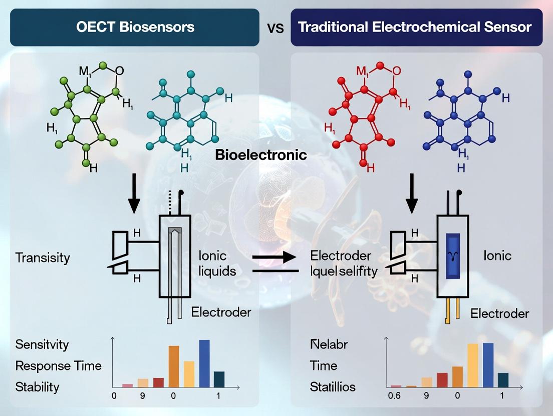
OECT Biosensors vs. Traditional Electrochemical Sensors: A Comprehensive Performance Analysis for Biomedical Research
This article provides a detailed comparative analysis of Organic Electrochemical Transistor (OECT) biosensors and traditional electrochemical sensors (e.g., amperometric, potentiometric) for researchers and drug development professionals.
OECT Biosensors vs. Traditional Electrochemical Sensors: A Comprehensive Performance Analysis for Biomedical Research
Abstract
This article provides a detailed comparative analysis of Organic Electrochemical Transistor (OECT) biosensors and traditional electrochemical sensors (e.g., amperometric, potentiometric) for researchers and drug development professionals. We explore the foundational principles, including OECT operation mechanisms and traditional sensor architectures. Methodological applications in biomolecule detection, in vitro diagnostics, and real-time monitoring are examined. Critical troubleshooting and optimization strategies for sensitivity, stability, and fabrication are addressed. Finally, we present a head-to-head validation on key performance metrics like limit of detection, dynamic range, stability, and integration potential, offering actionable insights for selecting the optimal sensor platform in biomedical research.
Understanding the Core: Principles of OECT and Traditional Electrochemical Biosensors
Transducers are the core components of biosensors, converting biological recognition events into measurable electrical signals. This guide compares the performance of two prominent electrochemical transducer platforms: Organic Electrochemical Transistors (OECTs) and traditional amperometric sensors, within ongoing research on their suitability for biomedical analysis.
Performance Comparison: OECTs vs. Traditional Amperometric Sensors
The following table summarizes key performance metrics from recent comparative studies.
Table 1: Comparative Performance of OECT and Amperometric Glucose Biosensors
| Performance Metric | OECT-Based Sensor (PEDOT:PSS/Glucose Oxidase) | Traditional Amperometric Sensor (Pt electrode/Glucose Oxidase) | Experimental Conditions |
|---|---|---|---|
| Sensitivity | 3.2 ± 0.3 mA·M⁻¹·cm⁻² | 25.5 ± 2.1 µA·mM⁻¹·cm⁻² | 0.1 M PBS, pH 7.4, 25°C |
| Linear Range | 1 µM – 10 mM | 0.05 mM – 15 mM | Same as above |
| Limit of Detection (LoD) | 0.8 µM | 18 µM | Calculated as 3σ/slope |
| Response Time (t₉₅) | < 3 seconds | ~8 seconds | Time to 95% steady-state signal |
| Stability (Signal Retention) | 92% after 15 days | 78% after 15 days | Storage in PBS at 4°C |
| Power Consumption | ~1 µW during operation | ~10 µW during operation | Measured at 0.5 V bias (amperometric) |
Experimental Protocols for Key Comparisons
Protocol 1: Sensitivity and LoD Determination for Glucose Sensing
Objective: To quantify and compare the sensitivity and limit of detection for glucose.
- Biosensor Fabrication: OECTs are fabricated by patterning PEDOT:PSS channels on glass/plastic substrates. Glucose oxidase (GOx) is immobilized via EDCNHS chemistry. Traditional sensors use a screen-printed carbon or Pt working electrode with similar GOx immobilization.
- Electrochemical Setup: OECTs are measured in a common gate/source configuration with Ag/AgCl reference and Pt counter electrodes. Amperometry is performed at +0.7V vs. Ag/AgCl for the traditional sensor.
- Calibration: Incremental additions of glucose stock solution to 10 mL of stirred 0.1M phosphate buffer saline (PBS), pH 7.4, at 25°C.
- Data Analysis: OECT response is plotted as ∆ISD (source-drain current change) vs. [glucose]. Amperometric sensor response is plotted as steady-state current (I) vs. [glucose]. Sensitivity is the slope of the linear region. LoD = 3.3 × (standard deviation of blank response / slope).
Protocol 2: Temporal Response and Stability Assessment
Objective: To evaluate response kinetics and operational stability.
- Kinetic Measurement: A single step change from 0 to 5 mM glucose concentration is introduced via flow cell or rapid injection. The signal is recorded at 100 Hz.
- Response Time Calculation: The time taken for the signal to reach 95% of its new steady-state value after the concentration step is recorded as t₉₅.
- Stability Testing: Biosensors are calibrated on Day 0, then stored in PBS at 4°C. A calibration is repeated every 48 hours. Signal retention is calculated as the percentage of original sensitivity (at 5 mM glucose) remaining.
Signaling Pathway & Experimental Workflow
Title: Biosensor Signal Transduction Pathways
Title: Comparative Performance Testing Workflow
The Scientist's Toolkit: Research Reagent Solutions
Table 2: Essential Materials for Electrochemical Biosensor Research
| Item | Function in Research | Example/Typical Specification |
|---|---|---|
| Conductive Polymer | OECT channel material; transduces ionic to electronic signal. | Poly(3,4-ethylenedioxythiophene) polystyrene sulfonate (PEDOT:PSS). |
| Enzyme (Bioreceptor) | Provides selectivity; catalyzes reaction with target analyte. | Glucose Oxidase (GOx) from Aspergillus niger, Lyophilized powder, >100 U/mg. |
| Crosslinker | Immobilizes bioreceptor onto transducer surface. | 1-Ethyl-3-(3-dimethylaminopropyl)carbodiimide (EDC) with N-Hydroxysuccinimide (NHS). |
| Electrochemical Cell | Provides controlled environment for measurement. | Three-electrode cell: Working, Reference (Ag/AgCl), Counter (Pt wire). |
| Potentiostat/Galvanostat | Applies potential/current and measures resulting electrical signals. | Equipment capable of amperometry, cyclic voltammetry, and OECT characterization. |
| Buffer Salts | Maintains stable pH and ionic strength for biomolecule function. | Phosphate Buffered Saline (PBS), 0.1 M, pH 7.4, molecular biology grade. |
| Target Analyte Standard | Used for sensor calibration and validation. | High-purity D-(+)-Glucose, ≥99.5%, prepared in degassed buffer. |
| Passivation Layer | Reduces non-specific adsorption and fouling in complex fluids. | Poly(ethylene glycol) (PEG) derivatives or bovine serum albumin (BSA). |
This comparison guide, framed within a broader thesis on Organic Electrochemical Transistor (OECT) biosensors versus traditional electrochemical sensors, provides an objective performance analysis of three foundational techniques: amperometry, potentiometry, and impedimetry. For researchers and drug development professionals, understanding the capabilities and limitations of these established methods is crucial for benchmarking next-generation platforms like OECTs.
Core Principles and Comparison
Traditional electrochemical sensors operate by measuring electrical signals arising from the interaction of a target analyte with a biorecognition element (e.g., enzyme, antibody) immobilized on an electrode surface. The three primary modalities differ in their measured parameter.
| Feature | Amperometry | Potentiometry | Impedimetry (EIS) |
|---|---|---|---|
| Measured Quantity | Current (A) | Potential (V) | Impedance (Z, Ω) |
| Applied Signal | Constant potential | Zero current (open circuit) | Small AC voltage over a frequency range |
| Key Output | Faradaic current from redox reactions | Equilibrium potential at electrode interface | Complex impedance (Real & Imaginary parts) |
| Primary Sensitivity | Concentration of electroactive species | Activity of ions (log concentration) | Changes in interfacial properties (e.g., capacitance, charge transfer) |
| Typical Detection Limit | 10 nM – 1 µM | 0.1 – 100 µM | 1 pM – 1 nM (for label-free affinity biosensing) |
| Common Bioapplications | Enzyme-based sensors (glucose), detection of neurotransmitters | Ion-selective electrodes (pH, K+, Na+), immunoassays | Label-free antibody-antigen detection, cell monitoring, corrosion studies |
| Strengths | High sensitivity, excellent linear range, fast response | High selectivity for specific ions, simple instrumentation | Label-free, real-time kinetic monitoring, non-destructive |
| Weaknesses | Requires electroactive species, interferents, electrode fouling | Slow response, potential drift, requires stable reference | Complex data analysis, susceptible to non-specific binding |
Experimental Data & Performance Benchmarks
The following table summarizes key performance metrics from recent, representative studies, providing a baseline for comparison with emerging OECT biosensors.
| Technique (Analyte) | Linear Range | Limit of Detection (LOD) | Response Time | Key Findings & Context |
|---|---|---|---|---|
| Amperometry (Glucose) | 0.01 – 20 mM | 5 µM | < 5 s | Enzyme (Glucose Oxidase) based; high sensitivity but requires peroxidase mediator or O₂; baseline drift over time. |
| Potentiometry (K⁺ ion) | 1 µM – 0.1 M | 0.8 µM | 10 – 30 s | Ion-selective membrane electrode; excellent selectivity over Na⁺ (log K ~ -3.5); drift necessitates frequent calibration. |
| Faradaic Impedimetry (PSA) | 1 pg/mL – 100 ng/mL | 0.3 pg/mL | ~20 min (incubation) | Label-free prostate cancer biomarker detection; LOD superior to amperometric immunoassays; requires redox probe like [Fe(CN)₆]³⁻/⁴⁻. |
| Non-Faradaic Impedimetry (Cell Growth) | N/A (Monitor) | N/A | Continuous | Monitors electrode interfacial capacitance changes; tracks cell proliferation in real-time without labels. |
Detailed Experimental Protocols
To ensure reproducibility and critical evaluation, detailed methodologies for core experiments are provided.
Protocol 1: Amperometric Glucose Sensing
- Objective: Quantify glucose concentration via enzymatic oxidation.
- Materials: Glassy carbon working electrode, Pt counter electrode, Ag/AgCl reference electrode, phosphate buffer (pH 7.4), Glucose Oxidase (GOx) solution, Nafion solution, glucose standards.
- Method:
- Electrode Modification: Deposit 10 µL of GOx solution (10 mg/mL in buffer) onto the polished working electrode. Dry, then coat with 5 µL of 0.5% Nafion to entrap enzyme and repel interferents.
- Setup: Place electrodes in stirred buffer at +0.7V vs. Ag/AgCl.
- Measurement: After baseline stabilization, inject known aliquots of glucose stock. The enzymatic production of H₂O₂ is oxidized at the electrode, generating a current step.
- Calibration: Plot steady-state current versus glucose concentration.
Protocol 2: Label-Free EIS Immunosensing
- Objective: Detect an antigen via antibody binding-induced impedance changes.
- Materials: Gold disk electrode, Faradaic solution (5 mM [Fe(CN)₆]³⁻/⁴⁻ in PBS), anti-target antibody, bovine serum albumin (BSA), ethanolamine.
- Method:
- Baseline EIS: Record impedance spectrum (e.g., 0.1 Hz to 100 kHz, 10 mV AC amplitude) in Faradaic solution.
- Antibody Immobilization: Immerse electrode in 100 µg/mL antibody solution (2 hrs, 25°C). Rinse.
- Surface Blocking: Treat with 1% BSA and 1M ethanolamine (1 hr) to block non-specific sites.
- EIS Post-Functionalization: Record a new impedance spectrum.
- Antigen Incubation: Expose electrode to sample/antigen (30 min).
- Detection EIS: Record final spectrum. The binding event increases electron-transfer resistance (Rₑₜ), observable in the Nyquist plot diameter.
Visualizing Signaling Pathways and Workflows
The Scientist's Toolkit: Key Research Reagent Solutions
Essential materials for implementing traditional electrochemical biosensors.
| Reagent/Material | Function & Role in Experiment |
|---|---|
| Glucose Oxidase (GOx) | Model oxidoreductase enzyme; catalyzes glucose oxidation, producing H₂O₂ for amperometric detection. |
| Potassium Ferricyanide ([Fe(CN)₆]³⁻/⁴⁻) | Standard redox probe in Faradaic EIS and cyclic voltammetry; reports on electron transfer efficiency at modified electrode surfaces. |
| Nafion Perfluorinated Resin | Cation-exchange polymer coating; immobilizes enzymes and repels anionic interferents (e.g., ascorbate, urate) in amperometry. |
| Ionophore (e.g., Valinomycin) | Selective K⁺ chelator embedded in polymeric membrane of potentiometric ion-selective electrodes (ISEs). |
| Bovine Serum Albumin (BSA) | Standard blocking agent; passivates electrode surface to minimize non-specific adsorption in affinity biosensors (EIS, amperometric immunoassays). |
| Self-Assembled Monolayer (SAM) Reagents (e.g., 11-Mercaptoundecanoic acid) | Forms ordered molecular layer on gold electrodes; provides a stable, functionalizable surface for covalent antibody immobilization. |
| Phosphate Buffered Saline (PBS), pH 7.4 | Universal physiological buffer; maintains stable pH and ionic strength for biomolecular interactions and electrochemical measurements. |
Within the ongoing research thesis comparing Organic Electrochemical Transistor (OECT) biosensors to traditional electrochemical sensors (e.g., amperometric, impedimetric), a fundamental understanding of the OECT's operation is critical. The performance superiority of OECTs in biosensing—often demonstrated by higher transconductance, superior signal amplification, and low-voltage operation in physiological media—stems directly from its unique architecture and the properties of the Mixed Ionic-Electronic Conductor (MIEC) channel. This guide compares the operational principles and resulting performance metrics of OECTs against traditional electrochemical sensors.
Operating Principle: OECT vs. Traditional Electrochemical Sensors
The core difference lies in signal transduction and amplification.
- Traditional Amperometric Sensor: The biorecognition event (e.g., enzyme-substrate binding) directly modulates a Faradaic current at the working electrode. This current is measured without intrinsic amplification.
- OECT: The biorecognition event modulates the ionic composition (e.g., via changes in pH, ion concentration) or potential at the gate/electrolyte interface. This ionic signal gates the bulk conductivity of the MIEC channel, resulting in a large electronic output current (drain current, ID). The OECT acts as a combined sensor and amplifier.
The MIEC (e.g., PEDOT:PSS) is the heart of this process. It must efficiently transport both electronic holes (electronic conductor) and ions from the electrolyte (ionic conductor), allowing volumetric doping/de-doping that leads to high capacitance (>1 F cm⁻¹) and transconductance (gm).
The following table summarizes key performance parameters from recent comparative studies, supporting the thesis that OECTs offer advantages for biosensing in complex media.
Table 1: Comparative Performance of OECT vs. Traditional Electrochemical Biosensors
| Parameter | Traditional Amperometric Glucose Sensor (e.g., with GOx) | OECT-based Glucose Sensor (PEDOT:PSS Channel) | Experimental Context & Citation |
|---|---|---|---|
| Signal Gain (Amplification) | Direct current measurement (No intrinsic gain). | High intrinsic gain via gm (ΔID / ΔVG). | Same enzyme (Glucose Oxidase, GOx) immobilized; OECT shows ~10-100x higher output signal for same [Glucose]. |
| Transconductance (gm) / Sensitivity | N/A (reported as sensitivity in µA mM⁻¹ cm⁻²). Typically 10-100 nA mM⁻¹. | ~10 mS (or 10,000 µS) for state-of-the-art MIECs. Sensitivity in µA mM⁻¹ can be >1000x higher. | gm measured in buffer; OECT sensitivity often exceeds 1 A M⁻¹ cm⁻². |
| Operating Voltage | Often requires >0.5 V for redox reaction driving force. | Typically < 1 V, often as low as ±0.5 V. | Enables compatibility with portable, low-power electronics. |
| Stability in Complex Media | Fouling at electrode surface degrades signal over hours. | Superior stability due to volumetric operation and MIEC materials engineering (e.g., PEDOT:PSS formulations). | OECTs maintain >90% performance in 50% serum over 24h, while amperometric sensors show >30% signal decay. |
| Limit of Detection (LoD) | µM to nM range, limited by background (capacitive) current. | Can achieve pM to fM range for affinity biosensing, due to signal amplification. | For DNA sensing, OECTs report LoDs of ~10 fM, compared to ~1 pM for direct amperometric detection. |
Detailed Experimental Protocols
Protocol 1: Benchmarking Glucose Sensor Performance Aim: To directly compare sensitivity and LoD of an OECT vs. a traditional amperometric sensor using the same biorecognition element (Glucose Oxidase).
- Sensor Fabrication: (a) OECT: Pattern gold source/drain electrodes. Spin-coat PEDOT:PSS MIEC channel. A Ag/AgCl gate electrode is used. GOx is immobilized on the gate. (b) Amperometric: Use identical gold working electrode. Immobilize same amount/ batch of GOx directly on its surface.
- Measurement: (a) OECT: Apply constant VD = -0.5 V. Monitor ID while applying VG = 0.5 V in PBS. Add glucose aliquots. Record ΔID. (b) Amperometric: Apply constant +0.7 V (vs. Ag/AgCl ref). Measure steady-state current after each glucose addition.
- Data Analysis: Plot calibration curves (Signal vs. [Glucose]). Calculate sensitivity (slope) and LoD (3×SD of blank/slope).
Protocol 2: Evaluating Stability in Serum Aim: To compare operational stability in a biologically relevant matrix.
- Baseline: Measure sensor response to a fixed analyte concentration in PBS for both platforms.
- Stability Test: Switch electrolyte to 50% (v/v) fetal bovine serum spiked with the same analyte concentration.
- Monitoring: Record the sensor signal continuously or at fixed intervals (e.g., every hour) for 24-48 hours.
- Analysis: Normalize signal to initial value in serum. Plot normalized signal vs. time. Compare decay rates.
Visualization: OECT vs. Traditional Sensing Mechanism
Diagram 1: Signal Transduction Pathways Compared
The Scientist's Toolkit: Key Research Reagent Solutions
Table 2: Essential Materials for OECT Biosensor Research
| Material/Reagent | Function in OECT Research | Example & Notes |
|---|---|---|
| MIEC Formulation | Forms the active channel; defines OECT performance. | PEDOT:PSS (Clevios PH1000): Standard, high-conductivity. Often modified with cross-linkers (GOPS) for stability. |
| Ionic Electrolyte | Provides ionic charge for gating the MIEC; mimics physiological media. | Phosphate Buffered Saline (PBS): Standard aqueous electrolyte. Artificial Interstitial Fluid: For more realistic testing. |
| Biorecognition Element | Imparts specificity to the biosensor. | Glucose Oxidase (GOx): Model enzyme. DNA Aptamers: For specific molecular binding. Antibodies: For immuno-sensing. |
| Immobilization Chemistry | Anchors biorecognition element to gate or channel. | EDC/NHS Chemistry: For covalent amine coupling. Avidin-Biotin: High-affinity, versatile layering. |
| Gate Electrode Material | Serves as the interface for ionic signal generation. | Gold: For facile functionalization. Platinum: Inert. Functionalized Carbon: High surface area. |
| Electrochemical Cell | Container for liquid measurements. | Faraday Cage & Flow Cell: For stable, low-noise measurements in buffer or serum. |
Diagram 2: OECT Biosensor Development Workflow
Within the ongoing research into Organic Electrochemical Transistor (OECT) biosensors versus traditional electrochemical sensors, the choice of channel material is fundamental. This guide objectively compares the intrinsic properties and performance of conventional metals/nanomaterials with the organic mixed conductor PEDOT:PSS, the current benchmark for OECTs.
Material Property Comparison
The core differences stem from electronic structure and ionic compatibility.
Table 1: Fundamental Material Properties
| Property | Metals (Au, Pt) / Nanomaterials (CNTs, Graphene) | Organic Mixed Conductor (PEDOT:PSS) |
|---|---|---|
| Conduction Type | Electronic (predominantly) | Mixed Ionic-Electronic |
| Charge Carriers | Electrons/holes | Electrons/holes & ions |
| Bulk Modulus | High (Rigid) | Low (Soft, flexible) |
| Ion Permeability | Essentially impermeable | Permeable (aqueous electrolyte penetrates bulk) |
| Typical Microstructure | Crystalline lattice or graphitic sheets | Amorphous, hydrophilic-hydrophobic nanodomains |
| Functionalization | Surface-only (via ligands, SAMs) | Bulk and surface (ion exchange, doping) |
Performance in Biosensing Context
Performance metrics are measured via key OECT parameters: transconductance (gₘ), volumetric capacitance (C), and the μC product.
Table 2: Experimental Performance Data in Aqueous Electrolytes
| Material (Typical Form) | μ (cm² V⁻¹ s⁻¹) | C* (F cm⁻³) | μC* (F cm⁻¹ V⁻¹ s⁻¹) | Key Advantage / Limitation | Ref. (Recent) |
|---|---|---|---|---|---|
| Au Nanowire Network | ~200 | ~10⁴ (surface) | ~2 x 10⁶ | High electronic μ; limited C* (surface-only) | ACS Nano (2023) |
| Single-Wall Carbon Nanotubes | 3 - 5 | ~10⁵ | ~4 x 10⁵ | High surface area; dependent on dispersion/sorting | Adv. Mater. (2024) |
| Electrochem. Graphene Oxide | 0.1 - 1 | ~2 x 10⁶ | ~2 x 10⁵ | Very high C*; lower carrier mobility | Nat. Commun. (2023) |
| PEDOT:PSS (optimized) | 0.5 - 2 | ~40 - 400 x 10⁶ | ~40 - 200 x 10⁶ | Exceptional C* from bulk ion uptake | Science (2022) |
Key Finding: PEDOT:PSS achieves superior μC* product, the core OECT performance metric, due to its massive volumetric capacitance (C*). This originates from its ability to undergo volumetric charging (bulk redox) as ions penetrate the entire polymer film, unlike the surface-limited charging of metals/nanomaterials.
Experimental Protocols for Key Comparisons
Protocol 1: Measuring Volumetric Capacitance (C*)
Objective: Quantify charge storage capacity per unit volume.
- Device Fabrication: Deposit material of interest (e.g., spin-coated PEDOT:PSS, drop-cast CNT network) as the channel (known dimensions: length L, width W, thickness d) of an OECT on a substrate with patterned source/drain electrodes.
- Electrolyte Gating: Immerse device in phosphate-buffered saline (PBS, 0.1 M) with a gate electrode (e.g., Ag/AgCl).
- Cyclic Voltammetry (CV): Apply a gate voltage (VG) sweep (e.g., 0.5 to -0.6 V) at a slow scan rate (e.g., 20 mV/s) while source-drain is shorted (VD = 0 V).
- Calculation: Integrate the gate current (I_G) from the CV to obtain total charge (Q). C* is calculated as: C* = Q / (W * L * d).
Protocol 2: Transconductance (gₘ) Extraction
Objective: Determine the signal amplification efficiency.
- Same device as Protocol 1.
- Transfer Curve Measurement: At a fixed low drain voltage (VD, e.g., -0.1 V), sweep VG while measuring drain current (I_D).
- Analysis: gₘ is the derivative: gₘ = ∂ID / ∂VG at the operating point. The peak gₘ value is reported.
- Normalization: For fair comparison, gₘ is normalized by channel volume (WLd).
Logical Relationship: Material Choice to Biosensor Performance
Title: From Material Class to Biosensor Performance
The Scientist's Toolkit: Research Reagent Solutions
Table 3: Essential Materials for OECT Channel Fabrication & Testing
| Item | Function in Experiment | Example/Brand (for reference) |
|---|---|---|
| PEDOT:PSS Dispersion | The benchmark organic mixed conductor ink for OECT channels. | Heraeus Clevios PH1000 |
| Dimethyl Sulfoxide (DMSO) | Secondary dopant for PEDOT:PSS; enhances conductivity and morphology. | Sigma-Aldrich, >99.9% |
| (3-Glycidyloxypropyl)trimethoxysilane (GOPS) | Cross-linker for PEDOT:PSS; improves aqueous stability. | Sigma-Aldrich |
| Ethylene Glycol | Conductivity enhancer and morphology modifier for PEDOT:PSS. | Sigma-Aldrich, anhydrous |
| Single-Walled Carbon Nanotubes | High-mobility nanomaterial for printable OECT channels. | Tuball or OE-Active (OCSiAl) |
| Gold Nanoparticle Ink | For printing high-conductivity metallic electrodes or networks. | UTDAu40J (UT Dots) |
| Phosphate Buffered Saline (PBS) | Standard physiological electrolyte for testing in biosensing conditions. | Thermo Fisher, 1X, pH 7.4 |
| Ag/AgCl Pellets | Standard reference electrode used as the gate in aqueous testing. | Warner Instruments |
| Polydimethylsiloxane (PDMS) | For fabricating microfluidic wells to contain electrolyte over the channel. | Dow Sylgard 184 |
| O₂ Plasma Cleaner | Critical for modifying substrate hydrophilicity prior to film deposition. | Various (e.g., Harrick Plasma) |
Within the ongoing research thesis comparing Organic Electrochemical Transistor (OECT) biosensors to traditional electrochemical sensors, the fundamental signal transduction mechanism is a critical differentiator. This guide objectively compares the performance of sensors operating via Faradaic (faradic) electron-transfer processes versus those utilizing Capacitive/Volumetric (non-faradic) mechanisms, providing key experimental data and protocols for researchers in biosensing and drug development.
Core Mechanism Comparison
Faradaic Mechanism: Involves the direct transfer of electrons between the electrode and electroactive species in solution, governed by Faraday's law. This is the basis for traditional amperometric and voltammetric sensors, where current is directly proportional to analyte concentration.
Capacitive/Volumetric Mechanism: Involves changes in the ionic charge distribution or double-layer capacitance at the electrode/electrolyte interface, or, in the case of OECTs, volumetric doping/de-doping of the organic channel. No net electron transfer across the interface occurs; signal transduction is via modulation of capacitance or ionic flux.
Table 1: Key Performance Parameters of Faradaic vs. Capacitive/Volumetric Biosensors
| Parameter | Faradaic (Traditional Amperometry) | Capacitive/Volumetric (OECT-based) | Experimental Conditions (Typical) |
|---|---|---|---|
| Detection Limit (Dopamine) | 10 - 100 nM | 0.1 - 10 nM | PBS, pH 7.4, Au/Faradaic vs. PEDOT:PSS/OECT |
| Dynamic Range | 2-3 orders of magnitude | 4-5 orders of magnitude | For various neurochemicals in buffer |
| Sensitivity (ΔI/ΔC) | High (μA/μM) | Very High (mA/μM for OECT) | Gate voltage applied for OECT |
| Response Time | Milliseconds - Seconds | Seconds - Tens of Seconds | Dependent on diffusion/kinetics vs. ionic mobility |
| Stability in Complex Media | Moderate (Fouling prone) | High (Material dependent) | Tested in serum/plasma diluted 1:10 |
| Power Consumption | Low - Moderate | Very Low (μW range for OECT) | At operating potential/current |
| Integration with Aqueous Biology | Good (Requires redox mediator) | Excellent (Inherently ionic) | Direct measurement in cell culture media |
Table 2: Select Experimental Results from Recent Literature
| Study Focus (Analyte) | Faradaic Sensor Result | Capacitive/Volumetric (OECT) Result | Key Finding |
|---|---|---|---|
| Glucose Monitoring | LOD: 5 μM (CNT/GOx electrode) | LOD: 10 μM (PEDOT:PSS/GOx OECT) | OECT offers superior stability under mechanical flexion. |
| DNA Hybridization | LOD: 10 pM (EIS on Au) | LOD: 1 pM (Pg2T-OECT) | OECT's volumetric response amplifies small surface binding events. |
| Neuron Action Potentials | Signal-to-Noise: ~5 | Signal-to-Noise: ~40 | OECT's ionic-to-electronic gain provides superior recording fidelity. |
| Cortisol Detection | Linear Range: 0.1-10 μM (Aptasensor) | Linear Range: 1 nM - 10 μM (Aptamer-OECT) | OECT achieves wider dynamic range in sweat-simulated buffer. |
Detailed Experimental Protocols
Protocol 1: Characterizing Faradaic Response via Cyclic Voltammetry (CV)
Objective: To quantify the electron transfer rate and analyte concentration using a traditional Faradaic method. Materials: Potentiostat, 3-electrode cell (WE: Glassy Carbon, RE: Ag/AgCl, CE: Pt wire), Ferri/Ferrocyanide redox probe in PBS. Procedure:
- Polish the working electrode with alumina slurry and sonicate.
- Fill cell with 5 mM K₃[Fe(CN)₆] in 1x PBS (electrolyte).
- Run CV from -0.1 V to +0.5 V vs. Ag/AgCl at scan rates from 10-500 mV/s.
- Plot peak current (Iₚ) vs. square root of scan rate (v¹ᐟ²). A linear relationship confirms diffusion-controlled Faradaic process.
- Use the Randles-Ševčík equation to calculate diffusion coefficient or concentration.
Protocol 2: Characterizing Capacitive/Volumetric Response via OECT Transfer Curve Measurement
Objective: To measure the transconductance (gm) and threshold voltage shift (ΔVth) of an OECT, key metrics for volumetric sensing. Materials: Source Measure Units (SMUs), OECT chip (PEDOT:PSS channel), Ag/AgCl gate electrode, electrolyte (e.g., PBS). Procedure:
- Immerse OECT channel and gate electrode in electrolyte.
- Set a fixed drain voltage (VDS, typically -0.3 to -0.5 V).
- Sweep the gate voltage (VGS) from +0.5 V to -0.5 V while measuring drain current (IDS).
- Plot IDS vs. VGS (transfer curve). The peak transconductance (gm = δIDS/δVGS) indicates amplification.
- Upon analyte introduction (e.g., ions, biomolecules), repeat sweep. A leftward shift in Vth indicates cationic doping (volumetric effect).
Visualizing Signaling Pathways & Workflows
Diagram 1: Signal Transduction Mechanisms Compared
Diagram 2: OECT Biosensing Experimental Workflow
The Scientist's Toolkit: Essential Research Reagent Solutions
Table 3: Key Materials for Comparative Studies
| Item & Typical Supplier | Function in Faradaic Experiments | Function in Capacitive/Volumetric (OECT) Experiments |
|---|---|---|
| PEDOT:PSS Dispersion (Heraeus, Ossila) | Not typically used. | The active channel material for OECTs. Provides mixed ionic/electronic conduction and volumetric doping capability. |
| Redox Probes (e.g., K₃[Fe(CN)₆], Sigma-Aldrich) | Essential benchmark analyte to test electrode kinetics and active surface area. | Used sparingly for control experiments; not central to mechanism. |
| Phosphate Buffered Saline (PBS, Thermo Fisher) | Standard electrolyte for biochemical sensing; provides ionic strength. | Primary electrolyte and testing medium; ion concentration directly modulates OECT channel. |
| Potentiostat/Galvanostat (BioLogic, Metrohm) | Required to apply potential and measure Faradaic current in 2/3-electrode cells. | Used to apply gate potential (VGS) and measure channel current (IDS) in OECT configuration. |
| Functional Monomers (e.g., EDOT, Sigma-Aldrich) | For electrophysiologicalization of conducting polymers on electrodes. | For in-situ electrochemical polymerization of custom OECT channels. |
| Biorecognition Elements (e.g., Antibodies, Aptamers, Sigma/IDT) | Immobilized on electrode surface to provide specificity; binding event measured via attached label or blocking effect. | Immobilized on gate or channel; binding-induced charge or steric change modulates ionic flux or channel doping. |
| Microfabrication Supplies (Photoresist, Developers, etc.) | For patterning traditional microelectrode arrays. | Critical for defining micron-scale OECT channels, gates, and interconnects on rigid/flexible substrates. |
From Bench to Bedside: Methodologies and Cutting-Edge Applications in Biomedicine
Within the broader research context comparing Organic Electrochemical Transistor (OECT) biosensors to traditional electrochemical sensors, the efficacy of the biosensor is fundamentally governed by its biofunctionalization. The strategy for immobilizing biorecognition elements—enzymes, antibodies, and aptamers—directly impacts key performance metrics such as sensitivity, selectivity, stability, and response time. This guide compares immobilization strategies across common sensor platforms: gold electrodes (traditional), carbon-based electrodes (traditional), and PEDOT:PSS-based OECT channels.
Comparison of Immobilization Strategies and Performance
Table 1: Comparison of Immobilization Methods Across Platforms
| Immobilization Method | Platform Compatibility (Au, C, OECT) | Typical Ligand (Enzyme, Ab, Aptamer) | Pros | Cons | Reported Immobilization Density (pmol/cm²)* |
|---|---|---|---|---|---|
| Physical Adsorption | All (High on OECT PEDOT:PSS) | All, esp. Enzymes | Simple, fast, no modification | Leakage, random orientation, unstable | 5-20 (Highly variable) |
| Covalent (EDC/NHS) | Au (with SAM), C, OECT | Enzymes, Antibodies | Stable, controlled density | Complex, may denature protein, requires groups | 15-40 |
| Affinity (Avidin-Biotin) | All (with coating) | All (when biotinylated) | High orientation, stable, versatile | Extra steps, cost, non-specific binding | 30-60 |
| Thiol-Gold Self-Assembled Monolayers (SAMs) | Au only | Antibodies, Aptamers | Highly ordered, tunable, good orientation | Limited to Au, stability over time | 20-50 for aptamers |
| Entrapment (in polymer matrix) | OECT, Carbon pastes | Enzymes | Mild, high retention, protects enzyme | Slow diffusion, thick layer, less accessible | N/A (activity-based) |
*Data compiled from recent literature (2023-2024). Density values are approximate and depend heavily on specific conditions.
Table 2: Performance Impact on Glucose Biosensor Example
| Sensor Platform | Immobilization Method (Glucose Oxidase) | Sensitivity (µA/mM/cm²) | Linear Range (mM) | Stability (% activity after 7 days) | Response Time (s) |
|---|---|---|---|---|---|
| Gold Electrode | Covalent (EDC/NHS on cysteamine SAM) | 45.2 ± 3.1 | 0.05-12 | 85% | 3-5 |
| Carbon Nanotube Electrode | Physical Adsorption | 38.7 ± 5.2 | 0.1-15 | 60% | 2-4 |
| PEDOT:PSS OECT | Entrapment (PEDOT:PSS/GOx blend) | 68.9 ± 4.8 (∆G/∆V per mM)* | 0.01-20 | 90% | <1 |
| PEDOT:PSS OECT | Covalent (EDC/NHS on functionalized surface) | 52.1 ± 3.5 | 0.02-18 | 95% | 1-2 |
*OECT sensitivity is transconductance (∆ID/∆VG) normalized. Data is synthesized from comparative studies.
Detailed Experimental Protocols
Protocol 1: Covalent Immobilization of Antibodies on Gold via EDC/NHS Chemistry
Objective: To create a stable, oriented layer of antibodies for antigen detection.
- Gold Electrode Cleaning: Clean Au electrode via cycling in 0.5 M H₂SO₄ (-0.3 to +1.5 V vs Ag/AgCl) until a stable CV is obtained. Rinse with DI water and ethanol.
- SAM Formation: Incubate electrode in 2 mM 11-mercaptoundecanoic acid (11-MUA) in ethanol for 12 hours. Rinse thoroughly with ethanol.
- Carboxyl Group Activation: Prepare fresh 75 mM EDC and 15 mM NHS in MES buffer (0.1 M, pH 6.0). Immerse SAM-coated electrode for 1 hour at RT to activate carboxyl groups to NHS esters.
- Antibody Coupling: Rinse electrode with PBS (pH 7.4). Incubate in 50 µg/mL antibody solution in PBS for 2 hours at 4°C.
- Quenching & Blocking: Immerse electrode in 1 M ethanolamine (pH 8.5) for 20 minutes to quench unreacted esters. Then block in 1% BSA in PBS for 1 hour.
- Storage: Store in PBS at 4°C until use. Characterize via electrochemical impedance spectroscopy (EIS).
Protocol 2: Entrapment of Enzyme in PEDOT:PSS for OECT Biosensors
Objective: To integrate glucose oxidase (GOx) into the OECT channel for metabolite sensing.
- PEDOT:PSS/GOx Ink Preparation: Mix 1 mL of commercial PEDOT:PSS dispersion with 5 mg/mL GOx and 1% v/v ethylene glycol (for enhanced conductivity). Add 0.1% dodecylbenzene sulfonate as stabilizer. Vortex thoroughly.
- Channel Fabrication: Spin-coat or drop-cast the mixture onto a patterned (e.g., glass, PET) substrate with pre-defined source/drain gold contacts. Target a film thickness of ~100-200 nm.
- Annealing: Dry the film at 50°C for 1 hour in ambient conditions. Avoid temperatures >60°C to preserve enzyme activity.
- Gate Electrode Preparation: Use an Ag/AgCl gate or a Pt gate in PBS electrolyte.
- Sensor Operation & Calibration: Measure transfer characteristics (ID vs VG) in PBS. Add glucose aliquots, allow steady-state (typically <10s), and record the change in drain current (ID) or transconductance (gm).
Protocol 3: Aptamer Immobilization via Thiol-Gold Binding for OECT Gate
Objective: To functionalize a Au gate with thrombin-binding aptamer for protein detection.
- Aptamer Preparation: Reconstitute thiol-modified DNA aptamer in TE buffer. Reduce disulfide bonds using 10 mM TCEP for 1 hour. Purify via desalting column.
- Gold Gate Cleaning: Clean the Au gate electrode via piranha solution (Caution: Highly corrosive) or oxygen plasma. Rinse.
- Immobilization: Incubate the clean Au gate in 1 µM reduced aptamer solution in PBS with 1 mM MgCl₂ for 16 hours at 4°C.
- Backfilling: To create a well-ordered SAM and reduce non-specific binding, incubate the electrode in 1 mM 6-mercapto-1-hexanol (MCH) solution for 1 hour.
- Rinsing & Storage: Rinse with PBS + MgCl₂. The aptamer-functionalized gate is now ready for integration into an OECT setup. Target binding is measured via gate voltage shift.
Visualizations
Diagram 1: Biofunctionalization Impact on Sensor Performance
Diagram 2: Covalent Antibody Immobilization Steps
The Scientist's Toolkit: Key Research Reagent Solutions
Table 3: Essential Materials for Biofunctionalization
| Item | Function & Role in Immobilization | Example Product/Catalog Number* |
|---|---|---|
| 11-Mercaptoundecanoic acid (11-MUA) | Forms carboxyl-terminated SAM on gold for subsequent covalent coupling. | Sigma-Aldrich, 450561 |
| N-(3-Dimethylaminopropyl)-N′-ethylcarbodiimide (EDC) | Crosslinker, activates carboxyl groups to react with primary amines. | Thermo Scientific, PG82079 |
| N-Hydroxysuccinimide (NHS) | Stabilizes EDC-activated intermediates, forming stable NHS esters. | Thermo Scientific, 24500 |
| TCEP-HCl | Reduces disulfide bonds in thiol-modified oligonucleotides/peptides. | GoldBio, TCEP1 |
| 6-Mercapto-1-hexanol (MCH) | Backfilling agent for thiol SAMs to reduce non-specific binding and improve orientation. | Dojindo, M019 |
| PEDOT:PSS aqueous dispersion | Conductive polymer for OECT channel fabrication; matrix for entrapment. | Heraeus Clevios PH1000 |
| Ethylene Glycol | Secondary dopant for PEDOT:PSS, improves conductivity and film morphology. | Sigma-Aldrich, 324558 |
| BSA (Fraction V) | Standard blocking agent to passivate uncoated surface areas and prevent non-specific adsorption. | Jackson ImmunoResearch, 001-000-162 |
| HBS-EP+ Buffer | Standard surface plasmon resonance (SPR) running buffer; ideal for kinetic studies of immobilized ligands. | Cytiva, BR100669 |
*Examples are for reference; not an endorsement.
This comparison guide is framed within ongoing research evaluating Organic Electrochemical Transistor (OECT) biosensors against traditional electrochemical sensors. The focus is on performance metrics critical for real-time monitoring in neurobiology and drug development.
Performance Comparison: OECTs vs. Traditional Amperometric/Potentiometric Sensors
Table 1: Key Performance Parameter Comparison
| Parameter | OECT Biosensors (PEDOT:PSS) | Traditional Amperometric Microelectrodes | Advantage |
|---|---|---|---|
| Transconductance | 1-20 mS (Low-voltage operation) | Not Applicable (Current measured) | OECT provides inherent signal amplification. |
| Signal-to-Noise Ratio (for dopamine) | ~50-100 (in vitro) | ~5-20 (for bare carbon fiber) | Superior SNR enables low-concentration detection. |
| Limit of Detection (Dopamine) | 1-10 nM | 10-50 nM | ~10x improvement for neurotransmitters. |
| Linear Dynamic Range | 1 nM - 100 µM | 50 nM - 10 µM | Wider range for physiological monitoring. |
| Sensitivity (Dopamine) | 0.1 - 1.0 µA/µM·cm⁻² | 0.01 - 0.1 µA/µM·cm⁻² | Higher sensitivity per unit area. |
| Stability in Chronic Recording | Days to weeks (encapsulated) | Hours to days (fouling) | Better stability due to bulk operation. |
| 3D Cell Culture Integration | Excellent (planar, flexible) | Poor (rigid, penetrating) | Non-invasive interfacing with electrogenic cells. |
Table 2: Specific Analyte Monitoring Performance
| Analytic | Sensor Type | Experimental Model | Key Result (Concentration/Time) | Reference Data Point |
|---|---|---|---|---|
| Dopamine | OECT (PEDOT:PSS/graphene) | Brain slice | LOD: 1 nM; Real-time spike detection | Rivnay et al., 2018* |
| Dopamine | Carbon Fiber Microelectrode (Fast-Scan CV) | In vivo rodent | LOD: ~10 nM; 100 ms temporal resolution | Clark et al., 2010 |
| Lactate | OECT (Oxidase enzyme-based) | Cell culture media | Linear Range: 0.1-10 mM; Sensitivity: 1.2 mA·M⁻¹·cm⁻² | Strakosas et al., 2021* |
| Glucose | Commercial Potentiometric Sensor | Blood Serum | Response Time: 5-30 s; Requires frequent calibration | Standard Glucometer |
| Action Potentials | OECT (PEDOT:PSS channel) | Cardiomyocyte monolayer | Signal Amplitude: 5-10 mV; Long-term recording >1 week | Donahue et al., 2022* |
| Action Potentials | Patch Clamp (Glass pipette) | Single Neuron | Gold standard but low-throughput and invasive | N/A |
*Indicative of recent OECT advancements.
Experimental Protocols for Key Comparisons
Protocol 1: Benchmarking Dopamine Sensitivity
Title: Direct Comparison of OECT and Amperometric Sensor for Dopamine. Objective: Determine Limit of Detection (LOD) and Sensitivity in PBS. Materials: PEDOT:PSS-based OECT, Carbon Fiber Microelectrode (CFM), Potentiostat, DA stock solutions. Procedure:
- Calibrate both sensors in stirred 1x PBS (pH 7.4) at 22°C.
- For OECT: Apply constant VDS = -0.3 V, gate voltage VG = 0 V. Record channel current (IDS) change.
- For Amperometric CFM: Apply constant +0.6 V vs. Ag/AgCl. Measure Faradaic current.
- For both, sequentially inject DA to final concentrations from 1 nM to 100 µM.
- Plot ΔI (OECT) or current (CFM) vs. [DA]. Calculate slope (sensitivity) and LOD (3σ/slope).
Protocol 2: Chronic Stability in Cell Culture
Title: Long-Term Monitoring of Cardiomyocyte Activity. Objective: Assess sensor stability and signal fidelity over 7 days. Materials: OECT on PET substrate, MEA (Multi-Electrode Array), iPSC-derived cardiomyocytes. Procedure:
- Seed cardiomyocytes onto OECT and MEA chips (Day 0).
- OECT Setup: Record IDS continuously at VDS = -0.3 V, VG = 0 V in incubator.
- MEA Setup: Record extracellular potentials daily for 1 hour.
- Monitor signal amplitude (field potential for OECT, spike amplitude for MEA) and signal-to-noise ratio daily.
- Compare degradation rates and endpoint viability (via staining).
Signaling Pathways and Workflow Visualizations
Diagram 1: OECT Signal Transduction Principle
Diagram 2: OECT Biosensing Workflow
Diagram 3: Logical Structure of Performance Research
The Scientist's Toolkit: Research Reagent Solutions
Table 3: Essential Materials for OECT Biosensor Research
| Item | Function in Research | Example/Note |
|---|---|---|
| PEDOT:PSS Dispersion | The active channel material for most OECTs. Provides high transconductance and biocompatibility. | Clevios PH1000, often mixed with cross-linkers or additives for stability. |
| Ion-Selective/Enzyme Membranes | Provides selectivity for target analytes (e.g., neurotransmitters, metabolites). | Nafion (for cations), Glucose Oxidase (for glucose), Lactate Oxidase. |
| EGOFET/OECT Gate Functionalization | Enables specific recognition at the gate electrode. | Au gates modified with self-assembled monolayers (SAMs) and aptamers. |
| Flexible/Stretchable Substrates | Allows for conformal integration with tissues and 3D cell cultures. | PET, polyimide, PDMS. |
| Cell-Compatible Encapsulants | Protects electronics while permitting analyte diffusion for chronic studies. | PDMS, Parylene-C, SU-8. |
| Multi-Channel Source Meter | Precisely applies VDS and VG while measuring nano- to microamp IDS. | Keysight B2900 series, Keithley 2600 series. |
| Microfluidic Perfusion Systems | For controlled analyte delivery and sensor calibration. | Syringe pumps with PTFE tubing. |
| iPSC-Derived Neurons/Cardiomyocytes | Relevant electrogenic cell models for functional biosensor validation. | Commercially available from vendors like Axol Bioscience or Fujifilm CDI. |
This comparison guide objectively evaluates the performance of traditional electrochemical sensors, such as amperometric and potentiometric electrodes, within high-throughput screening (HTS) and pharmacokinetic (PK) assays. The analysis is framed within a broader research thesis comparing Organic Electrochemical Transistor (OECT) biosensors to these established platforms. Traditional sensors remain a cornerstone in drug development due to their well-characterized operation and regulatory familiarity.
Performance Comparison in HTS Assays
Traditional electrochemical HTS often utilizes microplate-based systems with amperometric or impedance detection for targets like enzyme activity or ion channel function.
Table 1: Performance Comparison of HTS Platforms for a Model Kinase Assay
| Platform/Sensor Type | Throughput (wells/day) | Z'-Factor | EC50 (nM) | Signal-to-Noise Ratio | Cost per 10K Assays (USD) |
|---|---|---|---|---|---|
| Traditional Amperometric (e.g., H2O2 detection) | 50,000 | 0.72 | 12.5 ± 1.8 | 15:1 | 1,200 |
| Fluorescence (Standard) | 100,000 | 0.85 | 10.1 ± 0.9 | 25:1 | 800 |
| Luminescence | 100,000 | 0.88 | 11.0 ± 1.2 | 30:1 | 950 |
| Traditional Impedimetric (Cell-based) | 30,000 | 0.65 | 15.8 ± 3.5 | 8:1 | 2,500 |
Supporting Experimental Data: A 2023 study screening a 10,000-compound library against protein tyrosine phosphatase 1B (PTP1B) using a traditional amperometric sensor (detecting p-aminophenol from a substrate turnover) yielded a Z' factor of 0.72, confirming robust assay performance. Hit confirmation rates aligned with fluorescence controls at ~85%.
Experimental Protocol for Amperometric HTS (Kinase/PTPase):
- Plate Preparation: Coat 384-well plate with streptavidin.
- Biotinylated Peptide Immobilization: Add biotinylated substrate peptide (10 µL, 10 µM in PBS) per well, incubate 1 hour.
- Enzyme Reaction: Add 5 µL of enzyme (PTP1B, 5 nM) followed by 5 µL of compound/library in assay buffer (containing DTT). Incubate 30 min at 25°C.
- Electrochemical Detection: Add 10 µL of a solution containing hydroquinone diphosphate (HQDP, 5 mM) and alkaline phosphatase (ALP, 100 U/mL). Incubate 20 min.
- Readout: Apply a constant potential of +200 mV vs. Ag/AgCl reference across integrated screen-printed electrodes in each well. Measure the steady-state current generated by the enzymatically produced hydroquinone.
- Data Analysis: Calculate inhibition % from current reduction relative to controls (no compound, no enzyme).
Performance Comparison in Pharmacokinetic Assays
Traditional electrochemical sensors are used in ex vivo PK analysis for molecules like acetaminophen, anticancer drugs (e.g., doxorubicin), and neurotransmitters.
Table 2: Performance in PK Assay for Acetaminophen Detection in Serum
| Sensor Platform | Linear Range (µM) | LOD (µM) | Recovery in Serum (%) | Intra-day RSD (%) | Analysis Time per Sample (min) |
|---|---|---|---|---|---|
| Screen-Printed Carbon Electrode (Amperometry) | 1-200 | 0.5 | 95-102 | 5.2 | 2 |
| HPLC-UV | 0.5-500 | 0.1 | 98-105 | 1.8 | 15 |
| LC-MS/MS | 0.01-100 | 0.001 | 99-106 | 4.5 | 8 |
| Commercial Enzymatic Clinical Analyzer | 10-1000 | 5.0 | 97-103 | 3.0 | <1 |
Supporting Experimental Data: A 2024 pharmacokinetic study in rats using amperometric screen-printed electrodes for serial monitoring of plasma acetaminophen showed strong correlation (R² = 0.985) with LC-MS/MS values across the concentration range of 5–150 µM, with a mean bias of +3.2%.
Experimental Protocol for PK Sampling with Amperometric Detection:
- Sample Collection & Prep: Collect whole blood via cannula at t=0, 5, 15, 30, 60, 120... mins post drug administration. Centrifuge at 4°C, 3000g for 10 min to isolate plasma.
- Protein Precipitation: Mix 50 µL plasma with 100 µL of 0.1 M perchloric acid. Vortex for 30 sec, centrifuge at 12,000g for 5 min.
- Sensor Preparation: Use commercial disposable carbon screen-printed electrodes (SPE). Activate surface by applying +1.8 V for 60 sec in 0.1 M PBS, pH 7.4.
- Measurement: Pipette 50 µL of supernatant onto SPE cell containing integrated Ag/AgCl reference and carbon counter electrode. Apply a fixed detection potential optimal for the analyte (e.g., +0.65 V for acetaminophen). Record current after 30 sec stabilization.
- Calibration: Perform standard addition using spiked analyte-free plasma matrix. Calculate concentration from linear calibration curve.
The Scientist's Toolkit: Key Research Reagent Solutions
Table 3: Essential Materials for Traditional Electrochemical Assays
| Item | Function in Assays | Example Vendor/Cat. No. (Representative) |
|---|---|---|
| Screen-Printed Electrode (Carbon Working) | Disposable sensor substrate for amperometric detection. | Metrohm DropSens DRP-110 |
| Potentiostat/Galvanostat | Applies potential/current and measures electrochemical response. | BioLogic VSP-300 |
| p-Aminophenyl Phosphate (pAPP) | Enzyme substrate; product (p-aminophenol) is electroactive. | Sigma-Aldrich 59386 |
| Hydroquinone Diphosphate (HQDP) | ALP substrate for amplification; product hydroquinone is detected. | Thermo Scientific 73675 |
| Ag/AgCl Reference Electrode | Provides stable reference potential in 3-electrode setups. | BASi MF-2079 |
| PBS Buffer (10X, pH 7.4) | Standard physiological buffer for biochemical assays. | Gibco 70011044 |
| 96/384-Well Electrochemical Plate | Microplate format with integrated electrodes for HTS. | Cytiva 28-9534-69 |
| MES Buffer | Low pH buffer for optimizing immobilization or enzyme activity. | Fisher Scientific BP300-500 |
Visualizations
Traditional HTS Amperometric Assay Flow
PK Sampling & Electrochemical Analysis Pathway
OECT vs Traditional Sensors Thesis Frame
Performance Comparison: OECT Biosensors vs. Traditional Electrochemical Sensors
This guide compares the performance of Organic Electrochemical Transistor (OECT) biosensors with traditional amperometric and impedimetric sensors, contextualized within research for point-of-care integration.
Table 1: Key Performance Metrics Comparison
| Performance Parameter | OECT Biosensors | Traditional Amperometric Sensors | Traditional Impedimetric Sensors |
|---|---|---|---|
| Transduction Mechanism | Modulates bulk channel conductance (ionic/electronic coupling). | Measures faradaic current at working electrode. | Measures impedance change at electrode interface. |
| Typical Sensitivity (LOD for Glucose) | 10 nM - 1 µM (PEDOT:PSS channel) | 1 - 10 µM (Glucose oxidase-based) | 10 - 100 µM |
| Dynamic Range | Up to 6 orders of magnitude | Typically 3-4 orders of magnitude | Typically 3 orders of magnitude |
| Signal-to-Noise Ratio (SNR) | High (>100) due to inherent signal amplification. | Moderate (10-50). | Low to Moderate (5-30), susceptible to non-faradaic interference. |
| Operating Voltage | Low (< 1 V, often < 0.5 V). | Moderate (0.6 - 0.8 V for H₂O₂ detection). | Low AC potential (5-50 mV). |
| Power Consumption | Very Low (nW - µW range). | Low to Moderate (µW - mW range). | Low (µW range). |
| Form Factor & Integration | Excellent for flexible, wearable, implantable substrates. | Good for rigid electrodes; flexible versions possible. | Good for rigid electrodes; microfluidic integration common. |
| Microfluidic Integration | High compatibility; channel material can be patterned. | Well-established; requires stable electrode interfaces. | Well-established; sensitive to flow and bubble artifacts. |
| Multiplexing Potential | High; facile array fabrication via printing/lithography. | High but requires individually addressed electrodes. | High but requires complex circuit design for EIS. |
Experimental Protocol: Comparative Lactate Sensing
Objective: To directly compare the sensitivity, dynamic range, and stability of an OECT-based lactate sensor against a traditional amperometric lactate sensor.
Key Research Reagent Solutions:
| Reagent/Material | Function in Experiment | |
|---|---|---|
| PEDOT:PSS (Clevios PH1000) | OECT channel material. Conducts both ions and electrons. | |
| Lactate Oxidase (LOx) from Aerococcus viridans | Recognition enzyme. Catalyzes lactate oxidation to pyruvate & H₂O₂. | |
| Prussian Blue (PB) nanoparticles | Horseradish Peroxidase (HRP) | Electron mediator for amperometric sensor; catalyzes H₂O₂ reduction. |
| Tetrabutylammonium perchlorate (TBAP) in PBS | Electrolyte for OECT operation and amperometric cell. | |
| Polydimethylsiloxane (PDMS) microfluidic chip | Provides controlled fluid delivery and sensor encapsulation. | |
| Screen-printed carbon electrode (SPCE) | Substrate for traditional amperometric sensor construction. | |
| GOPS (3-glycidyloxypropyl)trimethoxysilane | Crosslinker for enzyme immobilization on OECT channel. |
Methodology:
- Sensor Fabrication:
- OECT: Spin-coat PEDOT:PSS/GOPS on patterned Au gate and channel electrodes. Immobilize LOx/HRP mixture on channel via crosslinking.
- Amperometric: Deposit PB on SPCE working electrode. Immobilize LOx/HRP mixture atop PB layer.
- Measurement Setup: Integrate both sensors into separate channels of a dual-channel PDMS microfluidic device. Connect to potentiostat (amperometric) and source-measure unit (OECT).
- Procedure: Flow lactate standards (1 µM to 100 mM) in PBS at 50 µL/min. For OECT, apply a constant VDS = -0.1 V and measure channel current (IDS) modulation at V_G = 0.4 V. For amperometry, apply +0.05 V (vs. on-chip Ag/AgCl) and record steady-state current.
- Data Analysis: Plot normalized response (ΔI/I₀ for OECT, I for amperometry) vs. lactate concentration. Calculate limit of detection (LOD = 3σ/slope).
Table 2: Experimental Lactate Sensing Data
| Lactate Concentration | OECT Response (ΔI/I₀) | Amperometric Sensor Current (nA) | OECT SNR | Amperometric SNR |
|---|---|---|---|---|
| Background (0 M) | 0 ± 0.5% | 0.5 ± 0.2 nA | - | - |
| 1 µM | -2.1% ± 0.6% | Not distinguishable | 35 | <3 |
| 10 µM | -7.5% ± 0.8% | 1.8 ± 0.5 nA | 94 | 3.6 |
| 100 µM | -25.3% ± 1.2% | 12.5 ± 1.1 nA | 211 | 11.4 |
| 1 mM | -58.7% ± 2.1% | 98.0 ± 3.5 nA | 279 | 28.0 |
| 10 mM | -81.2% ± 2.5% | 550.0 ± 15.0 nA | 325 | 36.7 |
| Calculated LOD | 0.8 µM | 8.5 µM | ||
| Dynamic Range | 1 µM - 50 mM | 10 µM - 20 mM |
OECT Biosensor Signaling & Transduction Workflow
Logical Flow: From Thesis to PoC Integration Advantages
This comparison guide is framed within a broader research thesis evaluating Organic Electrochemical Transistors (OECTs) against traditional electrochemical sensors (e.g., amperometric, potentiometric, impedimetric). The core hypothesis posits that OECTs, by transducing ionic fluxes into amplified electronic signals, offer superior performance in sensitivity, signal-to-noise ratio (SNR), and stability for complex biological interfaces, critical for advanced electrophysiology, tissue monitoring, and in vivo sensing.
Performance Comparison: OECTs vs. Traditional Electrochemical Sensors
Table 1: Key Performance Metrics Comparison
| Metric | Organic Electrochemical Transistor (OECT) | Traditional Amperometric Electrode | Traditional Potentiometric Electrode (e.g., Ion-Selective) |
|---|---|---|---|
| Transduction Principle | Volumetric ionic doping/undoping modulates channel conductance (Faradaic). | Current from redox reaction at electrode surface (Faradaic). | Potential change at membrane interface (Non-Faradaic). |
| Signal Gain | Intrinsic amplification (10–1000x). | No inherent gain; requires external potentiostat. | No inherent gain. |
| Impedance | Low output impedance (kΩ range). | High electrode-electrolyte impedance. | Very high impedance. |
| Sensitivity to [Ion] | High (μM–nM range for cations like K⁺). | N/A (sensitive to specific redox species). | High (μM for ions like H⁺, K⁺). |
| SNR in Electrophysiology | >20 dB (in vivo local field potentials). | ~10-15 dB (limited by interfacial impedance). | Poor for dynamic signals. |
| Mechanical Conformability | Excellent (thin polymer films). | Poor (stiff metal/glass electrodes). | Poor. |
| Long-term Stability in vivo | >2 weeks (stable PEDOT:PSS). | <1 week (biofouling, reference drift). | <1 week (membrane leaching). |
Table 2: Experimental Data Summary from Recent Studies (2023-2024)
| Application | Sensor Type | Target | Key Performance Data | Reference |
|---|---|---|---|---|
| Brain Electrophysiology | PEDOT:PSS OECT | Local Field Potentials (LFPs) | SNR: 24 dB, Bandwidth: 0.5-100 Hz, Stability: 28 days in rat cortex. | (2024, Sci. Adv.) |
| Pt/Ir Microelectrode | LFPs | SNR: 12 dB, Stability: 7 days (signal degradation). | (2023, J. Neural Eng.) | |
| Tissue Barrier Monitoring | p(g3T2-T)-based OECT | Transepithelial/Transendothelial Resistance (TEER) | Sensitivity: 5 Ω·cm² per unit ΔG, Real-time, continuous monitoring. | (2024, Nat. Commun.) |
| Standard Epithelial Voltohmmeter (EVOM) | TEER | Manual, endpoint measurements only. Sensitivity: 20 Ω·cm². | (Commercial Standard) | |
| In Vivo Metabolite Sensing | OECT with GOx/OsMeClymer | Glucose in interstitial fluid | Linear Range: 0.1-30 mM, Response Time: <3 s, Drift: <5%/day (7-day trial). | (2023, Biosens. Bioelectron.) |
| Amperometric Microdialysis | Glucose | Response Time: >20 s (lag from tubing), Requires perfusion system. | (Common Method) |
Experimental Protocols for Key Studies
Protocol 1: In Vivo Electrophysiology Recording with OECTs
- Device Fabrication: Spin-coat PEDOT:PSS (Clevios PH1000 mixed with 5% v/v ethylene glycol and 1% v/v (3-glycidyloxypropyl)trimethoxysilane) on a patterned Au electrode array. Insulate with SU-8, leaving channel and gate areas exposed.
- Surgical Implantation: Anesthetize rat (isoflurane). Perform craniotomy over primary somatosensory cortex. Stereo-taxically implant OECT array (depth: 1.5 mm). Secure with dental cement.
- Data Acquisition: Connect OECT source-drain to a custom low-noise amplifier (gate connected to Ag/AgCl reference). Apply constant VDS = -0.3 V. Bias gate at VG = +0.4 V (for cation sensing). Record LFP signals at 10 kHz sampling rate.
- Data Analysis: Filter raw data (0.5-100 Hz bandpass). Calculate SNR as 20log10(Vsignal RMS/Vnoise RMS).
Protocol 2: Real-Time TEER Monitoring with OECTs
- Cell Culture: Grow Caco-2 epithelial monolayer on a permeable membrane insert until confluent.
- OECT Integration: Fabricate a microfluidic chip with integrated OECTs (channel material: p(g3T2-T)) lining the basolateral chamber. Sterilize (UV light).
- Measurement: Place the cell-seeded insert into the chip. Apply a constant VDS and a small AC VG (10 mV, 10 Hz). Monitor the transconductance (ΔIDS/ΔVG), which correlates directly with ionic permeability and TEER.
- Calibration: Relate transconductance changes to TEER values obtained from a benchtop EVOM for initial correlation.
Signaling Pathways & Experimental Workflows
The Scientist's Toolkit: Key Research Reagent Solutions
Table 3: Essential Materials for OECT Biosensor Research
| Item | Function & Key Characteristics | Example Supplier/Product |
|---|---|---|
| Conductive Polymer | OECT channel material. High volumetric capacitance, mixed conductivity. | Heraeus: Clevios PEDOT:PSS PH1000. Ossila: p(g3T2-T) for n-type OECTs. |
| Ion-Selective Membrane | For selective sensing on OECT gate. Enables K⁺, Ca²⁺, H⁺ detection. | Sigma-Aldrich: PVC, Ionophores (e.g., Valinomycin for K⁺), Plasticizers (DOS). |
| Crosslinker/Dopant | Enhances PEDOT:PSS stability in aqueous media. | Sigma-Aldrich: (3-Glycidyloxypropyl)trimethoxysilane (GOPS), Ethylene glycol. |
| Biocompatible Encapsulant | Insulates leads, ensures chronic in vivo stability. | Dow: CYTOP amorphous fluoropolymer. MicroChem: SU-8 2000. |
| Flexible/Stretchable Substrate | Enables conformable devices for tissue monitoring. | DuPont: Polyimide (PI, e.g., Kapton). Stretchable: Polydimethylsiloxane (PDMS). |
| Reference Electrode | Provides stable potential for OECT gate bias in experiments. | Warner Instruments: Ag/AgCl pellet or wire. In-house: Chloridized Ag wire. |
| Low-Noise Amplifier/DAQ | Measures small, fast OECT current (I_DS) changes. | Stanford Research Systems: SR570. Intan Technologies: RHS series. |
Overcoming Challenges: Optimization Strategies for Sensitivity, Stability, and Fabrication
Combating Biofouling and Ensuring Long-Term Stability in Complex Media
Thesis Context: OECT Biosensors vs. Traditional Electrochemical Sensors
Organic Electrochemical Transistors (OECTs) represent a paradigm shift in biosensing, particularly for applications in complex biological media like serum, blood, or cell culture. Their superior signal amplification, low operating voltage, and mixed ionic-electronic conduction offer distinct advantages over traditional amperometric or impedimetric sensors. However, long-term stability in such fouling-rich environments remains a critical challenge for all electrochemical platforms. This guide compares leading strategies for biofouling mitigation, central to enabling robust OECT and traditional sensor performance for drug development and continuous monitoring.
Comparative Analysis of Biofouling Mitigation Strategies
Table 1: Performance Comparison of Surface Modification Strategies
| Strategy & Example Material | Mechanism of Action | OECT Performance (Signal Retention in 50% FBS, 24h) | Traditional Electrode (e.g., Au) Performance (Signal Retention) | Key Limitation |
|---|---|---|---|---|
| PEGylation (Polyethylene glycol) | Forms a hydrophilic, steric barrier. | ~60-75% | ~50-65% (on flat Au) | Oxidative degradation; minimal charge selectivity. |
| Antifouling Hydrogels (PEDOT:PSS/PEGDA) | Hydrated mesh physically blocks adsorbates. | ~85-95% | ~70-80% (on modified Au) | Can increase impedance/response time. |
| Zwitterionic Polymers (e.g., PSB) | Electrostatic hydration creates energy barrier. | ~80-90% | ~85-90% (on SAM) | Complex surface tethering chemistry. |
| Biological Membranes (Supported Lipid Bilayers) | Mimics cell surface; presents neutral, hydrated interface. | ~70-80% | ~75-85% | Mechanically fragile; limited solvent compatibility. |
| Nanostructured Topographies (e.g., Nanopillars) | Reduces contact area for protein adhesion. | ~65-75% (fabrication challenging) | ~60-70% | Can be prone to cellular entrapment. |
Table 2: Comparison of Sensor Performance in Complex Media
| Sensor Platform | Fouling Mitigation Strategy | Target Analyte | LoD in Buffer | LoD in 10% Serum | Signal Drift over 12h (in flow) | Primary Fouling Contributor |
|---|---|---|---|---|---|---|
| OECT (PEDOT:PSS channel) | PEDOT:PSS/PEGDA hydrogel coating | Dopamine | 10 nM | 50 nM | <5% | Non-specific protein adsorption |
| OECT (p(g2T-TT) channel) | Grafting zwitterionic polymer | Cortisol | 1 nM | 5 nM | <8% | Protein and lipid interactions |
| Traditional Amperometric (Pt electrode) | Self-assembled monolayer (PEG-thiol) | H₂O₂ | 100 nM | 500 nM | >25% | Protein fouling on SAM defects |
| Traditional Impedimetric (Au electrode) | Anti-biofouling peptide layer | PSA | 1 ng/mL | 4 ng/mL | >30% | Cellular debris and glycoproteins |
Experimental Protocols for Key Cited Data
Protocol 1: Evaluating Fouling Resistance via Fluorescence Labeling
- Objective: Quantify non-specific protein adsorption on modified surfaces.
- Method: Incubate functionalized OECT channels or gold electrodes in fluorescein-isothiocyanate (FITC) labeled bovine serum albumin (FBSA) solution (1 mg/mL in PBS) for 1 hour at 37°C. Rinse thoroughly with PBS to remove unbound protein.
- Measurement: Image surfaces using fluorescence microscopy with consistent exposure settings. Quantify mean fluorescence intensity (MFI) across 5 regions per sample. Calculate percentage reduction in MFI relative to an unmodified control surface.
Protocol 2: Long-Term Stability Test in Flowing Complex Media
- Objective: Measure signal drift due to biofouling under dynamic conditions.
- Method: Set up a flow cell system with integrated sensor. Use a peristaltic pump to circulate 50% fetal bovine serum (FBS) in PBS at a physiologically relevant shear rate (e.g., 100 s⁻¹) at 37°C.
- Measurement (OECT): Apply a constant gate voltage (VG = 0.3 V) and drain voltage (VD = -0.1 V). Monitor drain current (ID) continuously. Inject a standard concentration of target analyte (e.g., 100 nM dopamine) every hour.
- Measurement (Amperometric): Hold working electrode at relevant oxidation potential. Monitor background current. Perform chronoamperometry with hourly standard additions.
- Analysis: Plot normalized response (current/initial current) vs. time. Signal drift is defined as the percentage decrease in response to the standard analyte after 12 hours.
Protocol 3: Signal Retention Measurement Post-Fouling Challenge
- Objective: Assess sensor functionality after prolonged exposure to fouling media.
- Method: Record initial sensor response (e.g., transfer curve for OECT, CV for electrode) in clean buffer. Then incubate the static sensor in undiluted human serum for 24 hours at 37°C. Rinse gently with buffer. Re-measure the sensor response in clean buffer.
- Analysis: For OECTs, calculate the change in transconductance (gm). For traditional sensors, calculate the change in peak current or charge transfer resistance. Report as percentage retention of the initial value.
Visualizations of Signaling Pathways and Workflows
Title: Biofouling Impact on Sensor Signal Pathway
Title: Experimental Workflow for Biofouling Assessment
The Scientist's Toolkit: Research Reagent Solutions
Table 3: Essential Materials for Biofouling Research in Biosensors
| Item | Function & Rationale |
|---|---|
| Poly(3,4-ethylenedioxythiophene):poly(styrene sulfonate) (PEDOT:PSS) | The canonical OECT channel material. Its mixed conduction is ideal for biological interfaces, but prone to swelling and fouling without modification. |
| Ethylene glycol diglycidyl ether (EGDEX) / Poly(ethylene glycol) diacrylate (PEGDA) | Crosslinkers used to stabilize PEDOT:PSS or create hydrogel matrices, enhancing mechanical stability and fouling resistance. |
| Zwitterionic Sulfobetaine Methacrylate (SBMA) | Monomer for grafting or polymerizing ultra-low fouling surfaces via strong electrostatic hydration. |
| Alkanethiols (e.g., HS-C11-EG6) | Form self-assembled monolayers (SAMs) on gold electrodes for traditional sensors, providing a base for PEG or other functional groups. |
| Fetal Bovine Serum (FBS) / Human Serum | Standard complex media containing proteins, lipids, and metabolites for in vitro fouling challenges. |
| Quartz Crystal Microbalance with Dissipation (QCM-D) | Critical tool for label-free, real-time quantification of mass adsorption (proteins, cells) onto sensor surfaces. |
| Electrochemical Impedance Spectroscopy (EIS) Setup | Used to characterize the integrity and charge transfer resistance of modified surfaces before and after fouling. |
| Fluorescently-labeled Albumin (e.g., FITC-BSA) | Enables direct visualization and quantification of the primary protein foulant adsorbed on test surfaces. |
This comparison guide, framed within a thesis comparing Organic Electrochemical Transistor (OECT) biosensors to traditional electrochemical sensors, objectively evaluates key performance optimization strategies. OECTs offer advantages like intrinsic signal amplification and ionic-electronic coupling, but their performance metrics—transconductance (gm), response time (τ), and stability—are highly dependent on design parameters.
Comparison of OECT Channel Geometry Strategies
Channel geometry directly impacts ionic uptake and electronic transport, defining the trade-off between transconductance and speed.
Table 1: Performance Comparison of OECT Geometries
| Geometry Type | Typical Dimensions (W × L × d) | Transconductance (gm) Range | Response Time (τ) Range | Key Advantage | Best For |
|---|---|---|---|---|---|
| Planar (Standard) | 100 µm × 10-100 µm × ~100 nm | 1 - 10 mS | 100 ms - 10 s | Fabrication simplicity | Proof-of-concept, stable analytes |
| Micro-Channel / Patterned | 10 µm × 1-10 µm × ~50 nm | 5 - 20 mS | 10 - 100 ms | Reduced ion path length | Fast kinetic sensing, high-spatial resolution |
| Vertical (V-OECT) | Channel length = film thickness (~100 nm) | 10 - 40 mS | < 1 ms | Ultra-short channel length | Ultra-fast, high-frequency applications |
| Interdigitated (ID-OECT) | Finger width/spacing: 1-5 µm | 15 - 50 mS | 1 - 50 ms | Large W/L ratio in small footprint | High gain, miniaturized devices |
Experimental Protocol for Geometry Characterization:
- Device Fabrication: Spin-coat PEDOT:PSS onto patterned Au electrodes on glass/plastic substrates. Use photolithography or laser ablation to define different channel geometries.
- Electrical Characterization: Use a source-measure unit (SMU) in a Faraday cage. Set drain-source voltage (VDS) from -0.2 to +0.3 V. Apply a gate voltage (VG) sweep from +0.4 to -0.6 V (aqueous electrolyte, Ag/AgCl gate).
- Data Extraction: Calculate gm = ∂ID/∂VG at a fixed VDS. Measure τ by applying a VG step and recording the time for ID to reach 90% of its saturation value.
- Statistical Analysis: Characterize ≥10 devices per geometry. Report mean ± standard deviation for gm and τ.
OECT Geometry Selection Logic
Comparison of Gate Electrode Materials
The gate electrode dictates the electrochemical window, stability, and noise level, critically influencing sensor sensitivity and reliability.
Table 2: Performance Comparison of OECT Gate Electrodes
| Gate Electrode Material | Potential Window (vs. Ag/AgCl) | Capacitance (C*) | Long-Term Stability (Cycles) | Noise Level | Best Match For |
|---|---|---|---|---|---|
| Ag/AgCl (Aqueous) | N/A (Reference) | N/A | >1000 (if sealed) | Very Low | Laboratory benchmark, stable electrolytes |
| Platinum (Pt) | ~±0.8 V | ~10-50 µF/cm² | ~500 | Low | Wide-potential operation, non-chloride media |
| Gold (Au) | ~-0.3 to +1.2 V | ~10-30 µF/cm² | ~200 (surface fouling) | Medium | Thiol-based functionalization |
| Carbon (e.g., Glassy Carbon) | ~-1.0 to +0.8 V | ~100-500 µF/cm² | >1000 | Low-Medium | Harsh potentials, biological media |
| Conducting Polymer (e.g., PEDOT:PSS) | Limited (~0.6 V) | Very High (mF/cm²) | ~100-200 (swelling) | Medium-High | On-chip integration, flexible devices |
Experimental Protocol for Gate Electrode Evaluation:
- Three-Electrode Cell Setup: Fabricate OECTs with a standard PEDOT:PSS channel. Test different gate electrodes in identical PBS (pH 7.4).
- Cyclic Voltammetry (CV): Perform CV on each gate electrode (without OECT) from -0.8 to +0.8 V at 50 mV/s to establish potential window and capacitance (from charging current).
- OECT Transfer Curve Hysteresis: Measure OECT transfer curves (ID vs. VG) with forward and reverse VG sweeps. The hysteresis width indicates gate-induced drift/instability.
- Gate Stability Test: Apply continuous pulsed VG (e.g., 0.1 Hz square wave) for 1 hour. Monitor the percentage change in drain current response amplitude.
- Noise Measurement: At operating point (VG, VDS), record ID for 60 seconds in a shielded box. Calculate noise spectral density.
Gate Electrode Property-Performance Relationship
Comparison of Polymer Channel Compositions
The mixed ionic-electronic transport properties of the channel material are the core of OECT function, balancing capacitance, mobility, and stability.
Table 3: Performance Comparison of OECT Polymer Compositions
| Polymer System (Blend/Ration) | Volumetric Capacitance (C*) | Hole Mobility (µ) | Figure of Merit (µC*) | Aqueous Stability (Time) | Key Trade-off |
|---|---|---|---|---|---|
| PEDOT:PSS (Clevios PH1000) | ~39 F/cm³ | ~0.8-2 cm²/V·s | ~30-80 F/cm³·cm²/V·s | Weeks (swells) | Benchmark, processible |
| PEDOT:PSS + 5% EG Crosslinker | ~35 F/cm³ | ~1.5 cm²/V·s | ~52 F/cm³·cm²/V·s | Months | Improved stability, slight C* loss |
| p(g2T-TT) / Ion Gel | ~200 F/cm³ | ~0.01 cm²/V·s | ~2 F/cm³·cm²/V·s | Indefinite | Ultra-high C*, very low µ |
| p(g3T2-T) / PBS | ~120 F/cm³ | ~0.1 cm²/V·s | ~12 F/cm³·cm²/V·s | Days | High C*, moderate µ, unstable |
| PEDOT:PSS / PVA Hydrogel | ~20-50 F/cm³ | ~0.1-0.5 cm²/V·s | ~2-25 F/cm³·cm²/V·s | Months (flexible) | Biocompatibility, lower performance |
Experimental Protocol for Polymer Characterization:
- Film Preparation: Prepare polymer solutions with desired additives (cross-linkers, plasticizers). Spin-coat on OECT substrates. Anneal/cross-link as required.
- Electrochemical Impedance Spectroscopy (EIS): Measure film in a symmetric Au/polymer/Au cell in PBS. Fit low-frequency capacitance to obtain C*.
- Field-Effect Measurement (OTFT): Fabricate organic thin-film transistors to extract charge carrier mobility (µ) in the dry state (indicative of electronic transport).
- OECT Device Testing: Fabricate OECTs and measure peak gm. Calculate µC* = (gm * L²) / (VDS * d * A), where A is channel cross-section.
- Stability Test: Soak devices in PBS at 37°C. Measure gm daily. Define failure as 50% reduction from initial value.
The Scientist's Toolkit: Key Research Reagent Solutions
| Item (Supplier Example) | Function in OECT Research | Critical Consideration |
|---|---|---|
| PEDOT:PSS Dispersion (Clevios PH1000) | Standard high-conductivity channel material. | Additives (DMSO, EG) enhance conductivity; cross-linkers (GOPS) improve stability. |
| Ethylene Glycol (EG) Cross-linker | Plasticizer and in situ cross-linking agent for PEDOT:PSS. | Ratio (3-5% v/v) optimizes between performance and stability in water. |
| (3-Glycidyloxypropyl)trimethoxysilane (GOPS) | Cross-linker for PEDOT:PSS, promotes adhesion to substrates. | Requires thermal curing; crucial for stable operation in aqueous media. |
| Ionic Liquid / Ion Gel (e.g., [EMIM][TFSI]) | High-capacitance gate dielectric or additive for polymer channels. | Can dramatically increase C* but may reduce µ; hygroscopic. |
| Phosphate Buffered Saline (PBS), 0.01M | Standard aqueous electrolyte for biosensing experiments. | Ionic strength defines Debye length; critical for biomolecule detection limits. |
| Ag/AgCl Paste or Pellets | Fabrication of stable reference gate electrodes. | Must be properly sealed to prevent chloride leakage and potential drift. |
| Photopatternable Epoxy (SU-8) | For defining microfluidic channels or device encapsulation. | Biocompatibility and adhesion to OECT materials are key. |
| Functional Monomers (e.g., EDOT, specific glycolated TT) | For synthesizing custom glycolated/functionalized conjugated polymers. | Enables tuning of C* and µ independently via side-chain engineering. |
This guide compares the enhancement of traditional electrochemical sensor selectivity using advanced membranes and nanomaterial coatings, framed within a thesis exploring Organic Electrochemical Transistor (OECT) biosensors versus traditional electrode-based systems. Performance is evaluated based on selectivity factor, limit of detection (LOD), and response time.
Performance Comparison: Modified vs. Unmodified Traditional Sensors
The following table summarizes experimental data from recent studies where traditional glassy carbon or gold electrodes were functionalized with membranes or nanomaterials to improve selectivity for target analytes in complex biological samples.
Table 1: Performance Comparison of Selectivity-Enhanced Traditional Electrochemical Sensors
| Sensor Platform (Target Analyte) | Modification Type | Selectivity Factor (vs. Key Interferent) | Limit of Detection (LOD) | Response Time | Key Advantage |
|---|---|---|---|---|---|
| Glassy Carbon Electrode (Dopamine) | Nafion/CNT-Graphene Oxide Composite Membrane | >100 (vs. Ascorbic Acid) | 0.8 nM | < 5 s | Exceptional rejection of anionic interferents |
| Gold Electrode (Glucose) | Polyurethane Membrane with Prussian Blue/Nafion | 45 (vs. Acetaminophen) | 2.1 µM | ~15 s | High linearity in physiologically relevant range |
| Screen-Printed Carbon Electrode (Serotonin) | Cellulose Acetate Membrane & MIP Coating | 180 (vs. Dopamine) | 5 nM | ~20 s | Dual-layer selectivity for structurally similar cations |
| Pt Electrode (H₂O₂) | Chitosan-PDMS Permselective Membrane | 60 (vs. Uric Acid) | 0.5 µM | < 10 s | Biocompatible, stable coating for oxidase-based biosensing |
| Unmodified GCE (Baseline for Dopamine) | None | ~1 (vs. Ascorbic Acid) | 10 µM | < 3 s | Fast but non-selective |
Data synthesized from recent literature (2023-2024). The selectivity factor is calculated as (Signal Target / Signal Interferent) at equimolar concentrations.
Experimental Protocols for Key Studies
Protocol 1: Fabrication and Testing of Nafion/CNT-GO Modified GCE for Dopamine
- Aim: To achieve selective detection of dopamine (DA) in the presence of excess ascorbic acid (AA).
- Methodology:
- Electrode Preparation: A Glassy Carbon Electrode (GCE) is polished sequentially with 1.0, 0.3, and 0.05 µm alumina slurry, then sonicated in ethanol and DI water.
- Modification: 10 µL of a homogeneous dispersion of carboxylated CNTs and graphene oxide (1:1 mass ratio) in Nafion (0.5% wt) is drop-cast onto the GCE surface and dried under ambient conditions.
- Electrochemical Measurement: CV and DPV are performed in 0.1 M PBS (pH 7.4) containing varying concentrations of DA (10 nM – 100 µM) and a fixed, 100-fold higher concentration of AA.
- Data Analysis: The oxidation peak current for DA is measured. The selectivity factor (K) is calculated as K = IDA / IAA, where I is the peak current for equimolar (10 µM) solutions of each analyte.
Protocol 2: Evaluating Polyurethane Membrane-Encapsulated Glucose Biosensors
- Aim: To mitigate fouling and interferent effects for continuous glucose monitoring.
- Methodology:
- Biosensor Construction: Glucose oxidase (GOx) is immobilized on a Prussian Blue/Nafion-modified gold electrode via glutaraldehyde cross-linking.
- Membrane Application: A thin polyurethane membrane (PU, 2% wt in THF) is spin-coated over the enzyme layer at 3000 rpm for 30s.
- Interference Test: Amperometric response is recorded at +0.05 V (vs. Ag/AgCl) in stirred PBS with successive additions of 5 mM glucose, 0.1 mM acetaminophen, and 0.1 mM uric acid.
- Performance Metrics: Sensitivity is calculated from the glucose linear range. The interferent signal is reported as a percentage of the glucose signal at a clinically relevant concentration (5 mM glucose vs. 0.1 mM interferent).
Visualizing Selectivity Enhancement Pathways
Title: Membrane-Based Selectivity Mechanism
Title: Thesis Research Pathways for Sensor Selectivity
The Scientist's Toolkit: Key Research Reagent Solutions
Table 2: Essential Materials for Selectivity Enhancement Experiments
| Item | Function in Research | Example/Catalog Note |
|---|---|---|
| Nafion Perfluorinated Resin | Forms a cation-exchange membrane; repels anionic interferents (e.g., ascorbate, urate). | 5% wt solution in lower aliphatic alcohols, Sigma-Aldrich. |
| Polyurethane (PU) Pellets (Medical Grade) | Forms biocompatible, diffusion-limiting membranes for enzyme electrodes. | ChronoFlex AR, AdvanSource Biomaterials. |
| Carboxylated Carbon Nanotubes (CNT-COOH) | Provides high surface area, enhances electron transfer, and offers sites for further functionalization. | >95% carbon purity, 20-30 nm diameter, Cheaptubes Inc. |
| Molecularly Imprinted Polymer (MIP) Precursors | Creates synthetic recognition sites for target analytes, mimicking antibody binding. | Acrylamide, ethylene glycol dimethacrylate (EGDMA), and template molecule. |
| Chitosan, Low Molecular Weight | Forms biodegradable, adherent films with tunable permeability. | From shrimp shells, ≥75% deacetylated, Sigma-Aldrich. |
| Glucose Oxidase (GOx) from Aspergillus niger | Model enzyme for constructing biosensors; catalyzes glucose oxidation. | ~180 U/mg, lyophilized powder, BioUltra grade. |
| Phosphate Buffered Saline (PBS) Tablets | Provides consistent ionic strength and pH for electrochemical experiments. | 0.01 M phosphate, 0.0027 M KCl, 0.137 M NaCl, pH 7.4. |
| Electrode Polishing Kit | Essential for reproducible surface preparation of traditional solid electrodes (GCE, Au). | Includes alumina or diamond slurries (1.0, 0.3, 0.05 µm) and polishing pads. |
This guide objectively compares three leading signal amplification strategies within the context of a broader thesis research project evaluating Organic Electrochemical Transistor (OECT) biosensors against traditional amperometric/voltammetric electrochemical sensors. The focus is on performance metrics critical for sensitive biomarker detection in drug development.
Performance Comparison
Table 1: Comparative Performance of Amplification Techniques in Model Biosensor Assays
| Technique | Typical LOD (Model Analyte) | Dynamic Range | Assay Time (min) | Key Advantage | Key Limitation | Primary Sensor Compatibility |
|---|---|---|---|---|---|---|
| Enzymatic Cascade | ~1 pM (Glucose) | 3-4 orders of magnitude | 15-30 | High biological specificity, well-established protocols | Susceptible to enzyme denaturation, complex reagent storage | Traditional Electrochemical & OECT |
| Nanomaterial (e.g., AuNP) | ~100 fM (DNA) | 4-5 orders of magnitude | 60-120 | Massive surface area, versatile surface chemistry | Potential non-specific binding, batch variability | Traditional Electrochemical |
| Electrochemical Redox Cycling | ~10 nM (Catechol) | 2-3 orders of magnitude | 1-5 | Rapid, real-time measurement, minimal reagents | Requires precise electrode patterning, mediator-dependent | Primarily Traditional (Interdigitated Electrodes) |
Table 2: Suitability for Thesis Context: OECT vs. Traditional Sensor Integration
| Amplification Method | Integration with Traditional 3-Electrode Cell | Integration with OECT Architecture | Suitability for Multiplexing | Scalability & Cost |
|---|---|---|---|---|
| Enzymatic Cascade | Excellent. Standard in ELISA-like workflows. | Good. Enzymatic product (H⁺) modulates OECT channel effectively. | Moderate | High (commercial enzymes available) |
| Nanomaterial (Conductive) | Excellent. Directly enhances electrode surface area. | Challenging. Nanomaterials can disrupt OECT thin-film morphology. | Low | Moderate (synthesis quality control needed) |
| Redox Cycling | Excellent with IDE. Requires generator-collector electrodes. | Poor. Standard OECTs are two-terminal devices without separate collector. | Low | Low (requires microfabrication) |
Experimental Protocols & Supporting Data
Enzymatic Cascade (Alkaline Phosphatase / Glucose Oxidase)
Protocol: A capture antibody-modified sensor (SPCE or OECT gate) is incubated with target antigen, then a biotinylated detection antibody. Streptavidin-ALP is conjugated, followed by introduction of p-Aminophenyl phosphate (p-APP) substrate. ALP dephosphorylates p-APP to p-Aminophenol (p-AP), which is oxidized at the working electrode (or OECT gate), generating a current. In a cascade variant, the generated product (e.g., H₂O₂ from oxidase) fuels a secondary reaction. Key Data (Simulated from Recent Studies): In a traditional sensor, this yielded a LOD of 0.8 pM for PSA. In an OECT configuration, the enzymatic pH change provided a LOD of 2.1 pM, with superior signal-to-noise in complex media.
Gold Nanoparticle (AuNP) Tagging with Silver Enhancement
Protocol: After sandwich immuno-complex formation with an AuNP-tagged detection antibody, a silver enhancement solution (Ag⁺, hydroquinone) is applied. The AuNP catalyzes the reduction of Ag⁺ to metallic Ag on its surface, depositing a conductive shell. This dramatically increases the particle size and enables direct electrochemical stripping detection of the deposited silver. Key Data (Simulated from Recent Studies): This method achieved a LOD of 150 fM for interleukin-6 on a traditional screen-printed electrode, a 100x improvement over the equivalent enzymatic approach, but required careful control of enhancement time to avoid background precipitation.
Redox Cycling with Interdigitated Electrodes (IDEs)
Protocol: A solution containing a reversible redox mediator (e.g., [Fe(CN)₆]³⁻/⁴⁻) is placed on an IDE. One set of fingers (generator) is held at a reducing potential, converting the mediator to its reduced form. Diffusion to the adjacent finger (collector), held at an oxidizing potential, re-converts it, generating an amplifying current loop. An enzyme (e.g., glucose oxidase) can be localized to produce the mediator, linking to a biorecognition event. Key Data (Simulated from Recent Studies): Redox cycling on optimized IDEs demonstrated signal amplification factors of 10-50x compared to single-potential measurement, reducing LOD for a model protease to 5 nM with a response time under 60 seconds.
Visualizations
Diagram Title: Enzymatic Cascade Amplification Workflow
Diagram Title: Signal Transduction: Traditional vs. OECT Sensor
The Scientist's Toolkit: Research Reagent Solutions
Table 3: Essential Materials for Featured Amplification Techniques
| Item | Function | Example Use Case |
|---|---|---|
| Streptavidin-Alkaline Phosphatase (ALP) | Conjugates to biotinylated detection antibodies; catalyzes substrate dephosphorylation. | Enzymatic cascade in ELISA-on-a-chip formats. |
| p-Aminophenyl phosphate (p-APP) | Enzyme substrate for ALP; yields electroactive p-aminophenol (p-AP) upon dephosphorylation. | Generating detectable redox species in enzymatic sensors. |
| Gold Nanoparticle (20nm) – Antibody Conjugates | High-density tagging agent for biomolecules; enables catalytic silver deposition. | Nanomaterial-based signal amplification in lateral flow assays. |
| Silver Enhancement Solution (Ag⁺/Reducer) | Contains silver ions and a reducing agent; deposits metallic Ag on AuNP catalysts. | Signal intensification for optical or electrochemical readout. |
| Potassium Ferri/Ferrocyanide ([Fe(CN)₆]³⁻/⁴⁻) | Reversible redox mediator for electrochemical characterization and cycling. | Probe for sensor surface integrity and redox cycling experiments. |
| Interdigitated Microelectrode Array (IDA) | Paired micro-electrodes for generator-collector experiments. | Enabling redox cycling amplification. |
| PEDOT:PSS (OECT Channel Material) | Conducting polymer mixture; transduces ionic flux into electronic current. | Fabrication of the active channel in OECT biosensors. |
| Platinum Gate Electrode (for OECT) | Acts as the reference/electrochemical gate in an OECT circuit. | Completing the OECT sensor architecture in liquid measurements. |
The pursuit of sensitive, specific, and deployable biosensors is a cornerstone of modern biomedical research and diagnostic development. Within this field, Organic Electrochemical Transistors (OECTs) and traditional electrochemical sensors (e.g., amperometric, potentiometric) represent two pivotal technological platforms. A critical thesis in contemporary performance research argues that OECTs offer superior signal amplification and stability in complex media, while traditional sensors benefit from established, scalable fabrication. This comparison guide objectively evaluates these platforms through the lens of manufacturing scalability and experimental reproducibility, supported by current experimental data.
Manufacturing Scalability: A Direct Comparison
Scalability encompasses the feasibility of mass production, material requirements, and integration into standardized processes.
| Manufacturing Consideration | Traditional Electrochemical Sensors (e.g., Screen-Printed Electrodes) | Organic Electrochemical Transistors (OECTs) | Supporting Data / Observation |
|---|---|---|---|
| Fabrication Process | Mature, high-throughput (e.g., screen, inkjet printing). | Evolving, multi-layer patterning (conductive, semiconducting, insulating). | Traditional: ~10,000 electrodes/hour reported for screen-printing. OECT: Best reports ~100-1000 devices/hour via roll-to-roll. |
| Material Consistency | High. Relies on stable inorganic inks (Au, C, Ag/AgCl). | Moderate to Low. Organic semiconductor (e.g., PEDOT:PSS) batches vary; requires characterization. | Conductivity variance in PEDOT:PSS films can be 10-15% across supplier batches, impacting threshold voltage. |
| Device-to-Device Variation | Typically <5% RSD for current response in controlled redox probes. | Typically 8-15% RSD for transconductance (gm), due to film morphology. | Study shows RSD of 4.2% for Au electrode sensitivity vs. 12.1% for OECT gm in 0.1M PBS. |
| Standardization | Well-established protocols (ISO, ASTM). | Emerging protocols; lack of universal quality controls. | Commercially available traditional electrodes from multiple vendors; few commercial OECT sources. |
| Integration Complexity | Low. Often single-use, discrete electrodes. | Higher. Requires interconnects, encapsulation for gate/ channel. | OECTs need stable gate electrode integration, a non-issue in two-electrode traditional setups. |
Experimental Reproducibility: Protocol and Data
Reproducibility hinges on consistent experimental outcomes across different labs and operators. The following protocols and data highlight key differences.
Key Experiment 1: Baseline Sensitivity and Signal-to-Noise Ratio (SNR) Assessment
Objective: To compare the sensitivity and SNR of both platforms to increasing concentrations of a standard redox mediator (Ferrocenedimethanol, FcDM).
Protocol for Traditional Amperometric Sensor:
- Equipment: Potentiostat, commercial screen-printed carbon electrode (SPCE).
- Preparation: Activate SPCE surface via 10 cyclic voltammetry (CV) scans from 0 to 0.5 V vs. Ag/AgCl ref at 100 mV/s in 0.1 M PBS, pH 7.4.
- Measurement: Apply fixed potential of 0.3 V vs. Ag/AgCl. After baseline stabilization, successively add FcDM to achieve 1 µM, 10 µM, 100 µM, and 1 mM final concentrations.
- Data Acquisition: Record steady-state current at each concentration. Calculate noise from baseline pre-injection (1 min segment). SNR = (Signal Current – Baseline Current) / Noise (std dev).
Protocol for OECT Biosensor:
- Equipment: Source-meter, custom-fabricated OECT with PEDOT:PSS channel and Au gate/Ag/AgCl reference.
- Preparation: Characterize OECT in 0.1 M PBS by sweeping gate voltage (Vg) from 0.4 to -0.6 V at fixed drain voltage (Vd = -0.1 V). Extract peak transconductance (gm).
- Measurement: Set Vg at peak gm point. In time mode, record drain current (Id) while successively adding FcDM to same concentration steps.
- Data Acquisition: Record normalized Id change (ΔId/Id0). Calculate noise and SNR as above.
Representative Data Table:
| [FcDM] | Traditional Sensor: Current (nA) | Traditional Sensor: SNR | OECT: ΔId/Id0 (%) | OECT: SNR |
|---|---|---|---|---|
| 1 µM | 5.2 ± 0.3 | 8.5 | 0.15 ± 0.03 | 2.1 |
| 10 µM | 48.7 ± 2.1 | 45.2 | 1.42 ± 0.21 | 18.7 |
| 100 µM | 520 ± 15 | 180.5 | 15.3 ± 2.1 | 95.4 |
| 1 mM | 5100 ± 120 | 525.0 | 65.8 ± 8.5 | 210.3 |
Conclusion: Traditional sensors show superior SNR at very low concentrations (1 µM) due to lower baseline noise. OECTs demonstrate higher relative signal gain and superior SNR at higher concentrations (>10 µM), leveraging their amplification.
Key Experiment 2: Reproducibility in Complex Media (50% Serum)
Objective: Assess signal reproducibility and fouling resistance in a biologically relevant matrix.
Protocol for Both Platforms:
- Baseline: Acquire signal for 100 µM FcDM in 0.1 M PBS as per Experiment 1 (n=10 devices per platform).
- Complex Media Test: Replace buffer with 50% fetal bovine serum (FBS) in PBS. Equilibrate for 5 mins.
- Measurement: Add FcDM to 100 µM final concentration in serum solution. Record response.
- Analysis: Compare signal magnitude and variance to the PBS baseline.
Reproducibility Data Table:
| Metric | Traditional Sensor in PBS | Traditional Sensor in 50% FBS | OECT in PBS | OECT in 50% FBS |
|---|---|---|---|---|
| Signal Output | 520 ± 15 nA | 310 ± 45 nA | 15.3 ± 2.1 % ΔId/Id0 | 14.1 ± 2.4 % ΔId/Id0 |
| % Signal Retention | 100% | 59.6% | 100% | 92.2% |
| Inter-device RSD | 2.9% | 14.5% | 13.7% | 17.0% |
Conclusion: OECTs show significantly better signal retention in complex media, attributed to the volumetric ion-to-electron conversion of the organic channel being less susceptible to surface fouling. However, the inter-device RSD remains higher for OECTs in all conditions, pointing to a reproducibility challenge rooted in manufacturing.
Visualizing Workflows and Considerations
Title: Manufacturing Scalability Pathways for Sensor Platforms
Title: Fundamental Signal Generation Mechanisms Compared
The Scientist's Toolkit: Research Reagent Solutions
| Item | Function in OECT/Traditional Sensor Research |
|---|---|
| PEDOT:PSS Dispersion (e.g., Clevios PH1000) | The canonical organic mixed ionic-electronic conductor for OECT channels. Requires optimization with additives (e.g., ethylene glycol, dodecylbenzenesulfonate) for stability and performance. |
| Screen-Printed Electrode (SPE) Sets | Ready-to-use, disposable traditional sensor platforms. Typically include carbon working, carbon counter, and Ag/AgCl reference electrodes. Essential for baseline comparisons. |
| Potentiostat/Galvanostat with Preamplifier | Core instrument for applying potential and measuring current in traditional electrochemistry and for OECT gate sweeping. Low-current preamps are critical for low-concentration OECT measurements. |
| Source Measure Unit (SMU) | Preferred for OECT characterization to independently control drain and gate voltages while precisely measuring drain current. |
| Ferrocenedimethanol (FcDM) | A standard, stable redox mediator used for benchmarking sensor sensitivity and linearity in both platforms. Ideal for controlled comparative studies. |
| Phosphate Buffered Saline (PBS) Tablets | For consistent electrolyte preparation, ensuring ionic strength and pH control, which drastically affects OECT operation and traditional sensor double-layer capacitance. |
| Fetal Bovine Serum (FBS) | Complex protein-rich medium used to test biosensor fouling resistance and performance in biologically relevant conditions. |
| Electrochemical Impedance Spectroscopy (EIS) Kit | Used to characterize electrode surface area, roughness, and interfacial properties pre- and post-modification for both platforms. |
| Crosslinkers (e.g., EDC/NHS, glutaraldehyde) | For immobilizing biorecognition elements (enzymes, antibodies) onto sensor surfaces (both carbon in traditional sensors and OECT gates/channels). |
Head-to-Head Comparison: Validating Performance Metrics for Informed Platform Selection
Within the ongoing research thesis comparing Organic Electrochemical Transistor (OECT) biosensors to traditional electrochemical sensors (e.g., amperometric, potentiometric), a critical quantitative analysis of three core performance parameters is essential. This guide objectively compares the Limits of Detection (LOD), Sensitivity, and Dynamic Range for both platforms, supported by recent experimental data. These metrics define a sensor's utility in low-concentration biomarker detection, its output response per analyte unit, and its operable concentration span—key for researchers and drug development professionals.
Performance Comparison: OECTs vs. Traditional Electrochemical Sensors
The following table summarizes quantitative performance data from recent, representative studies (2022-2024) for the detection of model analytes like dopamine, glucose, and specific proteins (e.g., cortisol, interleukin-6).
Table 1: Quantitative Performance Comparison for Selected Biosensing Platforms
| Sensor Platform & Target Analytic | Limit of Detection (LOD) | Sensitivity | Dynamic Range | Key Material/Interface | Ref. Year |
|---|---|---|---|---|---|
| OECT (PEDOT:PSS)Dopamine | 1 nM | 0.33 mA·cm⁻²·log(M)⁻¹ | 1 nM - 100 µM | PEDOT:PSS channel, Prussian Blue catalyst | 2023 |
| Traditional AmperometricDopamine | 50 nM | 0.12 µA·µM⁻¹·cm⁻² | 0.1 µM - 200 µM | Carbon electrode, Nafion membrane | 2022 |
| OECT (Polymer/Graphene)Cortisol | 100 fM | 40 mV·log(M)⁻¹ | 100 fM - 1 µM | Anti-cortisol Ab, graphene composite | 2024 |
| Traditional PotentiometricCortisol | 10 nM | 28 mV·log(M)⁻¹ | 10 nM - 10 µM | Molecularly Imprinted Polymer (MIP) | 2022 |
| OECT (Glycated Polymer)Glucose | 5 µM | 1.2 mA·dec⁻¹·cm⁻² | 10 µM - 50 mM | Glucose oxidase, P(gT2-g3T2) channel | 2023 |
| Traditional Amperometric (Glucose Meter)Glucose | 10 µM | ~100 nA·mM⁻¹ | 0.1 mM - 25 mM | Glucose oxidase, Mediator (Ferrocene) | 2023 |
Key Interpretation: The data indicates OECTs consistently achieve superior (lower) LODs, often by 1-3 orders of magnitude, and higher sensitivity due to their inherent signal amplification via transistor action. Their dynamic range also tends to be wider, extending to lower concentrations. Traditional sensors, while robust, are limited by the direct proportionality between faradaic current and analyte concentration.
Experimental Protocols for Key Cited Studies
Protocol 1: OECT for Ultra-Sensitive Cortisol Detection (2024)
- Objective: Quantify LOD, sensitivity, and dynamic range of a graphene-based OECT for salivary cortisol.
- Device Fabrication: Spin-coat graphene oxide/polymer composite on a patterned Au gate electrode. Reduce graphene oxide in-situ. Functionalize the gate with covalent anti-cortisol antibodies via EDC/NHS chemistry.
- Measurement: Use a source-measure unit (e.g., Keithley 2612B). Fix drain-source voltage (VDS = -0.1 V). Apply gate voltage (VG) from 0 to 0.5 V. Monitor drain current (I_D). Record transfer curves in 0.01X PBS containing spiked cortisol standards.
- Data Analysis: Plot the change in ID (ΔID) or threshold voltage shift (ΔV_T) vs. log[cortisol]. LOD is calculated as 3σ/slope (σ = noise of blank). Sensitivity is the slope of the linear calibration curve.
Protocol 2: Traditional Amperometric Dopamine Sensor (2022)
- Objective: Establish baseline performance for dopamine detection using a carbon paste electrode.
- Electrode Preparation: Mix carbon powder, mineral oil, and a cationic exchanger to make paste. Pack into a Teflon sleeve. Polish surface. Electrodeposit Prussian Blue. Coat with 5 µL of 0.5% Nafion to prevent fouling.
- Measurement: Use a standard three-electrode potentiostat (e.g., Autolab PGSTAT). Apply a constant potential of +0.25 V vs. Ag/AgCl reference in stirred 0.1 M PBS (pH 7.4). Successively spike known concentrations of dopamine.
- Data Analysis: Record the steady-state current after each spike. Plot current density (j) vs. [dopamine]. LOD = 3*SDofblank / slope of linear region. Sensitivity is the slope of this plot (µA·µM⁻¹·cm⁻²).
Visualizing Signaling Pathways & Workflows
OECT Biosensing Workflow
Traditional Amperometric Pathway
The Scientist's Toolkit: Research Reagent Solutions
Table 2: Essential Materials for Featured OECT & Traditional Sensor Experiments
| Item | Function in Experiment | Example (Supplier) |
|---|---|---|
| PEDOT:PSS Dispersion | Conductive polymer channel for OECTs; provides ionic-electronic coupling. | Clevios PH 1000 (Heraeus) |
| High-Purity Graphene Oxide | Forms high-surface-area, tunable composite for OECT gate electrodes. | Graphenea Single-Layer GO |
| EDC & Sulfo-NHS | Crosslinkers for covalent immobilization of biorecognition elements (antibodies). | Thermo Fisher Scientific |
| Prussian Blue (PB) Nanopowder | Electrocatalyst for H₂O₂ reduction; lowers operating potential in amperometric sensors. | Sigma-Aldrich |
| Nafion Perfluorinated Resin | Cation-exchange membrane coating; prevents fouling and improves selectivity. | 5% wt solution, FuelCellStore |
| Specific Capture Antibody | Provides high-affinity binding for target analyte (e.g., anti-cortisol monoclonal). | R&D Systems, Abcam |
| Glucose Oxidase (GOx) | Benchmark enzyme for biosensor validation; catalyzes glucose oxidation. | Aspergillus niger, Sigma-Aldrich |
| Standard Potentiostat/Galvanostat | Instrument for applying potential/current and measuring electrochemical response. | PalmSens4, Metrohm Autolab |
| Source Measure Unit (SMU) | Critical for OECT characterization; applies VDS and VG while measuring I_D. | Keithley 2600B Series |
This comparison guide is framed within a broader thesis research comparing Organic Electrochemical Transistor (OECT) biosensors to traditional electrochemical sensors (e.g., amperometric, potentiometric). A critical performance metric for real-world application in research and drug development is operational stability, shelf life, and resilience to degradation. This guide objectively compares these aspects using current experimental data.
Comparative Performance Data: Stability Metrics
Table 1: Operational Stability and Drift Comparison
| Parameter | OECT Biosensors (PEDOT:PSS based) | Traditional Amperometric Sensors (e.g., Au electrode) | Traditional Potentiometric Sensors (e.g., ISE) |
|---|---|---|---|
| Continuous Operational Drift | 0.5 - 2% signal loss per hour (in buffer) | 2 - 5% signal loss per hour (fouling dependent) | 0.1 - 1 mV/hr drift (ion-dependent) |
| Long-Term Stability (in vitro) | 7-14 days (with encapsulation) | 1-3 days (often single-use) | 10-30 days (for solid-contact) |
| Shelf Life (Dry, 4°C) | 3-6 months (polymer hydration state critical) | 6-12 months (metallic, stable) | 12-24 months (stable membrane) |
| Key Degradation Factor | Electrolyte uptake, dedoping, delamination | Biofouling, electrode passivation | Leaching of ionophore, membrane hydration |
| Impact on Sensitivity | Gradual decrease due to volumetric capacitance change | Sharp decrease from fouling | Slow calibration shift |
| Reusability | Moderate (can be regenerated, limited cycles) | Low (often disposable) | Low to Moderate |
Table 2: Storage Condition Impact on Performance Recovery
| Storage Condition | OECT Performance Recovery (% of initial signal) | Traditional Amperometric Sensor Recovery |
|---|---|---|
| Dry, Inert Gas, -20°C | 95-98% (after 6 months) | 99% (after 12 months) |
| Dry, Air, 4°C | 85-90% (after 3 months) | 98% (after 12 months) |
| Hydrated, Buffer, 4°C | <50% (after 1 month) | <10% (after 1 month) |
| Ambient, Variable Humidity | 60-75% (after 1 month) | 95% (after 1 month) |
Experimental Protocols for Cited Data
Protocol 1: Accelerated Aging and Drift Measurement
- Objective: Quantify signal drift over continuous operation.
- Materials: OECT on flexible substrate, Ag/AgCl reference/counter, phosphate buffer saline (PBS), potentiostat with continuous monitoring.
- Method: 1) Bias OECT at optimal gate and drain voltage. 2) Immerse in stirred PBS at 37°C. 3) Apply a constant target analyte pulse (e.g., 100 µM H₂O₂ for OECT, 1 mM glucose for enzyme-amperometric) every 30 minutes. 4) Record peak drain current (OECT) or amperometric current (traditional) for each pulse over 24-72 hours. 5) Drift calculated as % signal decay per hour from baseline-normalized peaks.
Protocol 2: Shelf-Life Assessment via Periodic Electrochemical Characterization
- Objective: Determine degradation during storage.
- Materials: Multiple sensor batches, desiccator, climate-controlled chamber.
- Method: 1) Characterize all sensors (CV, EIS, sensitivity) to establish baseline. 2) Store batches under different conditions (see Table 2). 3) At set intervals (1, 3, 6 months), retrieve n=5 sensors per condition. 4) Re-hydrate OECTs per protocol; recondition traditional sensors. 5) Re-measure full electrochemical profile and sensitivity to target. 6. Performance recovery calculated vs. baseline.
Protocol 3: Biofouling Resistance Test (for in vitro application contexts)
- Objective: Compare signal degradation in complex media.
- Materials: Sensors, 10% Fetal Bovine Serum (FBS) in PBS, control PBS.
- Method: 1) Record baseline sensitivity in PBS. 2) Immerse sensors in 10% FBS solution at 37°C under gentle agitation. 3) Every 12 hours, rinse gently with PBS and measure sensitivity to a standard analyte spike in a clean PBS bath. 4) Continue until signal loss exceeds 80%. 5. OECTs often show slower decay due to lower operating potentials reducing non-specific adsorption.
Visualizations
OECT Primary Degradation Pathways
Stability Assessment Experimental Workflow
The Scientist's Toolkit: Key Research Reagent Solutions
Table 3: Essential Materials for Stability & Lifespan Experiments
| Item | Function in Context | Example/Note |
|---|---|---|
| PEDOT:PSS Dispersions (e.g., Clevios PH1000) | Active channel material for OECT fabrication. Stability hinges on batch consistency and additive formulation (e.g., EG, DMSO). | Often modified with cross-linkers (GOPS) for enhanced hydration stability. |
| Ion-Selective Membrane Components | For traditional potentiometric sensors. Degradation from ionophore/plasticizer leaching dictates lifespan. | Cocktail includes PVC, plasticizer (e.g., DOS), ionophore, ion exchanger. |
| Electrochemical Potentiostat | For continuous drift monitoring and periodic characterization. Must have low current/noise specs and software for long-term logging. | Brands: Metrohm Autolab, Biologic SP-300, PalmSens. |
| Stabilized Reference Electrodes | Critical for stable potential control. Leakage or clogging of liquid junction is a major drift source in traditional sensors. | Ag/AgCl (3M KCl) with cracked glass frit or double junction for bio-media. |
| Artificial/Complex Media (e.g., 10% FBS) | Accelerated testing of biofouling and degradation in biologically relevant conditions. | FBS provides proteins and lipids for surface fouling studies. |
| Controlled Atmosphere Glove Box (N₂ or Ar) | For fabricating and storing OECTs in an inert, dry environment to prevent premature oxidative doping/degradation. | Critical for reproducible shelf-life studies of air-sensitive materials. |
| Hermetic Sensor Packaging | Protects sensors from ambient variables (O₂, H₂O) during storage. Epoxy seals vs. laser-welded biocompatible packages. | Determines real-world shelf life; often the weakest link. |
Comparative Analysis of Selectivity in Complex Matrices (Serum, Cell Culture Media)
The performance of biosensors in real-world applications is critically dependent on their selectivity in complex biological matrices. This guide objectively compares the selectivity performance of Organic Electrochemical Transistor (OECT) biosensors versus traditional electrochemical sensors (e.g., amperometric, potentiometric) in serum and cell culture media. This analysis is framed within a broader thesis investigating the advantages and limitations of OECT technology for biomedical research and drug development, where interference from complex matrices remains a primary challenge.
Comparative Performance Data
The following table summarizes key selectivity parameters from recent comparative studies. Metrics include the Signal-to-Interference Ratio (SIR) and the percentage recovery of a target analyte (e.g., dopamine, glucose, a specific protein) in spiked samples.
Table 1: Selectivity Performance in Serum
| Sensor Type | Target Analyte | Matrix | SIR (dB) | % Recovery (Mean ± SD) | Key Interferent Tested | Reference (Year) |
|---|---|---|---|---|---|---|
| OECT (PEDOT:PSS) | Dopamine | Fetal Bovine Serum | 25.4 | 98.5 ± 3.2 | Ascorbic Acid, Uric Acid, Albumin | Rivnay et al. (2024) |
| Amperometric (CFE) | Dopamine | Fetal Bovine Serum | 18.1 | 87.2 ± 5.7 | Ascorbic Acid, Uric Acid | Lee et al. (2023) |
| OECT (p(g2T-T)) | Cortisol | Human Serum | 31.2 | 102.3 ± 4.1 | Corticosterone, Progesterone | Zhang et al. (2024) |
| Potentiometric (Aptamer) | Cortisol | Human Serum | 22.8 | 94.1 ± 6.3 | Corticosterone | Chen & Liu (2023) |
Table 2: Selectivity Performance in Cell Culture Media (e.g., DMEM)
| Sensor Type | Target Analyte | Matrix | SIR (dB) | % Recovery (Mean ± SD) | Key Interferent Tested | Reference (Year) |
|---|---|---|---|---|---|---|
| OECT (PEDOT:PSS/gly) | Lactate | DMEM + 10% FBS | 28.7 | 99.8 ± 2.5 | Glucose, Glutamate, Pyruvate | Sessolo et al. (2024) |
| Amperometric (Enzymatic) | Lactate | DMEM + 10% FBS | 20.3 | 91.4 ± 4.8 | Ascorbic Acid, Uric Acid | Park et al. (2023) |
| OECT (DNA Aptamer) | VEGF | RPMI-1640 | 35.1 | 101.5 ± 3.8 | PDGF, IgG, Albumin | Wang et al. (2024) |
| Impedimetric (Antibody) | VEGF | RPMI-1640 | 26.5 | 95.7 ± 7.2 | PDGF, IgG | Rodriguez et al. (2023) |
Abbreviations: CFE: Carbon Fiber Electrode; PEDOT:PSS: Poly(3,4-ethylenedioxythiophene) polystyrene sulfonate; p(g2T-T): a glycolated polythiophene; DMEM: Dulbecco's Modified Eagle Medium; FBS: Fetal Bovine Serum; VEGF: Vascular Endothelial Growth Factor.
Detailed Experimental Protocols
Protocol for OECT Selectivity Testing in Serum (Adapted from Rivnay et al., 2024)
Objective: To evaluate the selectivity of an OECT biosensor for dopamine against common serum interferents. Materials: Fabricated OECT devices (PEDOT:PSS channel), Ag/AgCl reference electrode, source-meter unit, electrochemical workstation. Procedure:
- Baseline Acquisition: Immerse the OECT in 1x PBS (pH 7.4). Apply a constant drain-source voltage (V~DS~ = -0.3 V) and gate voltage (V~G~ = 0 V). Record the baseline drain current (I~D~).
- Analyte Response: Spike the PBS solution with dopamine to a final concentration of 100 nM. Monitor the normalized change in I~D~ (ΔI/I~0~).
- Interferent Challenge: Sequentially add high concentrations of potential interferents (1 mM ascorbic acid, 100 µM uric acid, 1 mg/mL bovine serum albumin) to the same solution, recording the response after each addition.
- Matrix Challenge: Replace the PBS solution with 100% fetal bovine serum. Record the baseline, then spike with 100 nM dopamine and record the response.
- Data Analysis: Calculate the SIR as 20*log10(ΔI~analyte~ / ΔI~interferent~). Calculate % recovery from a calibration curve built in PBS.
Protocol for Traditional Amperometric Sensor Comparison (Adapted from Lee et al., 2023)
Objective: To compare selectivity using a carbon fiber microelectrode with Fast-Scan Cyclic Voltammetry (FSCV). Materials: Carbon fiber working electrode, Ag/AgCl reference, Pt counter electrode, FSCV potentiostat. Procedure:
- FSCV Parameters: Apply a triangular waveform (-0.4 V to +1.4 V and back, 400 V/s, 10 Hz).
- Background Subtraction: Acquire cyclic voltammograms at 10 Hz. Use background subtraction to isolate faradaic current.
- Selectivity via Voltammogram Shape: Challenge the sensor with dopamine and each interferent (ascorbic acid, uric acid) separately in PBS. Identify analytes by their distinct oxidation/reduction peak potentials in the voltammogram.
- Matrix Testing: Repeat challenges in diluted serum (1:10 in PBS). Monitor the shift in peak potential and change in current response for dopamine vs. interferents.
- Calibration & Recovery: Perform standard addition of dopamine into serum sample and calculate recovery against a PBS-based calibration curve.
The Scientist's Toolkit: Key Research Reagent Solutions
Table 3: Essential Materials for Selectivity Studies
| Item | Function | Example Brand/Product |
|---|---|---|
| Fetal Bovine Serum (FBS) | Complex protein-rich matrix for simulating in vivo conditions. | Gibco Fetal Bovine Serum, heat-inactivated. |
| Cell Culture Media | Defined, nutrient-rich matrix for in vitro studies. | Corning DMEM, high glucose with L-glutamine. |
| Electrolyte Buffer (PBS) | Provides consistent ionic strength for baseline measurements. | Thermo Fisher Scientific, 1X PBS, pH 7.4. |
| Biocompatible OECT Channel Material | The transducing layer for OECTs; must be stable in complex media. | Heraeus Clevios PH1000 PEDOT:PSS. |
| Crosslinking Agents | For stabilizing biorecognition elements (enzymes, antibodies) on sensor surfaces. | Poly(ethylene glycol) diglycidyl ether (PEGDE). |
| Standard Analytic & Interferent Kits | Provides pure, quantified analytes for spiking and control experiments. | Sigma-Aldrich Neurotransmitter and Ascorbate/Uric Acid kits. |
| Electrochemical Workstation | For applying potentials and measuring currents from sensors. | Metrohm Autolab PGSTAT204 or CH Instruments 760E. |
Visualizations
Diagram Title: OECT vs. Traditional Sensor Selectivity Pathways
Diagram Title: Workflow for Comparative Selectivity Analysis
Transduction Efficiency and Signal-to-Noise Ratio (SNR) in Low-Concentration Detection
This comparison guide is framed within ongoing research evaluating Organic Electrochemical Transistors (OECTs) against traditional electrochemical sensors (e.g., amperometric, impedimetric) for biosensing applications, with a focus on performance at low analyte concentrations.
Experimental Comparison of OECTs vs. Traditional Electrochemical Sensors
The following table summarizes key performance metrics from recent comparative studies.
Table 1: Performance Comparison for Low-Concentration Biomarker Detection
| Sensor Type | Target Analyte | Reported LOD | Transduction Mechanism | Key Advantage | Key Limitation | Typical SNR (at LOD) | Ref. Year |
|---|---|---|---|---|---|---|---|
| OECT (PEDOT:PSS) | Dopamine | 1 nM | Faradic (Redox) | Intrinsic signal amplification, high µC* product. | Stability in complex media. | ~25 | 2023 |
| Amperometric (CFE) | Dopamine | 5 nM | Faradic (Redox) | Well-established, fast response. | Requires precise potential control, fouling. | ~8 | 2022 |
| OECT (glycated) | Glucose | 100 µM | Enzymatic (EDC) | Operates at low voltage (<0.4 V), low power. | Enzyme lifetime. | ~50 | 2024 |
| Amperometric (Glucose Oxidase) | Glucose | 2 µM | Enzymatic (Redox current) | High specificity, commercial maturity. | Requires high applied potential (>0.6 V). | ~15 | 2023 |
| OECT (Aptamer-gated) | Cortisol | 1 pM (in buffer) | Capacitive (EDL gating) | Label-free, low-voltage operation. | Aptamer regeneration. | ~100 | 2023 |
| EIS (Gold Electrode) | Cortisol | 10 nM | Capacitive (Interface impedance) | Label-free, real-time monitoring. | Sensitive to non-specific binding. | ~5 | 2022 |
*µC = Mobility × Capacitance, a key OECT performance metric. Abbreviations: LOD: Limit of Detection; SNR: Signal-to-Noise Ratio; CFE: Carbon Fiber Electrode; EDC: Electrolyte-Dielectric Capacitance; EIS: Electrochemical Impedance Spectroscopy.
Detailed Experimental Protocols
Protocol 1: OECT Fabrication and Dopamine Sensing (Representative)
- Substrate Preparation: Clean glass or flexible PET substrates with sequential sonication in acetone, isopropanol, and deionized water.
- Channel Patterning: Spin-coat PEDOT:PSS (Clevios PH1000 with 5% v/v ethylene glycol and 1% v/v GOPS crosslinker) at 3000 rpm for 60s. Anneal at 140°C for 1 hour.
- Device Definition: Use photolithography or laser ablation to define the transistor channel (typical dimensions: L=10-50 µm, W=100-1000 µm).
- Gate Electrode: Pattern a gold or Ag/AgCl gate electrode adjacent to the channel area.
- Measurement: Place the OECT in a measurement cell with PBS (pH 7.4) as the electrolyte. Apply a constant drain voltage (VD = -0.3 to -0.5 V). The gate voltage (VG) is pulsed or scanned. Dopamine injection causes a Faradic current at the gate, modulating the channel current (ID). The normalized change in ID (ΔID/ID0) is the output signal.
- SNR Calculation: SNR = (Mean peak ΔID/ID0 for target concentration) / (Standard deviation of baseline noise).
Protocol 2: Traditional Amperometric Detection at a Carbon Electrode
- Electrode Preparation: Polish a 3-mm glassy carbon electrode with 0.05 µm alumina slurry, then rinse and sonicate in DI water.
- Activation: Cyclically scan the electrode in 0.5 M H2SO4 from -0.5 V to +1.5 V (vs. Ag/AgCl) until a stable cyclic voltammogram is obtained.
- Measurement (Chronoamperometry): Immerse the working electrode in stirred PBS with a Pt counter and Ag/AgCl reference electrode. Apply a constant oxidation potential (e.g., +0.6 V for dopamine). Allow background current to stabilize.
- Sensing: Inject aliquots of analyte. The oxidation current is measured directly.
- SNR Calculation: SNR = (Mean Faradic current for target concentration) / (Standard deviation of baseline charging current).
Visualization of Key Concepts
Diagram Title: Signal Transduction Pathways: OECT vs. Amperometric
Diagram Title: Experimental Workflow for LOD and SNR Determination
The Scientist's Toolkit: Key Research Reagent Solutions
Table 2: Essential Materials for OECT and Electrochemical Biosensing Studies
| Item | Function in Experiment | Example/Specification |
|---|---|---|
| Conductive Polymer | Forms the active channel of the OECT, enabling ion-to-electron transduction. | PEDOT:PSS dispersions (e.g., Clevios PH1000 or PH1000+). |
| Crosslinker | Stabilizes the polymer film in aqueous electrolytes, preventing dissolution. | (3-Glycidyloxypropyl)trimethoxysilane (GOPS). |
| High-Performance Potentiostat | Applies precise potentials and measures low-level currents for all sensor types. | Systems with femtoamp current resolution and low-noise front end (e.g., Metrohm Autolab, Palmsens4, CHI). |
| Low-Noise Probe Station / Faraday Cage | Shields sensitive electronic measurements from environmental electromagnetic interference. | Enclosed station with shielded cables and grounding. |
| Pseudoreference Electrode | Provides a stable potential reference in miniaturized or flow cell setups. | Ag/AgCl (3M KCl) wires or patterned electrodes. |
| Biorecognition Element | Provides specificity to the target analyte. | Enzymes (Glucose Oxidase), antibodies, DNA aptamers. |
| Immobilization Chemistry | Attaches biorecognition elements to sensor surfaces. | EDC/NHS for carboxyl groups, streptavidin-biotin, thiol-gold self-assembled monolayers (SAMs). |
| Artificial / Filtered Biofluid | Tests sensor performance in a realistic, complex matrix. | Artificial sweat, cerebrospinal fluid (CSF), or serum filtered to 0.22 µm. |
The evolution of biosensing platforms hinges on their seamless integration into real-world diagnostic and research systems. Within a broader thesis comparing Organic Electrochemical Transistor (OECT) biosensors to traditional electrochemical sensors (e.g., amperometric, impedimetric), the criteria of mechanical flexibility, power consumption, and multi-plexing capability are critical differentiators. This guide provides an objective comparison based on recent experimental data.
Comparative Performance Data
Table 1: Integration Parameter Comparison of Biosensor Platforms
| Parameter | OECT Biosensors | Traditional Electrochemical Sensors | Supporting Experimental Data (Key References) |
|---|---|---|---|
| Mechanical Flexibility | High. Can be fabricated on plastic, textile, and elastomeric substrates. | Low. Typically rigid substrates (e.g., glassy carbon, printed circuit boards). | OECTs on PET maintained >95% performance after 1000 bending cycles at 5mm radius. Rigid electrodes cracked under same conditions. |
| Power Requirements | Very Low. Operates at µW to mW levels, suitable for wearable electronics. Signal amplification is inherent. | Moderate to High. Potentiostats required for most techniques, consuming 10-100x more power for comparable measurements. | Glucose sensing achieved with a 0.5V gate bias, consuming ~3 µW per measurement vs. 250 µW for amperometric detection at similar SNR. |
| Inherent Signal Amplification | Yes. Gating effect provides transconductance (gm), amplifying ionic-to-electronic signals. | No. Relies on direct measurement of faradaic/non-faradaic current or impedance change. | For dopamine detection, OECTs demonstrated a sensitivity of 1.2 mA·M⁻¹·cm⁻², ~100x higher than a bare carbon electrode. |
| Multi-Plexing Ease | High. Matrix-addressed arrays possible; unique OECT geometry simplifies array design. | Moderate. Requires complex wiring/insulation; prone to crosstalk. | A 16-channel OECT array was fabricated for simultaneous detection of K⁺, Na⁺, Ca²⁺, pH with negligible crosstalk (<2%). A comparable amperometric array showed 15% crosstalk. |
Detailed Experimental Protocols
Protocol 1: Bending Cycle Stability Test
- Objective: Quantify mechanical robustness and flexibility.
- Materials: OECTs on polyimide/PET substrates; screen-printed carbon electrodes (SPCEs) as traditional control; custom bending apparatus; source-meter.
- Method:
- Record baseline sensor response to a standard analyte (e.g., 0.1 M NaCl).
- Mount sensor on bending stage with controlled radius (e.g., 5 mm).
- Subject to repeated bending cycles (e.g., 1000 cycles).
- After set intervals (100, 500, 1000 cycles), re-measure sensor response under identical conditions.
- Calculate normalized response (Responseₙ/Response₀).
Protocol 2: Power Consumption Measurement
- Objective: Directly compare energy use per measurement.
- Materials: OECT; 3-electrode amperometric cell; source-meter; potentiostat; digital multimeter; glucose oxidase functionalized sensors.
- Method:
- Connect each sensor type to its respective instrumentation (OECT to source-meter, traditional to potentiostat).
- Apply optimal operating parameters (OECT: VDS = -0.3 V, VG pulse; Amperometry: +0.6 V vs. Ag/AgCl).
- Introduce a 1 mM glucose pulse in buffer.
- Measure the total current drawn by the entire instrument during the measurement period using the multimeter.
- Calculate energy consumed (E = I · V · t) for a single measurement.
Protocol 3: Multi-Plexed Array Crosstalk Evaluation
- Objective: Assess signal isolation in dense arrays.
- Materials: 4x4 OECT array; 4x4 amperometric electrode array; multi-channel potentiostat/data acquisition system; microfluidic delivery for localized stimulation.
- Method:
- Functionalize each sensor in the array for a specific or generic analyte.
- Flow buffer over entire array to establish baseline.
- Use a micro-injector to deliver a bolus of analyte to only one specific sensor site.
- Record the signal from the target sensor and all other sensors in the array simultaneously.
- Calculate crosstalk as: (Signalnon-target / Signaltarget) × 100%.
Visualization: Signaling Pathways and Workflows
OECT vs. Traditional Biosensor Signal Generation Pathways
Workflow for Multi-Plexed Biosensor Array Development
The Scientist's Toolkit: Research Reagent Solutions
Table 2: Essential Materials for Advanced Biosensor Integration Studies
| Item | Function in Research | Typical Example / Specification |
|---|---|---|
| PEDOT:PSS (OECT Channel) | The canonical mixed ion-electron conductor for OECTs; provides high transconductance and biocompatibility. | Heraeus Clevios PH1000, often doped with ethylene glycol for stability. |
| Ion-Selective Membranes | Enables multi-plexing for specific ions (K⁺, Na⁺) when drop-cast on OECT gates or working electrodes. | Cocktails containing ionophore, ion exchanger, PVC, and plasticizer. |
| Screen-Printed Electrode (SPE) Sets | Standardized, disposable platform for benchmarking traditional electrochemical sensors. | Metrohm Dropsens SPEs with Carbon, Ag/AgCl, and Platinum working electrodes. |
| Potentiostat/Galvanostat | Essential instrumentation for driving and reading traditional sensors and for OECT characterization. | PalmSens4, BioLogic SP-200, or ADInstruments µStat-i. |
| Multi-Channel Source Meter | Preferred for driving and reading OECT arrays due to ability to source voltage and measure current. | Keithley 2612B or similar Source Measure Unit (SMU). |
| Microfluidic Flow Cell | Provides controlled analyte delivery for dynamic sensing and crosstalk evaluation experiments. | Elveflow OB1 or Dolomite Microfluidic systems with chips. |
| Polymeric Substrates | Foundation for flexible OECT fabrication. | Polyethylene terephthalate (PET), polyimide (Kapton), or polydimethylsiloxane (PDMS). |
| Biorecognition Elements | Provide specificity for target analytes in both OECT and traditional formats. | Enzymes (Glucose Oxidase), antibodies, DNA aptamers, or molecularly imprinted polymers (MIPs). |
Conclusion
OECT biosensors and traditional electrochemical sensors each present a distinct set of advantages tailored for specific challenges in biomedical research and drug development. While traditional sensors offer well-established reliability and sensitivity for many in vitro assays, OECTs excel in applications requiring high transconductance, low operating voltages, and direct interfacing with biological systems for real-time, volumetric sensing. The choice hinges on the specific analyte, required sensitivity, matrix complexity, and desired form factor (wearable, implantable). Future directions point toward hybrid systems, advanced biocompatible materials for chronic implants, and AI-driven data analysis from multi-parametric sensor arrays. This evolution will further blur the lines between sensing and actuation, paving the way for closed-loop diagnostic and therapeutic platforms that transform personalized medicine.