Mapping Tissue Biomechanics: A Comprehensive Guide to AFM Nanoindentation for Biological Elasticity Measurement
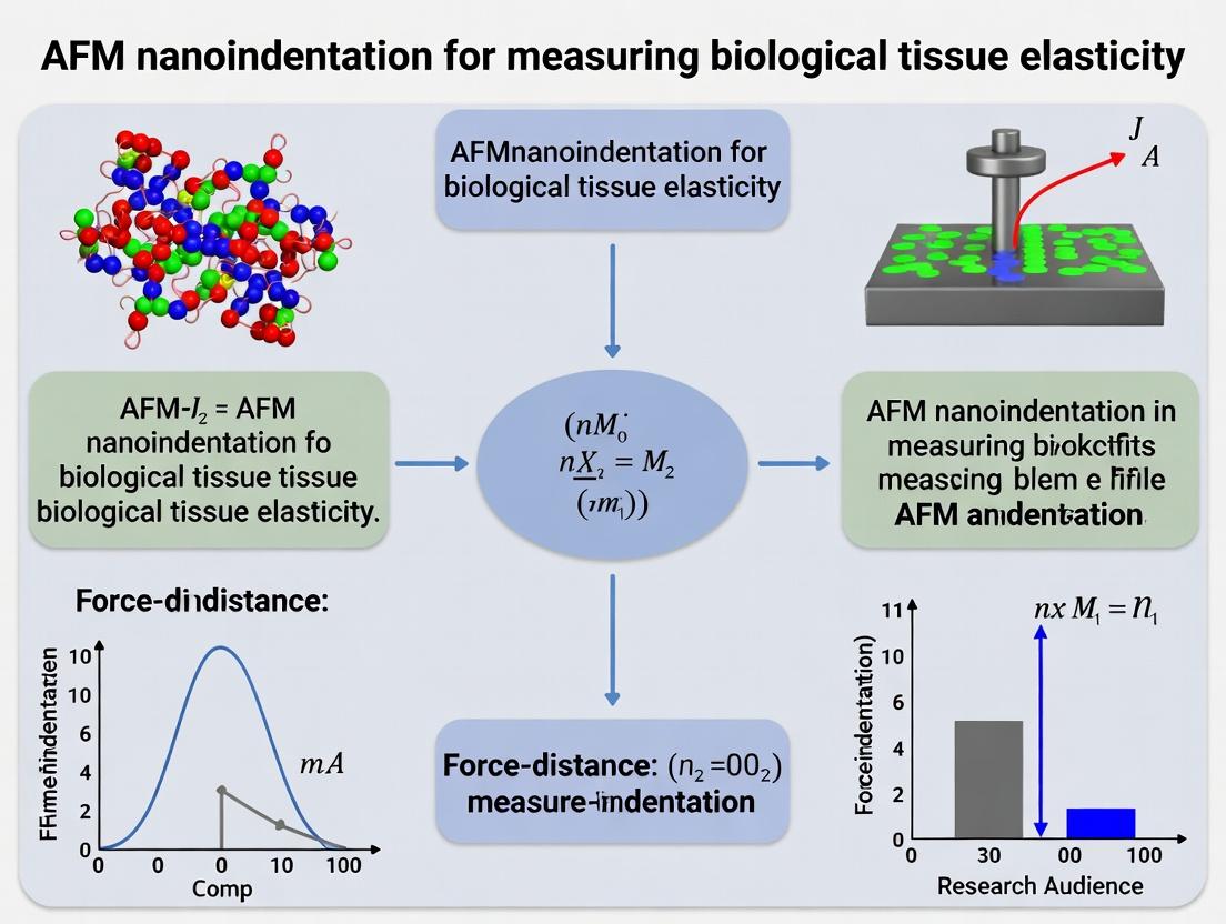
This article provides researchers, scientists, and drug development professionals with a detailed, current guide to Atomic Force Microscopy (AFM) nanoindentation for measuring the elasticity of biological tissues.
Mapping Tissue Biomechanics: A Comprehensive Guide to AFM Nanoindentation for Biological Elasticity Measurement
Abstract
This article provides researchers, scientists, and drug development professionals with a detailed, current guide to Atomic Force Microscopy (AFM) nanoindentation for measuring the elasticity of biological tissues. It covers foundational principles, from the role of tissue mechanics in physiology and disease to the core theory of AFM contact mechanics. The methodological section offers a step-by-step protocol for sample preparation, probe selection, and experimental execution. We address critical troubleshooting and optimization strategies for common challenges like sample hydration, surface detection, and data variability. Finally, the guide validates the technique through comparisons with bulk rheology, micropipette aspiration, and optical methods, highlighting its unique advantages and limitations. This resource synthesizes the latest research to empower robust, reproducible nanomechanical characterization in biomedical research.
Understanding Tissue Stiffness: Why Nanoscale Elasticity is Fundamental to Biology and Disease
Application Notes: AFM Nanoindentation in Mechanobiology
Atomic Force Microscopy (AFM) nanoindentation is a cornerstone technique for quantifying tissue elasticity (Young's modulus, E), providing direct correlation between mechanical properties and biological outcomes. Below are synthesized application notes and protocols derived from current research.
Table 1: Tissue Elasticity Ranges and Associated Biological States
| Tissue/Condition | Typical Young's Modulus (kPa) | Biological Context & Cell Fate Influence |
|---|---|---|
| Healthy Mammary Gland | 0.2 - 0.5 | Maintains epithelial homeostasis, luminal differentiation. |
| Malignant Breast Tumor | 4 - 12+ | Stromal stiffening promotes proliferation, invasion, EMT. |
| Early Stage Fibrosis (Liver/Lung) | 2 - 8 | Initial ECM cross-linking, activates pro-fibrotic signaling in fibroblasts. |
| Advanced Cirrhosis/Idiopathic Pulmonary Fibrosis | 15 - 50 | Severe tissue scarring, disrupts organ architecture, leads to failure. |
| Embryonic Mesenchyme | 0.1 - 1 | Permissive for rapid cell migration and morphogenetic movements. |
| Mature Bone | 10^4 - 10^6 | Provides mechanical support, regulates osteocyte activity via fluid shear. |
| Healthy Brain Tissue | 0.2 - 1 | Soft microenvironment essential for neurite outgrowth and astrocyte function. |
| Glioblastoma | 1 - 7 | Focal stiffening correlates with tumor grade and invasion propensity. |
Table 2: Key Mechanosensitive Pathways and Readouts
| Pathway Core | Primary Mechanosensor | Key Downstream Effector | Typical Functional Readout |
|---|---|---|---|
| YAP/TAZ | F-actin integrity, LINC complex | TEAD transcription factors | Nuclear YAP localization, CTGF expression |
| FAK-Src | Integrin clusters | Paxillin, ERK/MAPK | Paxillin phosphorylation (Y118), cell spreading area |
| TGF-β Activation | Force-dependent αv integrins | SMAD2/3 phosphorylation | Nuclear pSMAD2/3, α-SMA expression |
| Wnt/β-catenin | β-catenin stability via force | LEF1/TCF transcription | AXIN2 expression, β-catenin nuclear accumulation |
Protocol: AFM Nanoindentation of Fresh Tissue Sections for Elasticity Mapping
I. Sample Preparation
- Tissue Harvesting: For murine models (e.g., liver fibrosis, mammary tumors), perfuse with PBS, excise tissue, and embed in optimal cutting temperature (OCT) compound. Snap-freeze in liquid nitrogen-cooled isopentane.
- Sectioning: Cut 10-30 μm thick cryosections using a cryostat. Mount on Poly-L-Lysine-coated glass slides or Petri dishes. For live tissue slices (e.g., brain), use a vibratome to obtain 300-400 μm sections in ice-cold, oxygenated PBS.
- AFM Mounting: Place the slide/dish on the AFM stage. For hydrated measurement, immediately add appropriate physiological buffer (e.g., DMEM with HEPES, PBS) to cover the sample.
II. AFM Setup and Calibration
- Probe Selection: Use silicon nitride cantilevers with spherical polystyrene probes (diameter: 2-10 μm) for tissue. Typical spring constant (k): 0.01-0.1 N/m.
- Calibration: Calibrate the cantilever's spring constant using the thermal fluctuation method. Determine the optical lever sensitivity on a clean, rigid surface (e.g., glass).
- Programming: Define a measurement grid (e.g., 10x10 points over 100x100 μm²). Set approach/retract speed to 1-5 μm/s, indentation depth to 500-2000 nm (≤10% sample height), and trigger force to 1-5 nN.
III. Data Acquisition & Analysis
- Force Curve Acquisition: Automatically acquire force-indentation curves at each grid point in force spectroscopy mode.
- Model Fitting: Fit the retract curve's contact region with the Hertz/Sneddon contact mechanics model for a spherical indenter. The model relates force (F) to indentation (δ): F = (4/3) * (E/(1-ν²)) * √R * δ^(3/2), where R is tip radius, ν is Poisson's ratio (assumed 0.5 for incompressible tissue).
- Elasticity Map Generation: Compile calculated Young's modulus (E) values at each point to generate a spatial stiffness map co-registered with optical microscopy.
The Scientist's Toolkit: Research Reagent Solutions
| Item | Function in Mechanobiology Studies |
|---|---|
| Collagen I, High Concentration (≥5 mg/mL) | Polymerizes to form tunable 3D hydrogels for cell culture, mimicking stromal stiffness. |
| Polyacrylamide Hydrogel Kits | Provides substrata with precisely tunable elasticity (0.1-50 kPa) for 2D cell culture. |
| YAP/TAZ Inhibitor (Verteporfin) | Disrupts YAP-TEAD interaction, used to probe mechanotransduction pathway dependence. |
| FAK Inhibitor (PF-562271) | Targets ATP-binding site of FAK, used to inhibit integrin-mediated mechanosignaling. |
| TGF-β Receptor I Kinase Inhibitor (SB-431542) | Blocks Smad2/3 phosphorylation, used to dissect matrix-driven TGF-β activation. |
| Actin Polymerization Inhibitor (Latrunculin A) | Disrupts F-actin, used to decouple nuclear mechanotransduction (YAP/TAZ). |
| CellMask or SiR-Actin Stains | Live-cell compatible dyes for visualizing cell morphology and cytoskeletal dynamics. |
| Anti-paxillin (pY118) Antibody | Readout for integrin-mediated adhesion complex activation via immunofluorescence. |
Visualizations
Diagram 1: Core Mechanotransduction Pathway from ECM to Nucleus
Diagram 2: AFM Nanoindentation Workflow for Tissues
Diagram 3: Tissue Stiffness Feedback in Disease Progression
Biological tissues, such as cartilage, arterial walls, and tumors, are quintessentially heterogeneous, exhibiting significant spatial and hierarchical variations in mechanical properties from the organ scale down to the cellular and extracellular matrix (ECM) level. Traditional macro- or micro-scale mechanical tests (e.g., tensile testers, rheometers) provide bulk-averaged data that obscures these critical local variations. This application note, framed within a thesis on Atomic Force Microscopy (AFM) nanoindentation for biological tissue elasticity, elucidates why traditional methods fail and details protocols for AFM-based nanomechanical mapping to capture the true mechanical heterogeneity of biological samples.
The Failure of Traditional Mechanical Tests: A Quantitative Analysis
Traditional mechanical testing assumes material homogeneity and continuum behavior, assumptions grossly violated by biological tissues. The table below summarizes key limitations.
Table 1: Limitations of Traditional Mechanical Tests for Heterogeneous Biological Samples
| Test Method | Typical Scale | Key Assumption | Why It Fails for Heterogeneous Tissues | Representative Data Gap |
|---|---|---|---|---|
| Uniaxial/Biaxial Tensile | Macro (mm-cm) | Homogeneous strain, continuum material. | Averages over multiple tissue layers and cell/ECM domains. Misses local modulus variations critical to function (e.g., in osteochondral interface). | Reports a single Elastic Modulus (E) of ~0.1-1 MPa for cartilage, hiding the 0.5 kPa (pericellular) to 2 GPa (calcified cartilage) range. |
| Bulk Compression/Rheology | Macro/Micro (mm) | Uniform stress distribution, sample isotropy. | Indenter size (>mm²) is larger than microstructural features (cells, fibers). Measures composite response, not individual components. | Measures aggregate complex modulus, cannot resolve stiffness differences between collagen fibers (~GPa) and proteoglycan matrix (~kPa). |
| Microindentation | Micro (10-100 µm) | Semi-infinite half-space, homogeneous sub-surface. | Tip radius (≥5µm) is too large to probe single cells or fine ECM fibers. Contact area encompasses multiple heterogeneities. | May detect a "tissue-level" modulus but cannot map the stiffness gradient from a tumor's core (stiff) to its invasive front (soft). |
Core Principle: AFM Nanoindentation for Nanomechanical Mapping
AFM nanoindentation overcomes these limitations by using a sharp, nanoscale probe (tip radius: 5-50 nm) to apply pico- to nano-Newton forces and measure local indentation depth. By fitting force-distance curves to contact mechanics models (e.g., Hertz, Sneddon), a spatially resolved map of the Young's modulus (Elasticity) is generated, correlating mechanical properties with underlying biological structures.
Diagram 1: AFM Nanoindentation Workflow for Elasticity Mapping
Detailed Experimental Protocols
Protocol 4.1: Sample Preparation for AFM Nanoindentation of Soft Tissues
Objective: To immobilize fresh or fixed biological tissue sections without altering native mechanical properties. Materials: See "The Scientist's Toolkit" below. Procedure:
- Tissue Sectioning: Using a vibratome or cryostat, prepare tissue slices of 10-50 µm thickness onto glass coverslips. Optimal thickness ensures substrate stiffness does not influence measurement.
- Chemical Fixation (Optional): For stable, long-term measurements, immerse sample in 4% paraformaldehyde (PFA) in PBS for 20 minutes at 4°C. Rinse thoroughly (3x5 mins) with PBS. Note: Fixation can cross-link and stiffen tissue; live/fresh samples are preferred for physiological relevance.
- Immobilization: Apply a thin layer of medical-grade adhesive (e.g., Poly-L-Lysine) to a magnetic AFM specimen disk. Gently place the tissue-covered coverslip onto the adhesive, ensuring no air bubbles. Apply minimal pressure.
- Hydration: Place the mounted sample in the AFM liquid cell. Immediately submerge in appropriate physiological buffer (e.g., PBS, DMEM). Never allow the sample to dry out.
Protocol 4.2: AFM Nanoindentation & Elasticity Measurement
Objective: To acquire spatially correlated topographical and nanomechanical data. Materials: AFM with liquid-capable scanner, cantilevers (see Toolkit), fluid cell, analytical software (e.g., JPKSPM, Asylum, Bruker). Procedure:
- Cantilever Selection & Calibration:
- Select a sharp, nominal spring constant (k) cantilever (0.01-0.6 N/m for soft tissues).
- Thermal Tune Method: In fluid, acquire power spectral density of thermal noise. Fit the resonance peak to calculate the precise k.
- Determine the optical lever sensitivity (InvOLS) by recording a force curve on a rigid, non-compliant surface (e.g., clean glass).
- Engage & Topography Scan: Engage the tip onto the sample surface in fluid. Perform a contact-mode scan at low force (<0.5 nN) to obtain a height image and identify regions of interest (ROIs).
- Define Measurement Grid: Overlay a grid of indentation points (e.g., 32x32 to 64x64) on the ROI, ensuring spacing is smaller than the feature size of interest.
- Force Volume Acquisition:
- Set trigger point (e.g., 0.5-2 nN) and approach/retract speed (0.5-5 µm/s). Slow speeds minimize viscous effects.
- Initiate automated acquisition. At each grid point, the probe approaches, indents the sample until the trigger force is reached, and retracts, recording a full F-D curve.
- Data Analysis & Elasticity Mapping:
- For each F-D curve, subtract the baseline and convert deflection to force (using k and InvOLS).
- Identify the contact point. Fit the indentation segment (typically 10-90% of trigger point) to the Hertz model for a parabolic tip:
F = (4/3) * (E/(1-ν²)) * √R * δ^(3/2)where F=force, E=Young's modulus, ν=Poisson's ratio (assume 0.5 for incompressible tissue), R=tip radius, δ=indentation. - Use a scripting environment (e.g., MATLAB, Python with Nanite package) to batch-process all curves, extract E, and generate a 2D spatial elasticity map co-registered with topography.
Key Signaling Pathways in Mechanobiology
The nanomechanical properties measured by AFM are not passive traits; they are dynamically regulated by cellular mechanotransduction pathways. These pathways convert mechanical cues into biochemical signals.
Diagram 2: Core Mechanotransduction Pathway from ECM Stiffness
The Scientist's Toolkit: Research Reagent Solutions
Table 2: Essential Materials for AFM Nanoindentation of Biological Tissues
| Item | Example Product/Catalog # | Function & Critical Notes |
|---|---|---|
| AFM Cantilevers | Bruker PNPL (Pt-coated), ScanAsyst-Fluid+, Novascan qp-Bio-AC | Sharp, low spring constant probes for soft samples. Coating enhances reflectivity. Spring constant MUST be calibrated in-situ. |
| Bio-Adhesive | Poly-L-Lysine (0.1% w/v), Cell-Tak, APTES | Immobilizes tissue sections to substrate without significant chemical modification that alters mechanics. |
| Physiological Buffer | Phosphate Buffered Saline (PBS), Dulbecco's Modified Eagle Medium (DMEM) | Maintains sample hydration and, for live samples, physiological ionic balance and pH. |
| Fixative (Optional) | Paraformaldehyde (4% in PBS), Glutaraldehyde | Stabilizes tissue structure for prolonged measurement. Warning: Can artificially increase stiffness. Always compare to live/unfixed controls. |
| Calibration Grid | Bruker PG-GRID-10M, Ted Pella 600-50nm | Provides a standard with known pitch and height for verifying AFM scanner and tip geometry accuracy. |
| Rigidity Reference | Polydimethylsiloxane (PDMS) kits of known modulus (e.g., 1-100 kPa) | Essential for validating the accuracy of the entire force curve acquisition and analysis pipeline. |
| Analysis Software | JPKSPM Data Processing, Asylum Igor Pro, Open-source (Nanite in Python) | Converts raw deflection/position data into force-distance curves and performs model fitting to extract modulus. |
This document provides detailed application notes and protocols for Atomic Force Microscopy (AFM) nanoindentation, contextualized within a broader thesis research on measuring the elastic properties of biological tissues. The measurement of tissue elasticity is a critical parameter in understanding disease progression (e.g., fibrosis, cancer) and evaluating the efficacy of therapeutic interventions. AFM nanoindentation offers a unique capability to map mechanical properties at the nanometer-to-micrometer scale in near-physiological conditions.
Core Principles
Contact Mode AFM & Tip-Sample Interaction
In contact mode nanoindentation, a sharp tip mounted on a flexible cantilever is brought into controlled contact with the sample surface. The fundamental interaction is governed by Hooke's Law (F = -k * d), where the force (F) applied to the sample is calculated from the cantilever deflection (d) and its known spring constant (k). The tip-sample interaction force includes contributions from repulsive atomic forces, adhesion, and capillary forces (in air). For biological samples in liquid, capillary forces are minimized, allowing measurement of intrinsic mechanical properties.
Force-Distance Curves: The Fundamental Data
A force-distance (F-D) curve is a plot of cantilever deflection vs. piezoelectric actuator position (or tip-sample separation). A complete cycle consists of:
- Approach: Tip approaches until contact.
- Indentation: Tip applies load onto the sample.
- Withdrawal: Tip retracts, often showing adhesion "pull-off" forces.
The slope of the contact portion during loading is related to sample stiffness. Analysis of the indentation segment using contact mechanics models (e.g., Hertz, Sneddon, Oliver-Pharr) yields quantitative elastic modulus.
Table 1: Common AFM Cantilevers for Biological Nanoindentation
| Cantilever Type | Typical Spring Constant (k) | Typical Tip Radius | Typical Application | Key Considerations |
|---|---|---|---|---|
| Silicon Nitride (Sharp) | 0.01 - 0.1 N/m | 20 - 60 nm | High-resolution mapping of single cells | Soft, risk of bottoming out on stiff tissue. |
| Silicon Nitride (Colloidal) | 0.01 - 0.5 N/m | 1 - 10 μm | Bulk tissue elasticity, averaged measurement | Larger radius requires Hertz/Sneddon model adjustment. |
| Silicon (Sphere-tipped) | 1 - 50 N/m | 1 - 20 μm | Stiff tissues (bone, cartilage, fibrotic liver) | High k prevents excessive deflection on hard samples. |
| qp-BioAC (Aqua) | ~0.1 N/m | 20 nm | Standardized measurements in fluid | Thermally excited for in-situ k calibration. |
Table 2: Elastic Moduli of Representative Biological Tissues via AFM
| Tissue/Cell Type | Approximate Elastic Modulus (E) | Experimental Conditions (Model) | Biological Significance |
|---|---|---|---|
| Brain Tissue (Healthy) | 0.1 - 1 kPa | In PBS, Spherical tip (Hertz) | Baseline for neurological studies. |
| Breast Tissue (Carcinoma) | 1 - 5 kPa | In media, Spherical tip (Sneddon) | 5-10x stiffer than normal/benign tissue. |
| Cardiac Tissue (Fibrotic) | 20 - 100 kPa | In buffer, Spherical tip (Oliver-Pharr) | Indicator of heart disease severity. |
| Articular Cartilage | 0.1 - 1 MPa | In saline, Sharp tip (JKR for adhesion) | Degrades in osteoarthritis. |
| Liver (Fibrotic) | 5 - 25 kPa | In perfusate, Colloidal probe (Hertz) | Correlates with collagen deposition stage. |
Experimental Protocols
Protocol 4.1: Preparation of Hydrated Biological Tissue Sections for AFM
Objective: To prepare thin, hydrated, and intact tissue sections for nanoindentation measurements. Materials: Fresh or snap-frozen tissue, Optimal Cutting Temperature (OCT) compound, cryostat, phosphate-buffered saline (PBS), Petri dishes, substrate (glass slide or plastic dish). Procedure:
- Embed a small tissue piece (~5mm³) in OCT compound and freeze at -20°C.
- Using a cryostat, section tissue to 10-30 μm thickness at -20°C.
- Transfer section onto a clean glass slide or Petri dish. Gently melt OCT onto substrate by briefly touching the slide underside.
- Immediately immerse the section in PBS to remove OCT compound. Rinse 3x with fresh PBS.
- Keep the sample submerged in PBS at 4°C until AFM measurement (within 6 hours for best results).
Protocol 4.2: Acquisition and Analysis of Force-Volume Maps
Objective: To spatially map the elastic modulus of a tissue sample. Materials: AFM with fluid cell, cantilever (see Table 1), calibrated calibration grating, analysis software (e.g., NanoScope Analysis, JPKSPM, Gwyddion). Procedure:
- Cantilever Calibration: In air, determine the optical lever sensitivity (InvOLS) by acquiring F-D curves on a rigid sapphire or glass surface. In fluid, thermally tune the cantilever to determine its spring constant (k).
- Sample Mounting: Secure the prepared sample dish onto the AFM stage. Pipette sufficient PBS to submerge the tip.
- Engagement: Align the laser and engage the tip to the surface in contact mode.
- Force-Volume Setup: Define a grid (e.g., 16x16 points) over the region of interest. Set trigger threshold force (typically 0.5-5 nN) and Z-length (1-5 μm).
- Acquisition: Initiate automated acquisition. The system will collect an F-D curve at each grid point.
- Analysis:
- For each curve, align the baseline and convert deflection to force.
- Identify the point of contact.
- Fit the indentation segment (approach curve) with the Hertz/Sneddon model for a spherical/paraboloid tip:
- F = (4/3) * (E/(1-ν²)) * √R * δ^(3/2) (Hertz for sphere)
- where F is force, E is reduced modulus, ν is Poisson's ratio (assume ~0.5 for soft tissue), R is tip radius, and δ is indentation depth.
- Extract the apparent elastic modulus (E) from the fit.
- Compile moduli from all points into a 2D elasticity map.
The Scientist's Toolkit
Table 3: Essential Research Reagent Solutions for AFM Nanoindentation of Tissues
| Item | Function & Rationale |
|---|---|
| Phosphate-Buffered Saline (PBS), pH 7.4 | Maintains physiological osmolarity and pH, preventing tissue swelling or shrinkage during measurement. |
| Protease Inhibitor Cocktail | Added to PBS to prevent tissue degradation by endogenous proteases during long scans, preserving mechanical integrity. |
| Bovine Serum Albumin (BSA), 1% w/v | Used to passivate tips and substrates, minimizing non-specific adhesive forces that distort F-D curves. |
| Cell Viability Dyes (e.g., Calcein AM/Propidium Iodide) | For live tissue assessments, confirms region being probed is comprised of viable cells. |
| Optimal Cutting Temperature (OCT) Compound | Water-soluble embedding medium for cryo-sectioning; must be thoroughly rinsed to avoid contaminating measurements. |
| Calibration Gratings (TGZ1, TGX1) | For lateral (XY) calibration of the piezoelectric scanner, ensuring accurate spatial mapping. |
Visualization Diagrams
Title: AFM Force-Volume Mapping Workflow for Tissues
Title: Force-Distance Curve Analysis Segments
Application Notes
Atomic Force Microscopy (AFM) nanoindentation is a cornerstone technique for quantifying the nanomechanical properties of biological tissues, critical for understanding disease progression, tissue engineering, and drug efficacy. Central to this analysis is the extraction of Young's modulus (E) from force-distance curves, which relies on selecting an appropriate contact mechanics model. The Hertz, Sneddon, and Johnson-Kendall-Roberts (JKR) models form a foundational hierarchy, each with specific assumptions and applicability to biological samples.
- Hertzian Contact Theory: The baseline model for purely elastic, non-adhesive contact between two isotropic, homogeneous solids. It assumes small strains and infinitesimal contact adhesion. Its simplicity makes it widely used for preliminary stiffness mapping of cells and tissue sections, though its neglect of adhesion is a significant limitation for soft, adhesive biological matter.
- Sneddon's Extensions: Sneddon provided generalized solutions for different indenter geometries (e.g., conical, pyramidal) within the Hertzian framework (no adhesion). These are directly applicable to AFM where sharp tips are used to probe local heterogeneity or achieve higher spatial resolution.
- JKR Theory: Incorporates the effect of short-range surface adhesion within the contact area, which becomes dominant for very soft, compliant materials like hydrogels, single cells, and most hydrated tissues. It provides a more physically accurate description for biological systems where adhesive forces are non-negligible.
The choice of model directly impacts the derived Young's modulus, with significant deviations observed when adhesion is present but ignored. The following table summarizes the core force-indentation relationships and key parameters.
Table 1: Quantitative Comparison of Key Contact Models for AFM Nanoindentation
| Model & Core Assumption | Force (F) - Indentation (δ) Relationship | Key Parameters for Fitting | Primary Application in Bio-AFM |
|---|---|---|---|
| Hertz (Spherical Tip)Elastic, non-adhesive, spherical contact | ( F = \frac{4}{3} E_{eff} \sqrt{R} \delta^{3/2} ) | (E_{eff}): Effective Modulus(R): Tip Radius | Stiff tissue sections (bone, cartilage), preliminary cell mapping. |
| Sneddon (Conical Tip)Elastic, non-adhesive, conical contact | ( F = \frac{2}{\pi} E_{eff} \tan(\alpha) \delta^{2} ) | (E_{eff}): Effective Modulus(\alpha): Half-opening angle | High-res mapping with sharp tips, probing superficial lamina. |
| JKR (Spherical Tip)Elastic, adhesive contact | ( F = \frac{4E{eff}\sqrt{R}}{3} a^{3} - \sqrt{8\pi\gamma E{eff} a^{3}} )(with (a) as contact radius) | (E_{eff}): Effective Modulus(\gamma): Work of Adhesion(R): Tip Radius | Soft, adhesive samples: living cells, hydrogels, most hydrated tissues. |
Note: (E_{eff} = \frac{E}{1-\nu^2}), where E is the sample's Young's modulus and ν is its Poisson's ratio (often assumed ~0.5 for incompressible biological materials).
Experimental Protocols
Protocol 1: AFM Nanoindentation for Tissue Elasticity Using Hertz/Sneddon Models
Objective: To map the apparent Young's modulus of a fixed or stiff biological tissue sample while neglecting adhesive forces.
Materials (Research Reagent Solutions & Essential Materials):
- AFM with Liquid Cell: Enables measurement in physiological buffer.
- Cantilevers (Spherical or Sharp Tip):
- Spherical Probes: Colloidal probes (silica/glass beads, 2-10µm diameter) for Hertz analysis on heterogeneous tissues.
- Sharp Probes: Silicon nitride tips (pyramidal/conical, nominal k ~0.01-0.6 N/m) for Sneddon-based high-resolution mapping.
- Calibration Beams/Grid: For cantilever spring constant (k) calibration via thermal tune or Sader method.
- PBS (1X) or Appropriate Culture Buffer: Maintains sample hydration and ionic strength.
- Tissue Sample: Freshly harvested or fixed, mounted on a rigid substrate (e.g., glass slide, plastic dish) using a thin layer of biocompatible adhesive or collagen.
- Vibration Isolation Table: Critical for nano-scale measurement stability.
Procedure:
- Cantilever Calibration: In air or liquid, calibrate the inverse optical lever sensitivity (InvOLS) and the spring constant (k) of the cantilever using standard thermal fluctuation or force curve on a rigid surface methods.
- Sample Mounting: Securely affix the tissue sample within the AFM liquid cell. Immerse in appropriate buffer to prevent dehydration.
- Approach & Force Curve Acquisition: Using the AFM software, approach the probe to the sample surface and program the acquisition of force-distance curves. Typical parameters:
- Trigger Point: 1-10 nN (set to avoid excessive indentation).
- Approach/Retract Velocity: 0.5-5 µm/s (quasi-static condition).
- Spatial Resolution: For mapping, define a grid (e.g., 32x32 points) over the region of interest.
- Data Processing & Model Fitting (Hertz): a. Convert the raw deflection (V) vs. position (m) data to Force (F = k * deflection) vs. Indentation (δ = position - deflection) for each curve. b. For a spherical probe, fit the approach segment of the processed curve to the Hertz model: ( F = \frac{4}{3} \frac{E}{1-\nu^2} \sqrt{R} \delta^{3/2} ). c. Use a fixed, assumed Poisson's ratio (ν = 0.5) and the known tip radius (R). Extract the Young's modulus (E) as the fitting parameter.
- Validation: Perform indentation to a depth <10% of sample thickness to avoid substrate effect. Repeat over multiple locations (n>50) for statistical significance.
Protocol 2: Adhesion-Inclusive Modulus Measurement via JKR Model Fitting
Objective: To accurately determine the Young's modulus of soft, adhesive living cells or tissue constructs.
Materials: Items 1, 3, 4, 5, 6 from Protocol 1, plus:
- Soft, Spherical Probes: Colloidal probes with low spring constant (k ~0.01-0.1 N/m) to ensure measurable adhesion and prevent sample damage.
- Live Cell/Tissue Culture Medium: Maintains viability during measurement.
Procedure:
- Probe & System Preparation: Calibrate a soft colloidal probe as in Protocol 1. Ensure the AFM stage and liquid cell are sterile if working with live samples.
- Sample Equilibration: Allow live samples to equilibrate in the AFM stage-top incubator or medium for ≥30 minutes to stabilize temperature and pH.
- Adhesive Force Curve Acquisition: Approach the probe to the sample surface at low velocity (0.1-1 µm/s) to allow adhesive interactions. Use a higher trigger point or limit to ensure a clear retract adhesion signature.
- JKR Analysis Workflow: a. Process raw data to Force vs. Indentation (δ) and Force vs. Piezo displacement. b. Identify key parameters from the retract curve: Pull-off Force (F_{ad}) and Contact Radius at Zero Load (a₀) inferred from the snap-out point and contact mechanics equations. c. Implement a JKR fitting routine, either by: * Directly fitting the entire loading-unloading curve using the JKR equation. * Using the JKR adhesion map method: Calculate E and γ by simultaneously solving the equations for load-dependent contact radius (if available via contact stiffness) and the pull-off force.
- Critical Controls: Perform measurements rapidly to minimize cellular remodeling. Include multiple cells/tissue areas and biological replicates.
Workflow Diagram: Model Selection for AFM Nanoindentation
Atomic Force Microscopy (AFM) nanoindentation has become a cornerstone technique for quantifying tissue and cellular elasticity (Young's modulus) in the field of mechanobiology. This application note synthesizes recent discoveries that link alterations in tissue stiffness to disease progression in oncology, fibrotic disorders, and neurodegeneration, providing a detailed methodological framework for researchers.
Key Quantitative Discoveries
Table 1: Quantified Tissue Stiffness in Pathological States
| Disease / Tissue Type | Healthy Stiffness Range (kPa) | Diseased Stiffness Range (kPa) | Key Pathological Finding | Primary Measurement Technique | Citation (Recent) |
|---|---|---|---|---|---|
| Pancreatic Ductal Adenocarcinoma (PDAC) | 0.5 - 1.5 | 4.0 - 12.0 | Stromal fibrosis drives stiffness, promoting invasion and chemoresistance. | AFM nanoindentation (spherical tip, 5-10 μm) | Wei et al., Nature Cell Biology, 2024 |
| Idiopathic Pulmonary Fibrosis (IPF) | 1.0 - 2.5 | 15.0 - 30.0+ | ECM cross-linking and collagen deposition directly impair lung function. | AFM force mapping on ex vivo tissue sections | Liu & Herrera, Science Translational Medicine, 2023 |
| Alzheimer's Disease (Hippocampus) | ~0.2 - 0.5 | Increased by 2-4 fold | Amyloid-β plaques and neuroinflammation lead to measurable tissue stiffening. | AFM on murine brain slices (3 μm spherical tip) | Fernandez et al., Nature Neuroscience, 2023 |
| Liver Fibrosis (Cirrhosis) | 0.3 - 0.8 | 5.0 - 25.0 | Stiffness activates hepatic stellate cells, creating a progressive feedback loop. | Multifrequency AFM on biopsy samples | Carter & Ma, Cell Reports, 2024 |
| Breast Carcinoma | 0.1 - 0.5 (normal parenchyma) | 1.5 - 10.0 (tumor) | Tumors are stiffer, but peritumoral stiffness predicts metastasis risk. | In vivo and ex vivo AFM | Park et al., Advanced Science, 2024 |
Detailed Experimental Protocols
Protocol 1: AFM Nanoindentation of Fresh Tissue Sections for Stiffness Mapping
Application: Quantifying spatial heterogeneity in fibrosis and solid tumors.
Materials:
- Fresh or optimally preserved tissue (OCT embedded, not fixed).
- Atomic Force Microscope with temperature/humidity control chamber.
- Spherical probe tips (5-10 μm diameter, SiO₂ or polystyrene, k ~0.1 N/m).
- Fluid cell for immersion in appropriate physiological buffer (e.g., PBS).
- Calibration grating (e.g., TGXYZ series).
Procedure:
- Sample Preparation: Cryosection tissue to 20-30 μm thickness. Mount on glass bottom dish. Keep hydrated with buffer. Do not allow to dry.
- AFM & Probe Calibration:
- Perform thermal tune in air to determine spring constant (k).
- Determine inverse optical lever sensitivity (InvOLS) on a rigid surface (glass) in buffer.
- Force Mapping:
- Define a grid (e.g., 32x32 points) over the region of interest (ROI).
- Set trigger force: 1-2 nN (to avoid damaging soft tissue).
- Approach/retract speed: 5-10 μm/s.
- Acquire 2-3 force curves per point for statistical reliability.
- Data Analysis:
- Fit the retraction curve's contact region (typically 10-30% of indentation) with the Hertz contact model for a spherical indenter.
- Generate spatial elasticity maps (Young's modulus, E) correlated with histological markers from adjacent slices.
Protocol 2: Correlating Stiffness with Cellular Signaling viaIn SituImmunofluorescence
Application: Linking measured local stiffness to activation of mechanotransduction pathways (e.g., YAP/TAZ, MRTF).
Procedure:
- AFM Measurement First: Perform force mapping on a live cell culture or fresh tissue section as in Protocol 1. Record precise XY coordinates.
- Immediate Fixation: Gently fix the sample with 4% PFA for 15 min immediately after AFM scan.
- Immunostaining:
- Permeabilize with 0.1% Triton X-100 for 10 min.
- Block with 5% BSA for 1 hour.
- Incubate with primary antibodies (e.g., anti-YAP/TAZ, anti-phospho-Myosin Light Chain, anti-α-SMA for myofibroblasts) overnight at 4°C.
- Incubate with fluorescent secondary antibodies and nuclear stain (DAPI) for 1 hour.
- Correlative Microscopy:
- Use the recorded XY coordinates to relocate AFM measurement points on a confocal microscope.
- Quantify fluorescence intensity (nuclear/cytoplasmic ratio of YAP) and correlate directly with the Young's modulus value at each corresponding point.
Signaling Pathway Diagrams
Title: Mechanotransduction in Cancer and Fibrosis
Title: Stiffness in Neural Degeneration Pathways
The Scientist's Toolkit: Key Research Reagent Solutions
Table 2: Essential Materials for Mechanobiology Studies
| Item | Function/Application | Example Product/Type |
|---|---|---|
| AFM with Environmental Control | High-resolution force mapping in physiological conditions. | Bruker BioResolve, JPK NanoWizard with BioCell. |
| Spherical AFM Probes | Gentle, Hertz-model-compliant indentation of soft tissues. | Novascan SiO₂ beads (5-10 μm), Polystyrene beads. |
| Tunable Hydrogels (2D & 3D) | To in vitro model specific tissue stiffness for cell culture. | BioGel XP (Matrigen), PEG-based hydrogels, collagen I of defined concentration. |
| Mechanosensitive Pathway Inhibitors/Activators | To perturb and study stiffness-sensing pathways. | Blebbistatin (myosin II), Y27632 (ROCK), Verteporfin (YAP). |
| Live-Cell Tension Reporters | Visualize cellular forces in real-time. | FRET-based tension biosensors (e.g., VinTS, E-cadherin TSMod). |
| Antibodies for Mechanotransduction | Detect activation of stiffness-related signaling. | Phospho-FAK (Tyr397), Nuclear YAP/TAZ, α-SMA. |
| Cross-linking/Softening Enzymes | To modulate stiffness of ex vivo samples. | LOXL2 inhibitor (PXS-5153A), Collagenase (for softening). |
| Correlative Microscopy Software | To align AFM elasticity maps with fluorescence images. | Bruker PeakForce QI-LI, JPK DP, open-source Fiji/ICY plugins. |
Step-by-Step Protocol: Executing Robust AFM Nanoindentation on Biological Tissues
Application Notes for AFM Nanoindentation Research
Accurate measurement of tissue elasticity via Atomic Force Microscopy (AFM) nanoindentation is fundamentally dependent on sample preparation. This protocol details methods for embedding, sectioning, and immobilizing diverse biological tissues to preserve native mechanical properties and ensure reliable, repeatable indentation.
Core Principle: The preparation must minimize mechanical and structural artifacts while providing a stable, flat surface for AFM probing. The chosen method varies significantly based on tissue viability (live vs. fixed) and origin (native vs. engineered).
Comparative Data of Preparation Methods
Table 1: Quantitative Impact of Preparation Methods on Apparent Elastic Modulus
| Tissue Type | Preparation Method | Typical Measured Modulus (kPa)* | Key Artifact/Risk | Best For AFM? |
|---|---|---|---|---|
| Live Soft Tissue (e.g., Cell Sheet) | Hydrated Immobilization in Agarose Well | 1 - 10 | Over-constraint, hypoxia | Yes, for viability |
| Fixed Soft Tissue (e.g., Liver) | FFPE Sectioning & Adhesive Immobilization | 5 - 20 | Cross-linking hardening | No, for absolute modulus |
| Fixed Soft Tissue | Optimal Cutting Temperature (O.C.T.) Embedding, Cryosectioning | 2 - 15 | Ice crystal damage | Yes, for histology correlation |
| Fixed Hard Tissue (e.g., Bone) | Polymethylmethacrylate (PMMA) Embedding, Grinding/Polishing | 10,000 - 20,000 | Dehydration shrinkage | Yes, for hard materials |
| Engineered Hydrogel | Direct Adhesive Immobilization | 0.5 - 50 | Swelling/desiccation | Yes, with humidity control |
| Decellularized ECM | Critical Point Drying, Adhesive Mounting | 50 - 500 | Drying-induced stiffening | No, for hydrated properties |
*Apparent Elastic Modulus range is method-dependent and illustrative. Values are influenced by fixation, embedding medium, and hydration.
Table 2: Protocol Selection Guide for AFM Samples
| Parameter | Live Tissue | Chemically Fixed Tissue | Engineered Tissue (Hydrogel) |
|---|---|---|---|
| Primary Goal | Measure dynamic, physiologically relevant mechanics | Measure structure-linked mechanics, archive samples | Measure designed matrix properties |
| Optimal Embedding | None, or low-melt agarose for restraint | O.C.T. (cryo) or Paraffin (FFPE) for sectioning | Often direct mounting, possible agarose embedding |
| Sectioning Requirement | Minimal; tissue explant < 200 µm thick | Cryostat (5-30 µm) or Microtome (3-10 µm) | Microtome with cryo-cooling (if stiff) |
| Immobilization Method | Bio-adhesive (e.g., Cell-Tak) in fluid cell | Poly-L-Lysine or APTES-coated glass/PDMS | Cyanoacrylate or epoxy to rigid substrate |
| Critical Control | Temperature, CO₂, culture medium perfusion | Hydration level during measurement | Swelling equilibrium in measurement buffer |
Detailed Experimental Protocols
Protocol A: Cryo-Embedding and Sectioning of Fixed Tissues for AFM-Correlative Mechanics/Histology
Objective: Prepare thin, hydrated tissue sections from fixed samples for AFM nanoindentation, allowing subsequent histological staining.
Materials:
- Fresh or fixed tissue specimen (< 5mm³)
- 4% Paraformaldehyde (PFA) or preferred fixative
- Phosphate Buffered Saline (PBS)
- Sucrose gradients (10%, 20%, 30% in PBS)
- Optimal Cutting Temperature (O.C.T.) compound
- Isopentane chilled by liquid nitrogen
- Cryostat
- Poly-L-Lysine or APTES-coated glass slides/dishes
- AFM-compatible fluid cell
Procedure:
- Fixation: Immerse tissue in 4% PFA for 24 hours at 4°C.
- Cryoprotection: Sequentially immerse fixed tissue in 10%, 20%, and 30% sucrose solutions (12-24 hours each) until it sinks.
- Embedding: a. Fill a cryomold with a layer of O.C.T. b. Orient tissue specimen in the mold. c. Completely cover with O.C.T., avoiding bubbles. d. Slowly lower the mold into isopentane chilled by liquid nitrogen until fully solidified. Store at -80°C.
- Sectioning: a. Equilibrate block to cryostat chamber temperature (-20°C). b. Cut sections at 10-40 µm thickness, collecting on poly-L-lysine-coated AFM dishes. c. Allow sections to air-dry and adhere for 30-60 minutes.
- Rehydration & Immobilization: Rehydrate sections in PBS for 15 minutes prior to AFM. Ensure sections are fully covered by measurement buffer in the fluid cell.
Protocol B: Immobilization of Live, Engineered Tissue Constructs
Objective: Securely mount a compliant, hydrated engineered tissue (e.g., collagen hydrogel) without inducing pre-stress or compromising viability.
Materials:
- Engineered tissue construct
- AFM-compatible rigid substrate (e.g., glass-bottom dish, plastic petri)
- Fibrin-based bio-adhesive or medical-grade cyanoacrylate (thin layer)
- Measurement buffer (e.g., cell culture medium)
- Low-melting-point agarose (2%)
Procedure:
- Substrate Preparation: Clean substrate with ethanol and air-dry.
- Adhesive Application: Apply minimal, sparse dots of adhesive to the substrate. For fibrin glue, follow manufacturer's gelation instructions.
- Construct Placement: a. Gently blot excess liquid from the construct. b. Carefully lower the construct onto the adhesive dots, allowing one edge to contact first. c. Apply gentle pressure with a flat tool for 30 seconds.
- Lateral Restraint (Optional for very soft constructs): a. Create a shallow well around the construct using a silicone isolator. b. Fill the well with warm (37°C) 2% low-melt agarose and let it gel at 4°C for 5 minutes. This restrains lateral slippage without compressing the sample vertically.
- Hydration: Gently flood the dish with pre-warmed measurement buffer. Proceed with AFM immediately.
Visualization Diagrams
Diagram 1: Tissue Prep Workflow for AFM
Diagram 2: Immobilization Method Comparison
The Scientist's Toolkit: Key Research Reagent Solutions
Table 3: Essential Materials for AFM Tissue Sample Preparation
| Item | Function in Preparation | Key Considerations for AFM |
|---|---|---|
| Optimal Cutting Temperature (O.C.T.) Compound | Water-soluble embedding medium for cryosectioning. Provides support during cutting. | Must be fully hydrated/rehydrated before AFM to avoid contaminating the tip with polymer. |
| Poly-L-Lysine Solution (0.1% w/v) | Positively charged adhesive coating for glass/substrates. Binds negatively charged tissue sections. | Standard for fixed tissue sections. Ensure coating is thin and even to avoid a measurable adhesive layer. |
| Cell-Tak | Bio-adhesive protein mixture derived from mussels. Bonds tissue to substrate in aqueous environments. | Critical for live tissue immobilization. Use minimal amount; spot application is better than a continuous film. |
| APTES (3-Aminopropyl)triethoxysilane) | Silane coupling agent that functionalizes glass/silica with amine groups for covalent bonding. | Used with glutaraldehyde crosslinker for ultra-stable mounting. Can increase local substrate stiffness. |
| Low-Melting-Point Agarose (2-4%) | Thermoreversible gel used to create a physical restraint well around soft samples. | Gelation temperature ~25-30°C. Provides lateral support without vertical compression if applied correctly. |
| Critical Point Dryer | Instrument that removes water from fixed samples using liquid CO₂, preserving delicate structures. | Caveat: Drying dramatically alters mechanics. Only use if measuring dry, architectural properties is the goal. |
| Cryostat | Precision microtome in a freezing chamber for sectioning frozen, embedded tissues. | Section thickness must be >> AFM indentation depth (typically 5-10x) to avoid substrate effect. |
This guide, framed within a broader thesis on AFM nanoindentation for biological tissue elasticity measurement research, details the selection and application of atomic force microscopy (AFM) probes for quantifying the mechanical properties of diverse biological tissues. The choice of probe geometry—spherical, pyramidal, or colloidal—is critical for obtaining accurate, reproducible, and biologically relevant nanomechanical data, directly impacting research in tissue engineering, disease pathophysiology, and drug development.
Probe Geometry Characteristics and Selection Criteria
Table 1: Comparative Characteristics of AFM Probe Geometries for Tissue Nanoindentation
| Feature | Spherical Tip (Colloidal) | Pyramidal Tip (Sharp) | Colloidal Tip (Custom) |
|---|---|---|---|
| Typical Radius | 1 - 25 µm | < 20 nm (apex) | 0.5 - 10 µm |
| Contact Mechanics Model | Hertz (spherical) | Sneddon (pyramidal/conical) | Hertz (spherical) |
| Effective Elastic Modulus Range | 100 Pa - 100 kPa (soft tissues) | 1 kPa - 10 GPa (stiffer tissues/cells) | 10 Pa - 50 kPa (very soft tissues) |
| Spatial Resolution | Low (bulk property) | High (sub-cellular) | Low to Medium (local property) |
| Tissue Penetration Depth | Deep (>500 nm) | Shallow (<200 nm) | Tunable (200-1000 nm) |
| Primary Tissue Applications | Brain, adipose, lung, intact organs, hydrogels | Bone, cartilage, dense ECM, cellular stiffness | Engineered soft matrices, lymph nodes, liver sinusoids, spheroids |
| Key Advantage | Minimizes damage; well-defined contact; averages over heterogeneities | High spatial resolution; standard & calibrated probes | Tunable size and surface chemistry; precise functionalization |
| Main Limitation | Low lateral resolution; may obscure local features | Can induce tissue damage/penetration; sensitive to topography | Attachment robustness; potential for off-axis indentation |
Application Notes & Protocols
Protocol 1: Spherical Tip Indentation for Soft Brain Tissue Slices
Application Note: Spherical probes (5-20 µm radius) are optimal for measuring the bulk viscoelasticity of soft, heterogeneous neural tissues, providing data relevant to traumatic brain injury and neurodegenerative disease studies.
- Sample Preparation: Prepare 300 µm thick coronal brain slices from murine models using a vibratome in ice-cold, oxygenated artificial cerebrospinal fluid (aCSF). Adhere slices to poly-D-lysine coated Petri dishes.
- AFM Setup: Mount a polystyrene or silica spherical colloidal probe (R=10 µm) on a tipless cantilever (k ≈ 0.1 N/m). Calibrate the spring constant via thermal tune method.
- Hydration & Environment: Perform all measurements in a fluid cell filled with aCSF at 37°C.
- Indentation Parameters: Set approach velocity to 2 µm/s, trigger force to 1 nN, and maximum indentation depth to 1000 nm. Acquire a 10x10 grid of force curves over a 50x50 µm area in the cortex region.
- Data Analysis: Fit the approach curve using the spherical Hertz model, assuming a Poisson's ratio of 0.5 for incompressible tissue. Report the reduced elastic modulus (Er).
Protocol 2: Pyramidal Tip Indentation for Articular Cartilage
Application Note: Sharp pyramidal probes (BL-TR400PB, tip radius < 20 nm) are used to map the spatial gradient of stiffness in cartilage, from the superficial zone to the deep zone, assessing osteoarthritis progression.
- Sample Preparation: Obtain fresh articular cartilage explants (≈ 2x2 mm) from bovine femoral condyles. Secure explant with the articular surface facing up in a dish containing phosphate-buffered saline (PBS) with protease inhibitors.
- AFM Setup: Use a silicon nitride pyramidal tip on a triangular cantilever (k ≈ 0.02 N/m). Calibrate the deflection sensitivity on a clean glass slide in PBS.
- Hydration & Environment: Maintain full sample hydration with PBS during measurement.
- Indentation Parameters: Use a 1 µm/s approach rate, 2 nN trigger force, and 300 nm max indentation. Perform line scans (20 indents, 10 µm spacing) from the surface inward.
- Data Analysis: Fit force curves using the pyramidal (Sneddon) model. Plot elastic modulus versus distance from the articular surface to reveal the stiffness gradient.
Protocol 3: Functionalized Colloidal Tip Adhesion Mapping on Liver Sinusoids
Application Note: Colloidal probes functionalized with specific ligands (e.g., collagen, laminin) can measure both localized elasticity and specific adhesion forces in vascular or sinusoidal tissues, relevant for metastasis research.
- Probe Functionalization: Glue a 5 µm silica microsphere to a tipless cantilever. Activate with oxygen plasma. Silanize with (3-Aminopropyl)triethoxysilane (APTES). Crosslink recombinant laminin protein via BS(PEG)9 linker.
- Sample Preparation: Prepare cryosections (10 µm thick) of perfuse-fixed liver tissue. Use within 24 hours.
- AFM Setup: Mount functionalized probe. Thermal tune spring constant (≈ 0.3 N/m).
- Measurement: In PBS buffer, first perform a standard force-volume map (5x5, 20x20 µm) to establish baseline elasticity. Then, at selected points, perform force-distance curves with a 2-second contact hold to allow ligand-receptor binding, followed by retraction at 500 nm/s to quantify unbinding adhesion forces.
- Data Analysis: Derive elasticity from the approach curve (Hertz model). Quantify adhesion from the retraction curve peaks. Correlate adhesion events with tissue histology landmarks.
The Scientist's Toolkit
Table 2: Essential Research Reagent Solutions for AFM Tissue Nanoindentation
| Item | Function in Experiment |
|---|---|
| Poly-D-Lysine / Poly-L-Lysine | Coats substrate to firmly anchor tissue slices or cells during fluid imaging. |
| Protease/Phosphatase Inhibitor Cocktail | Preserves native tissue mechanical properties by preventing post-extraction degradation. |
| BS(PEG)n Crosslinkers (e.g., BS(PEG)9) | Spacer for covalent attachment of specific proteins (ligands, antibodies) to colloidal probes. |
| (3-Aminopropyl)triethoxysilane (APTES) | Silanizing agent for creating an amine-reactive surface on silica probes for biofunctionalization. |
| Oxygen Plasma Cleaner | Activates probe and sample surfaces to ensure clean, hydrophilic conditions for bonding and imaging. |
| Calibration Grid (TGZ series) | Reference sample with known pitch and height for lateral and vertical AFM scanner calibration. |
Visualization of Experimental Workflow
Title: AFM Probe Selection and Analysis Workflow for Tissues
Within the broader thesis on Atomic Force Microscopy (AFM) nanoindentation for measuring the elasticity of biological tissues, precise calibration is the cornerstone of quantitative data. The accuracy of derived Young's modulus values is directly dependent on the rigorous calibration of three interdependent parameters: the cantilever spring constant (k), the deflection sensitivity (InvOLS), and the tip geometry. This document provides detailed application notes and protocols for these essential calibrations, tailored for research on soft, hydrated biological samples.
Cantilever Spring Constant Calibration
The spring constant must be measured, not taken from manufacturer specifications, which can have >100% error. The thermal tune method is recommended for soft cantilevers (0.01 - 10 N/m) used in bio-indentation.
Protocol: Thermal Tune Method
- Preparation: Mount the cantilever in the AFM liquid cell with an appropriate physiological buffer (e.g., PBS). Allow thermal equilibrium (≥20 mins).
- Data Acquisition: With the tip freely oscillating in fluid (no contact), record the deflection signal (V) at a high sampling rate (≥50 kHz) for 5-10 seconds.
- Spectral Analysis: Compute the Power Spectral Density (PSD) of the deflection signal.
- Fitting: Fit the resonant peak region of the PSD to a simple harmonic oscillator model, excluding the low-frequency "1/f" noise.
- Calculation: Apply the Equipartition Theorem: ( k = kB T / <δ^2> ), where ( kB ) is Boltzmann's constant, T is absolute temperature (K), and ( <δ^2> ) is the mean square deflection in meters, derived from the integral of the fitted PSD.
Table 1: Typical Spring Constant Values for Common Bio-AFM Cantilevers
| Cantilever Type | Nominal k (N/m) | Measured k Range (N/m) | Ideal Indentation Depth (Biological Tissue) |
|---|---|---|---|
| Silicon Nitride (MLCT) | 0.01 | 0.005 - 0.02 | 100 nm - 2 µm (soft cells, gels) |
| Silicon (PNP-TR) | 0.08 | 0.05 - 0.15 | 50 nm - 1 µm (most cells, tissues) |
| Soft Silicon (SCION) | 0.25 | 0.15 - 0.4 | 20 nm - 500 nm (stiffer ECM, cartilage) |
Deflection Sensitivity Calibration
Deflection Sensitivity (InvOLS) converts the photodetector voltage to cantilever deflection in meters. It must be measured for each tip/surface/liquid combination.
Protocol: Force-Distance Curve on a Rigid Surface
- Surface Selection: Use an atomically flat, rigid substrate (e.g., clean sapphire, freshly cleaved mica) immersed in the same buffer as experimental samples.
- Approach: Engage the tip and obtain a standard force-distance curve.
- Slope Measurement: Identify the region of constant compliance where the tip is in hard contact with the rigid surface. The slope of the deflection (V) vs. piezo displacement (nm) curve in this region is the InvOLS (nm/V or m/V).
- Averaging: Repeat the measurement at 5-10 different locations on the rigid surface and average the slope values.
Tip Geometry Characterization
The tip shape determines the contact mechanics model (Hertz, Sneddon, etc.) used for modulus calculation. Tip broadening from wear or contamination is a major source of error.
Protocol: Tip Characterization via Reference Sample Imaging
- Reference Sample: Image a tip characterization grating (e.g., TGZ01 or TGXYZ02, NT-MDT) or sharp spike array (e.g., HS-100MG, NanoWorld).
- Imaging Parameters: Use standard tapping mode in air or fluid. Ensure the scan size is large enough to capture the tip's influence on the image of sharp features.
- Reconstruction/Estimation: Use dedicated tip reconstruction software (e.g., WSxM, Gwyddion) or assume a shape model. For biological indentation, a parabolic or spherical tip shape is commonly assumed. The effective radius (R) is estimated from the reconstruction.
- Validation: Periodically re-image the reference sample, especially after contacting hard surfaces or if data inconsistency arises.
Table 2: Impact of Tip Geometry Assumption on Calculated Modulus (Example Data)
| Assumed Tip Shape | Estimated Radius (nm) | Calculated E (kPa) for Synthetic Gel | % Error vs. Known Standard |
|---|---|---|---|
| Paraboloid | 20 | 10.2 ± 1.1 | +2% |
| Sphere | 20 | 9.8 ± 1.3 | -2% |
| Cone (30° half-angle) | N/A | 15.5 ± 2.0 | +55% |
| Blunted Cone (R=100nm) | 100 | 7.1 ± 0.9 | -29% |
Integrated Calibration Workflow
The following diagram illustrates the logical sequence and interdependence of the calibration steps.
Title: Integrated AFM Calibration Workflow for Nanoindentation
The Scientist's Toolkit: Essential Research Reagent Solutions
Table 3: Key Materials and Reagents for AFM Bio-Indentation Calibration
| Item | Function & Importance | Example Product/Specification |
|---|---|---|
| Calibration Cantilevers | Pre-calibrated levers for validating the thermal tune method. Provides a secondary check. | BL-TR2250-B (Bruker), k~0.25 N/m, with quoted uncertainty. |
| Rigid Calibration Substrate | Provides an atomically flat, non-deformable surface for accurate InvOLS measurement in liquid. | Sapphire disc (RMS roughness <0.5 nm), Freshly cleaved Mica. |
| Tip Characterization Grid | Standard sample with sharp, known features for imaging to reconstruct tip shape and estimate radius. | TGZ01 (NT-MDT) - Silicon grating with sharp tips. |
| Soft Polymer Gel Standard | Reference material with known, homogeneous elastic modulus for end-to-end validation of the entire calibration chain. | PDMS or Polyacrylamide gels (e.g., 10 kPa or 50 kPa standards). |
| Bio-Compatible Buffer | Hydration medium that maintains cantilever and sample stability, prevents salt crystallization. | 1x Phosphate Buffered Saline (PBS), pH 7.4, 0.22 µm filtered. |
| Plasma Cleaner | Critical for cleaning cantilevers and substrates to remove organic contaminants, ensuring consistent contact. | Harrick Plasma, Oxygen/Argon gas. |
| Vibration Isolation Table | Essential for stable thermal spectra and high-resolution tip imaging. Minimizes low-frequency noise. | Active or passive isolation system with >6 dB attenuation above 5 Hz. |
| AFM Software with Thermal Analysis | Enables accurate PSD fitting and k calculation. Advanced packages offer automated scripts. | NanoScope Analysis (Bruker), JPK SPM, Asylum Research Igor Pro. |
Within the broader thesis on AFM Nanoindentation for Biological Tissue Elasticity Measurement Research, the precise optimization of acquisition parameters is not merely a procedural step but a foundational scientific requirement. The accurate determination of Young’s modulus (E) in heterogeneous, viscoelastic biological tissues—from cancerous biopsies to engineered cartilage—directly hinges on the controlled application of force at the nanoscale. This document provides detailed Application Notes and Protocols for optimizing the four critical parameters that govern data fidelity: Approach Speed, Force Setpoint, Indentation Depth, and Spatial Mapping Grid design. The goal is to minimize artifacts, account for tissue time-dependent properties, and generate statistically robust spatial elasticity maps for biomedical research and drug development.
Core Parameter Optimization: Principles & Quantitative Guidelines
Approach Speed & Rate-Dependency
Biological tissues exhibit viscoelasticity, meaning their measured modulus depends on the rate of loading. A high approach speed can lead to overestimation of elasticity due to viscous drag and hydrodynamic effects, while a very slow speed increases drift and experiment duration.
Current Search-Derived Guidelines:
- Typical Range: 0.5 - 10 µm/s for approach phase.
- Critical Rule: The indentation velocity (often during retraction for analysis) must be reported and consistent for comparative studies. For soft tissues (< 10 kPa), velocities of 1-10 µm/s are common to approach quasi-static conditions.
- Protocol: To test rate-dependency, perform force spectroscopy on a representative tissue location across a range of velocities (e.g., 0.5, 2, 5, 10 µm/s). Plot apparent Young's modulus vs. log(indentation velocity) to identify a plateau region suitable for your study.
Table 1: Optimized Parameter Ranges for Biological Tissues
| Parameter | Recommended Range | Rationale & Impact | Tissue-Specific Consideration |
|---|---|---|---|
| Approach Speed | 1 - 5 µm/s | Balances viscoelastic effects, drift, and data acquisition time. | Use lower end for very soft/hydrated tissues (e.g., brain); higher end for stiffer tissues (e.g., cartilage). |
| Force Setpoint | 0.5 - 10 nN | Ensures sufficient indentation depth for analysis while minimizing substrate effect and tissue damage. | Scale with tissue stiffness. Use 0.5-2 nN for single cells; 2-10 nN for dense tissues. |
| Indentation Depth | 300 - 1000 nm | Must be within the linear elastic regime and typically ≤ 10% of sample thickness to avoid substrate effect. | For thin tissue sections (< 10 µm), limit depth to 5-10% of thickness. |
| Mapping Grid Density | 32x32 to 64x64 points | Provides a statistical representation of heterogeneity. Higher density increases resolution and time. | Use 64x64+ for highly heterogeneous samples (e.g., tumor margin); 32x32 for more homogeneous areas. |
Force Setpoint & Indentation Depth
These are intrinsically linked parameters. The Force Setpoint is the trigger value that defines the maximum load applied, which results in a specific Indentation Depth.
Optimization Protocol:
- Initial Calibration: On a representative area, perform a series of single indentations with increasing force setpoints (e.g., 1, 2, 5, 10 nN).
- Depth Analysis: Record the resulting indentation depth (δ) from the force-distance curve for each setpoint.
- Substrate Effect Check: Calculate the ratio δ / sample thickness. If >0.1, the measured modulus may be artificially high due to the underlying stiff substrate (glass, plastic).
- Linear Regime Validation: Ensure the force-indentation data fits the Hertz/Sneddon model well (high R² value). A poor fit may indicate excessive plastic deformation or an inappropriate model.
- Setpoint Selection: Choose a Force Setpoint that yields an indentation depth between 300-1000 nm while satisfying the substrate-effect rule (δ/thickness ≤ 0.1).
Spatial Mapping Grids
Elasticity mapping transforms point measurements into topographical modulus maps.
Optimization Protocol:
- Define Region of Interest (ROI): Use optical or AFM topographical imaging to identify key structures.
- Select Grid Density: Balance resolution with acquisition time and sample longevity. A 64x64 grid over 50x50 µm² yields a lateral resolution of ~0.78 µm.
- Dwell Time: Set a 50-200 ms dwell time at the maximum force (setpoint) to allow for stress relaxation, crucial for viscoelastic tissues.
- Ordering: Use a random or meander pattern for point acquisition to minimize the effect of temporal drift on the spatial map.
Detailed Experimental Protocol: AFM Nanoindentation Mapping of Tissue Elasticity
Title: Protocol for Spatial Elasticity Mapping of Fresh Biological Tissue Sections via AFM Nanoindentation.
Materials & Reagents:
- Fresh or preserved tissue sample (e.g., biopsy, cryosection).
- Suitable buffer (e.g., PBS, culture medium) to maintain tissue hydration.
- Petri dish or fluid cell compatible with AFM stage.
- Cyanoacrylate glue or optimal cutting temperature (OCT) compound for immobilization (avoid for measurement surface).
- Calibrated AFM with temperature control if possible.
- Probe Selection: Silicon nitride cantilevers with colloidal spherical tips (diameter 2.5-10 µm) are preferred for tissues to provide well-defined Hertzian contact and avoid sharp tip damage.
The Scientist's Toolkit: Key Research Reagent Solutions
| Item | Function & Rationale |
|---|---|
| Colloidal AFM Probes (e.g., 5 µm silica sphere) | Spherical geometry enables application of Hertz contact mechanics model, crucial for accurate modulus calculation on soft, deformable tissues. |
| Phosphate-Buffered Saline (PBS) | Standard physiological buffer used to hydrate tissues during measurement, preventing dehydration artifacts and maintaining native mechanical state. |
| Bovine Serum Albumin (BSA) | Often added (0.1-1% w/v) to measurement buffer to passivate surfaces and reduce non-specific adhesive forces between tip and tissue. |
| Calibration Kit (PS & PDMS) | Polystyrene (PS, ~3 GPa) and Polydimethylsiloxane (PDMS, ~2 MPa) reference samples for daily cantilever sensitivity and spring constant verification. |
| Low-Melting-Point Agarose | Used to gently embed very soft tissues for lateral stability during mapping without significantly altering local stiffness at the measurement surface. |
Procedure:
- Sample Preparation: Immobilize tissue firmly in petri dish using a minimal amount of glue at the base only. Flood with appropriate buffer. Ensure measurement surface is clean and unobstructed.
- AFM Setup: Mount colloidal probe. Perform thermal tuning in air/liquid to determine cantilever's spring constant (k). Calibrate the optical lever sensitivity (InvOLS) on a clean, rigid surface in fluid.
- Optical Navigation: Use the AFM's integrated optical microscope to locate the ROI for mapping.
- Parameter Input: Input optimized parameters into the AFM software:
- Cantilever: Spring constant (k, typically 0.01 - 0.1 N/m for tissues).
- Approach: Speed = 2 µm/s, Setpoint = 5 nN.
- Map Definition: Grid = 64x64 points, Area = 50x50 µm², Dwell time = 100 ms.
- Trigger Mode: Force (setpoint).
- Engage & Map: Initiate automatic mapping. Monitor first few curves for consistency.
- Data Processing: For each force-distance curve:
- Apply baseline correction.
- Fit the extended Hertz model (for a spherical tip) to the approaching curve: F = (4/3) * (E/(1-ν²)) * √R * δ^(3/2), where ν is Poisson's ratio (assumed 0.5 for incompressible tissue).
- Extract Young's modulus (E) for each grid point.
- Spatial Analysis: Generate 2D and 3D elasticity maps histogram distribution of modulus values. Co-register with topography if available.
Visualized Workflows & Relationships
Diagram Title: Optimization Workflow for AFM Tissue Elasticity Thesis
Diagram Title: Single AFM Indentation Cycle with Dwell
Table 2: Troubleshooting Common Artifacts
| Observed Issue | Potential Cause | Corrective Action |
|---|---|---|
| Modulus increases with depth | Substrate effect (sample too thin). | Reduce Force Setpoint; ensure indentation depth ≤ 10% of sample thickness. |
| High adhesion 'pull-off' force | Non-specific tip-sample adhesion. | Increase retract speed; add BSA to buffer; ensure clean probe. |
| Noisy force curves | Contaminated probe or sample debris. | Clean/replace probe; rinse sample gently. |
| Drift in map over time | Thermal drift or sample relaxation. | Equilibrate system longer; use temperature control; reduce map time or grid points. |
| Poor Hertz model fit | Excessive plastic deformation or incorrect tip shape model. | Reduce Force Setpoint; verify tip shape and model (sphere vs. pyramid). |
This article details the application of Atomic Force Microscopy (AFM) nanoindentation for measuring the elastic properties of diverse biological tissues. The work is framed within a broader thesis on the critical role of tissue micromechanics in physiology and disease, providing a comparative analysis across four key areas: articular cartilage, vascular walls, solid tumors, and cerebral organoids. The protocols and data herein are designed to guide researchers and drug development professionals in implementing these techniques.
Table 1: Comparative Elastic Modulus (Young's Modulus) of Biological Tissues
| Tissue Type / Sample | Condition / Region | Approx. Elastic Modulus (kPa) | Key Biomechanical Implication |
|---|---|---|---|
| Articular Cartilage | Superficial Zone | 500 - 1000 | Resists shear forces during joint movement. |
| Middle/Deep Zone | 800 - 2000 | Provides compressive stiffness and load support. | |
| Osteoarthritic (Degraded) | 50 - 300 | Loss of stiffness correlates with proteoglycan loss and clinical severity. | |
| Vascular Wall | Healthy Aorta (Tunica Media) | 50 - 150 | Optimal compliance for blood pressure buffering. |
| Atherosclerotic Plaque | 1000 - 5000+ | Increased stiffness promotes rupture risk; lipid core is softer (~10-50 kPa). | |
| Cerebral Aneurysm Wall | 300 - 800 | Focal weakening and remodeling of vessel structure. | |
| Solid Tumors | Tumor Stroma (Desmoplastic) | 4000 - 15000 | High stiffness promotes oncogenic signaling and metastasis. |
| Tumor Cell Nucleus | 200 - 1000 | Softer nuclei correlate with invasive potential in some cancers. | |
| Metastatic Niche (Liver/Lung) | 3000 - 8000 | Stiff microenvironment supports disseminated tumor cell growth. | |
| Cerebral Organoids | Neuroepithelium (Day 30) | 0.5 - 2 | Very soft, mimicking early embryonic brain tissue. |
| Cortical Plate (Day 60-90) | 1 - 5 | Increasing stiffness with neuronal maturation and neurite outgrowth. | |
| Organoid with Gliosis | 5 - 15 | Reactive glial cells and ECM deposition increase stiffness. |
Table 2: Key Experimental Parameters for AFM Nanoindentation
| Parameter | Articular Cartilage | Vascular Walls | Solid Tumors | Cerebral Organoids |
|---|---|---|---|---|
| Recommended Cantilever | Pyrex-Nitride, spherical tip (R=2.5-5µm) | Sharp Silicon Nitride (k~0.01 N/m) or spherical | Sharp Silicon Nitride (k~0.1 N/m) for cells, spherical for matrix | Ultra-soft cantilevers (k~0.006 N/m), spherical tip (R=2.5µm) |
| Indentation Depth | 500 - 1000 nm | 300 - 500 nm (cells), 1000-2000 nm (matrix) | 200 - 500 nm (cells), 1000 nm (matrix) | 300 - 800 nm |
| Loading Rate | 0.5 - 1 µm/s | 0.5 - 1 µm/s | 0.5 - 1 µm/s | 0.3 - 0.5 µm/s |
| Analysis Model | Hertz (Spherical) | Hertz (Spherical or Paraboloid) | Hertz or Sneddon (pyramidal) | Hertz (Spherical), accounting for finite thickness |
| Critical Buffer | PBS with protease inhibitors | Physiological saline (e.g., Hanks' Buffer) | Cell culture medium at 37°C, 5% CO2 | Neural basal medium, low vibration |
Detailed Experimental Protocols
Protocol 1: AFM Nanoindentation of Articular Cartilage Explants
Objective: To map the spatial variation of elastic modulus in healthy and osteoarthritic cartilage.
Materials & Reagents:
- Fresh or frozen osteochondral plugs.
- Phosphate-Buffered Saline (PBS) with 1x protease inhibitor cocktail.
- 4% Paraformaldehyde (PFA) for fixation (optional for post-measurement histology).
- AFM system with fluid cell and temperature control (preferably 37°C).
- Spherical colloidal probe (polystyrene, 5µm diameter) mounted on a soft cantilever (k~0.01 N/m).
Procedure:
- Sample Preparation: Isolate cartilage from subchondral bone using a scalpel. Rinse in PBS+inhibitors. Secure explant (articular surface up) in a petri dish using a thin layer of cyanoacrylate glue.
- AFM Calibration: Perform thermal tune method in air to determine the spring constant (k) of the cantilever. Calibrate the sensitivity on a rigid glass slide in PBS.
- Measurement Grid: Define a 10x10 grid (100x100 µm area) spanning from superficial to deep zones relative to the surface.
- Indentation: In contact mode, approach each grid point at 0.5 µm/s. Record force-distance curves with a trigger force of 1-2 nN and indentation depth of 500 nm.
- Data Analysis: Fit the retract curve's contact region with the spherical Hertz model: F = (4/3) * (E/(1-ν²)) * √R * δ^(3/2), where E is Young's modulus, ν is Poisson's ratio (assume 0.5), R is tip radius, and δ is indentation.
- Validation: Correlate modulus maps with adjacent sections stained with Safranin-O for proteoglycan content.
Protocol 2: Mapping Stiffness in Atherosclerotic Plaque Cross-Sections
Objective: To identify mechanically heterogeneous regions (fibrous cap, lipid core, calcifications) in arterial plaques.
Materials & Reagents:
- Cryosections of human or murine artery (10-20 µm thickness) on Poly-L-Lysine coated slides.
- Hanks' Balanced Salt Solution (HBSS) at 37°C.
- Sharp silicon nitride cantilever (k~0.1 N/m, tip radius ~20 nm).
- Fluorescent dyes (e.g., Oil Red O, DAPI) for post-AFM staining correlation.
Procedure:
- Sectioning: Cut OCT-embedded vessels in a cryostat. Thaw sections at room temperature for 5 min and hydrate in HBSS.
- AFM Setup: Mount slide on AFM stage with fluid cell. Use an optical microscope to locate the plaque region.
- High-Resolution Mapping: Perform a force-volume map over a region of interest (e.g., 50x50 µm, 64x64 points). Use a trigger force of 0.5 nN to avoid sample damage.
- Curve Fitting: Use the Sneddon model (pyramidal tip) for fitting: F = (2/π) * (E/(1-ν²)) * tan(θ) * δ², where θ is the half-opening angle of the tip.
- Histological Correlation: After AFM, fix, stain sections, and overlay stiffness maps with histology images using fiduciary marks.
Protocol 3: Probing Tumor Cell and Stromal Mechanics in 3D Culture
Objective: To quantify the influence of the extracellular matrix on cancer cell stiffness in a 3D tumor model.
Materials & Reagents:
- MDA-MB-231 breast cancer cells.
- Rat tail Collagen I (high concentration, ~8 mg/mL).
- AFM-compatible cell culture medium (e.g., DMEM without phenol red).
- Spherical tip (R=2.5µm) on a soft cantilever (k~0.06 N/m).
Procedure:
- 3D Culture Preparation: Mix cells with neutralized collagen I solution to a final density of 5x10^5 cells/mL and collagen at 4 mg/mL. Polymerize in a 35 mm dish at 37°C for 30 min.
- AFM in Incubator: Place dish on a heated AFM stage (37°C, 5% CO2). Allow cells to acclimate for 1 hour.
- Targeted Indentation: Use integrated optical microscopy to select individual cells embedded in the matrix.
- Measurement: Approach the cell body at 0.5 µm/s. Perform 5-10 indents per cell, with a maximum indentation of 300 nm (≤10% of cell height).
- Stromal Measurement: Move the tip to an adjacent acellular region to measure the local matrix stiffness.
- Analysis: Fit curves with the Hertz model. Compare cell vs. matrix modulus for correlation analysis.
Protocol 4: Characterizing Developing Cerebral Organoids
Objective: To track the temporal evolution of tissue stiffness during neural differentiation and maturation.
Materials & Reagents:
- Human iPSC-derived cerebral organoids (day 30-120).
- Neural basal medium.
- Ultra-soft tipless cantilevers (k~0.006 N/m) with attached 2.5µm silica microsphere.
- Agarose molds to immobilize organoids.
Procedure:
- Immobilization: Gently place an organoid in a custom agarose mold submerged in neural basal medium. Ensure the region of interest (e.g., cortical bud) is accessible.
- Cantilever Selection: Critical due to organoid softness. Calibrate the ultra-soft cantilever carefully using the thermal method.
- Low-Force Mapping: Perform a low-resolution map (e.g., 10x10 points over 200x200 µm) with a very low trigger force (~0.1 nN) and slow approach (0.3 µm/s).
- Depth Control: Limit indentation to 500 nm to avoid substrate effects and tissue damage.
- Longitudinal Studies: Measure the same organoid line at different time points, using distinct organoids for endpoint histological analysis (e.g., immunofluorescence for neurons, glia, and ECM).
- Data Processing: Apply the Hertz model with a correction for finite sample thickness if the indentation depth is >10% of the sample height.
Diagrams
Title: Mechano-Inflammatory Pathway in Cartilage Degradation
Title: Generic AFM Nanoindentation Workflow for Tissues
Title: Tumor Stiffness Drives Pro-Metastatic Signaling
The Scientist's Toolkit: Research Reagent Solutions
Table 3: Essential Materials for AFM-based Tissue Mechanobiology
| Item | Function / Application | Example Product / Specification |
|---|---|---|
| Colloidal AFM Probes | Spherical tips for Hertz model fitting on soft tissues; minimize sample damage. | Novascan PS-A (5µm sphere), sQUBE (SiO2 spheres, 2.5-10µm). |
| Sharp AFM Probes | High-resolution mapping of thin sections or subcellular structures. | Bruker MLCT-Bio-DC (triangular, k~0.03 N/m), Olympus RC800PB. |
| Ultra-Soft Cantilevers | Essential for measuring extremely soft samples like organoids or single cells. | Bruker CellTak (k~0.01 N/m), NanoAndMore USC-F0.3-k0.06. |
| Protease Inhibitor Cocktail | Preserves ECM integrity in fresh tissue explants during measurement. | Sigma-Aldrich P8340 (aqueous solution). |
| Matrigel / Collagen I | For constructing 3D cell culture models with tunable stiffness. | Corning Matrigel GFR, Rat Tail Collagen I, high concentration. |
| Temperature & CO2 Control | Maintains tissue viability and physiological conditions during live AFM. | BioCell or PetriPer heater from JPK, stage-top incubators. |
| AFM Calibration Kit | For precise spring constant and sensitivity calibration. | Bruker PFQNM-LC-A Calibration Kit, MikroMasch CSC38. |
| Analysis Software | Processes force-distance curves and fits mechanical models. | JPK DP, Bruker Nanoscope, Open-source: AtomicJ, PyJibe. |
Solving Common Pitfalls: Optimizing AFM Nanoindentation for Accuracy and Reproducibility
Within the broader thesis on Atomic Force Microscopy (AFM) nanoindentation for quantifying biological tissue elasticity, maintaining true physiological conditions is paramount. The mechanical properties measured are highly sensitive to hydration, temperature, and ionic environment. This document provides detailed application notes and protocols for implementing robust hydration and environmental control within fluid cells, ensuring that nanoindentation data reflects in vivo-like tissue mechanics.
Core Principles and Quantitative Benchmarks
Successful physiological maintenance hinges on controlling specific parameters. The following table summarizes the critical targets for mammalian soft tissue studies.
Table 1: Target Physiological Parameters for AFM Nanoindentation of Biological Tissues
| Parameter | Target Physiological Range | Impact on Elasticity Measurement | Common AFM Fluid Cell Challenge |
|---|---|---|---|
| Temperature | 36.5 - 37.5°C for mammalian tissues | ↓ Temperature increases membrane viscosity & cytoskeletal rigidity, elevating apparent modulus. | Heat dissipation from scanner, ambient fluctuations. |
| pH | 7.35 - 7.45 (physiological buffer) | Deviations alter protein charge & conformation, affecting cell-cell and cell-ECM adhesion stiffness. | CO2 outgassing from buffer raises pH in open systems. |
| Osmolarity | 280 - 310 mOsm/kg for most tissues | Hypo-osmotic conditions cause swelling & softening; hyper-osmotic causes shrinkage & stiffening. | Evaporation in poorly sealed cells increases osmolarity. |
| Humidity | >95% to prevent evaporation (closed cell) | Evaporation concentrates salts, alters osmolarity, and dehydrates samples, drastically increasing stiffness. | Open fluid meniscus at cantilever insertion point. |
| Ionic Composition | [Ca2+] ~1.2 mM, [Mg2+] ~0.8 mM | Divalent cations are critical for integrin-mediated adhesion & tissue layer integrity. | Use of simple saline over complex culture media. |
Detailed Protocols
Protocol 3.1: Assembly and Priming of a Sealed, Temperature-Controlled Fluid Cell
Objective: To prepare an AFM fluid cell that maintains a stable 37°C, hydrated, and pH-buffered environment for >1 hour of indentation mapping.
Materials (Scientist's Toolkit):
- AFM with liquid cantilever holder and sealed fluid cell kit: (e.g., Bruker BioCell, JPK BioMAT). Provides a chamber with inlet/outlet ports and integrated heating.
- Circulating water bath or Peltier heater: Maintains precise temperature of the fluid cell body.
- Pre-warmed physiological buffer: (e.g., HEPES-buffered DMEM, PBS with Ca2+/Mg2+, or Ringer's solution). Maintains pH without CO2.
- Syringe pump or peristaltic pump (optional): For continuous perfusion to replenish nutrients/remove waste.
- Two 10 mL syringes with compliant tubing: For manual buffer exchange and priming.
- Temperature sensor (micro-thermocouple): For independent verification of sample plane temperature.
Procedure:
- Pre-warming: Connect the fluid cell's heating lines to the circulating bath set to 38.5°C. Allow the empty cell to equilibrate for 15 minutes. Pre-warm buffer in a 37°C incubator.
- Sample Mounting: Mount the tissue sample (e.g., cryosection on glass slide, engineered hydrogel) onto the magnetic disk. Apply a thin layer of vacuum grease to the sample mount rim to ensure a seal.
- Cell Assembly: Carefully lower the pre-warmed fluid cell onto the sample mount, ensuring a seal. Hand-tighten the retaining screws evenly.
- Priming: Using a syringe filled with warm buffer, connect tubing to the fluid cell's outlet port. Gently inject buffer until it flows from the inlet port, ensuring no air bubbles are trapped. Reverse flow direction several times to purge all bubbles.
- Final Connection: Connect inlet tubing to a reservoir of warm buffer. Maintain a slight positive pressure or close both ports if using a static volume. Insert the AFM cantilever into its holder.
- Temperature Calibration: Insert a micro-thermocouple through the fluid port onto the sample surface. Record temperature. Adjust bath setpoint until the sample plane reads 37.0 ± 0.2°C (typically requires a bath setpoint 1-1.5°C above target).
Protocol 3.2: Continuous Perfusion System for Long-Term Viability Studies
Objective: To enable AFM nanoindentation over multiple hours by continuously supplying fresh nutrients and stabilizing the environment.
Procedure:
- Setup: Connect the fluid cell inlet to a reservoir of pre-warmed, pre-gassed (5% CO2 if using bicarbonate buffer) media via a peristaltic pump using gas-impermeable tubing (e.g., Norprene). Connect the outlet to a waste collection flask.
- Flow Rate Calibration: Set a very low flow rate (50-100 µL/min). Excessive flow causes hydrodynamic drag on the cantilever.
- Equilibration: Allow the system to perfuse for at least 30 minutes before engaging the tip, ensuring thermal and chemical equilibrium.
- Validation: Periodically measure the effluent pH if using bicarbonate buffers to confirm CO2 tension is maintained.
Visualization of Experimental Workflow
Diagram 1: Fluid Cell Setup & Nanoindentation Workflow
Diagram 2: Impact of Environment on Measured Elasticity
Research Reagent Solutions & Essential Materials
Table 2: Essential Materials for Physiological AFM Nanoindentation
| Item | Function & Rationale |
|---|---|
| HEPES-buffered Cell Culture Media (e.g., DMEM) | Maintains physiological pH (7.4) without requiring a controlled CO2 atmosphere, ideal for short-term sealed fluid cells. |
| Dulbecco's Phosphate-Buffered Saline (DPBS) with Ca2+ & Mg2+ | Provides essential ionic balance and divalent cations necessary for maintaining tissue integrity and cell adhesion mechanics. |
| Norprene or Gas-Impermeable Tubing | Minimizes gas exchange (O2 in, CO2 out) in perfusion lines, preserving medium pH and dissolved gas concentrations. |
| Micro-thermocouple (100 µm bead) | Directly measures temperature at the sample surface, enabling calibration of the fluid cell heater offset. |
| Syringe Pump with Low Flow Rate Capability (<100 µL/min) | Enables continuous, laminar perfusion without inducing hydrodynamic forces on the AFM cantilever. |
| Sealed Fluid Cell with Luer-Lock Ports | Allows bubble-free priming and connection to perfusion systems while preventing evaporation. |
| High-Vacuum Grease (Non-toxic) | Creates a watertight seal between the fluid cell and sample substrate, crucial for preventing osmotic shifts. |
| Pre-Calibrated Osmometer | Validates the osmolarity of prepared buffers and used media, ensuring it remains within the 290 ± 10 mOsm/kg range. |
Accurate determination of the contact point (CP) between the atomic force microscope (AFM) probe and a soft, viscous sample is the critical first step for reliable nanoindentation and subsequent elasticity measurement. Biological tissues present unique challenges: low elastic modulus (E < 10 kPa), high adhesion, time-dependent viscoelastic creep, and surface hydration. This application note, framed within a thesis on AFM nanoindentation for biological tissue elasticity research, details current strategies to overcome surface detection drift and ensure robust CP determination for researchers and drug development professionals.
Core Challenges in Contact Point Detection on Soft Matter
The primary obstacles are:
- Drift: Thermal and mechanical drift in the Z-axis leads to a continuously shifting baseline force, causing premature or delayed CP identification.
- Liquid Meniscus & Adhesion: A thick hydration layer or culture medium can form a viscous bridge, generating long-range attractive forces that obscure the true mechanical contact.
- Surface Compliance: The soft sample deforms under negligible load, creating a poorly defined transition from non-contact to contact in the force-distance (F-D) curve.
- Sample Topography: Native tissue is often rough and heterogeneous, requiring local CP determination for each measurement point.
Quantitative Comparison of Contact Point Detection Methods
The following table summarizes the performance of current methods against key metrics relevant to soft, hydrated biological tissues.
Table 1: Comparative Analysis of Contact Point Detection Methods for Soft, Viscous Samples
| Method | Core Principle | Typical Drift Compensation | Best for Sample Type | Key Advantage | Key Limitation | Reported Accuracy (Z) on Soft Hydrogels* |
|---|---|---|---|---|---|---|
| Threshold-Based | Triggers CP at a user-defined absolute force or slope. | Poor | Stiffer tissues, cells | Simplicity, speed | Highly sensitive to drift and adhesion | > 50 nm |
| Extrapolation | Fits linear contact region back to zero deflection. | Moderate | Homogeneous, linear-elastic samples | Removes adhesion offset | Assumes linearity from first contact | ~20-30 nm |
| Stress-Relaxation Hold | Holds piezo position post-trigger; CP at force plateau. | Good | Highly viscous, viscoelastic tissues | Mitigates creep effects; identifies true mechanical equilibrium | Increases measurement time | ~10-20 nm |
| Thermal Noise / Dynamic | Monitors change in probe oscillation (amplitude, frequency) upon approach. | Excellent | Ultra-soft gels, hydrated surfaces | Long-range (non-contact) detection; low force | Complex setup; sensitive to fluid environment | < 10 nm |
| Machine Learning (CNN) | Trained network identifies CP from raw F-D curve morphology. | Good to Excellent | All types, esp. heterogeneous tissues | Robust to noise and variable curve shapes | Requires large, labeled training dataset | ~5-15 nm |
*Accuracy is defined as the reproducibility (standard deviation) of CP determination on a homogeneous polyacrylamide gel (E ≈ 1 kPa) in liquid, including contributions from instrumental drift. Data synthesized from recent literature (2023-2024).
Detailed Experimental Protocols
Protocol 1: Stress-Relaxation Hold for Viscoelastic Tissue Sections
This protocol minimizes the effect of viscous creep during CP determination on fresh or fixed tissue slices.
Materials:
- AFM with liquid cell and temperature control (if possible).
- Soft colloidal probe (e.g., 10-20 µm diameter silica sphere, nominal k ≈ 0.1 N/m).
- Sample: Tissue slice (e.g., liver, brain) submerged in appropriate buffer (PBS, DMEM).
- Calibrated cantilever (spring constant, inverse optical lever sensitivity).
Procedure:
- Approach and Coarse Trigger: Engage the probe towards the surface at a moderate speed (2-5 µm/s). Use a low, negative force threshold (-0.2 to -0.5 nN) to stop the coarse approach just after a noticeable deflection change.
- Fine Approach & Hold: Switch to a fine approach (0.5-1 µm/s). Program the piezo to continue extending a further 500-1000 nm beyond the initial trigger point and then hold its position for a defined period (5-15 s).
- Data Acquisition: Record the cantilever deflection (force) versus time during the entire hold period.
- Contact Point Analysis: In the post-processing software, plot the force relaxation curve. The true mechanical CP is defined as the piezo position at the start of the hold, minus the initial elastic deformation. This deformation is calculated from the initial force jump and an estimated sample modulus (from prior knowledge). The force will relax to a steady-state value, confirming stable contact.
- Indentation: Retract the piezo slightly to the calculated CP position, then begin the nanoindentation ramp for elasticity measurement.
Protocol 2: Dynamic "Tapping-Mode" Approach for Hydrated Surfaces
This protocol uses frequency shift detection to identify surface proximity before mechanical contact, ideal for thick fluid layers.
Materials:
- AFM capable of dynamic (intermittent contact) mode in liquid.
- Soft, sharp tip (e.g., SNL, k ≈ 0.3 N/m) for potential subsequent imaging.
- Frequency detection module (PLL or similar).
- Sample: Engineered tissue model or mucosal surface with thick hydration layer.
Procedure:
- Cantilever Tuning: In fluid, far from the surface, excite the cantilever and identify its fundamental resonance frequency (f0) and quality factor (Q).
- Approach with Frequency Monitoring: Initiate an approach curve while driving the cantilever at a fixed amplitude (A) slightly below f0 (e.g., f0 - 200 Hz). Continuously monitor the oscillation amplitude and/or frequency shift (Δf).
- Non-Contact Detection: A significant drop in amplitude or shift in frequency, caused by hydrodynamic damping or non-contact interactions, signals proximity to the fluid boundary or surface meniscus. Set a "pre-trigger" at this event.
- Final Contact: Continue the slow approach (0.2-0.5 µm/s). The CP is defined as the point where the amplitude drops to a set fraction (e.g., 10-20%) of its free-air value, indicating sustained mechanical contact, or where the phase angle shows a sharp change.
- Transition to Force Mode: Hold the piezo at the identified CP, switch off the oscillation drive, and transition to static force mode for indentation.
The Scientist's Toolkit: Essential Research Reagents & Materials
Table 2: Key Research Reagent Solutions for AFM Nanoindentation of Soft Tissues
| Item | Function & Rationale |
|---|---|
| Functionalized Colloidal Probes (e.g., SiO₂, PS beads) | Provide a well-defined, spherical geometry for Hertzian analysis; can be coated with ligands (e.g., collagen, RGD peptides) to modulate adhesion. |
| Bio-Inert Coating (e.g., PEG-Silane, mPEG-NHS) | Coats the cantilever/probe to minimize non-specific adhesion and protein fouling in biological fluids. |
| Temperature-Stabilized Fluid Cell | Maintains sample at physiological temperature (37°C), reducing thermal drift and preserving tissue viability. |
| Calibration Gratings (Soft) (e.g., PDMS with pillars) | Provides a soft, periodic structure for in-situ verification of tip radius and sensitivity on compliant surfaces. |
| Viscoelastic Reference Gels (e.g., PAAm, Agarose of known E, τ) | Essential for validating CP protocols and calibrating material models under conditions mimicking tissue. |
| Anti-Evaporation System (e.g., Petri dish lid, humidified chamber) | Prevents buffer evaporation during long scans, a major source of thermal and mechanical drift. |
| Drift-Compensation Software Module | Real-time algorithm that tracks a reference point (e.g., on a rigid substrate spot) to actively correct for XY and Z drift. |
Visualized Workflows and Strategies
Decision Tree for CP Method Selection on Soft Tissue
Stress Relaxation Hold Protocol Steps
Primary Sources of Contact Point Error
Within the broader thesis on Atomic Force Microscopy (AFM) nanoindentation for biological tissue elasticity research, a central challenge is sample heterogeneity. Tissues are hierarchical composites where the mechanical properties of the extracellular matrix (ECM), individual cells, and intracellular organelles like the nucleus are interdependent yet distinct. Accurately deconvolving these contributions is critical for understanding disease mechanisms, such as fibrosis and cancer metastasis, and for evaluating drug efficacy. These Application Notes detail protocols and analytical approaches to isolate and measure the mechanics of each compartment.
Comparative Techniques for Multi-Scale Biomechanics
| Technique | Target Structure | Typical Modulus Range | Spatial Resolution | Key Advantage for Heterogeneity |
|---|---|---|---|---|
| Macro/Micro-Indentation | Bulk Tissue / Matrix | 0.1 kPa - 1 MPa | 100s µm | Measures ensemble properties; good for intact tissue. |
| AFM on Tissue Sections | ECM & Pericellular Matrix | 1 kPa - 100 kPa | 1-10 µm | Maps matrix heterogeneity; can avoid cells. |
| AFM on Live Cells | Cell Cortex & Cytoskeleton | 0.1 kPa - 10 kPa | 100-500 nm | Probes cell-scale mechanics; requires decellularization control. |
| AFM on Isolated Nuclei | Nuclear Envelope & Lamina | 0.5 kPa - 5 kPa | 200-500 nm | Isolates nuclear contribution; requires subcellular fractionation. |
| AFM with Sub-10nm Tips | Nuclear Pore Complexes | 100 MPa - 1 GPa | ~10 nm | Resolves sub-nuclear structures; technically challenging. |
Representative Elastic Moduli from Literature
| Biological Sample | Reported Elastic Modulus (E) | Measurement Condition & Note |
|---|---|---|
| Soft Tissue (e.g., Brain) | 0.1 - 1 kPa | AFM on live tissue slice, spherical tip (Ø5µm). |
| Collagen-Rich Dermis | 10 - 100 kPa | AFM on decellularized/lysed tissue section. |
| Mammalian Cell (Cytoplasm) | 0.5 - 2 kPa | AFM on live cell, physiological condition. |
| Cell Nucleus | 1 - 5 kPa | AFM on isolated nucleus or via intracellular indentation. |
| Nuclear Lamina | 10 - 25 kPa | AFM with sharp tip on isolated nucleus. |
Detailed Protocols
Protocol A: Decoupling Matrix Mechanics via Tissue Sectioning and Decellularization
Objective: To measure the intrinsic mechanical properties of the ECM, devoid of cellular contributions.
- Tissue Preparation: Flash-freeze fresh tissue in OCT compound. Cryosection at 10-20 µm thickness. Mount on poly-L-lysine coated glass slides.
- Decellularization: Treat sections with 0.5% Triton X-100 in PBS (with protease inhibitors) for 10 minutes. Rinse thoroughly with PBS.
- AFM Setup: Mount slide in liquid cell with PBS. Use a pyramidal (BL-TR400PB, k~0.02 N/m) or spherical (Ø5µm, k~0.1 N/m) tip.
- Indentation Mapping: Perform a force-volume map over a region of interest (e.g., 50x50 µm², 32x32 points). Maintain loading rate at 1 µm/s, max force 1-2 nN.
- Data Analysis: Fit force-indentation curves using the Hertz model for the respective tip geometry. Exclude points where curves indicate contact with underlying glass.
Protocol B: Isolating Single-Cell Mechanics on Hydrogels of Defined Stiffness
Objective: To measure the mechanics of individual cells, controlling for substrate/matrix effects.
- Substrate Preparation: Prepare polyacrylamide hydrogels with stiffness matching native tissue (e.g., 1 kPa, 10 kPa) using a well-established protocol. Functionalize with 0.2 mg/mL collagen I.
- Cell Seeding: Seed cells at low density (500-1000 cells/cm²) and allow to adhere for 6-18 hours in complete media.
- AFM Setup: Use a colloidal probe (Ø5-10µm silica bead, k~0.1 N/m). Perform measurements in cell culture medium at 37°C/5% CO₂.
- Measurement: Indent the perinuclear region of spread cells. Use a 1 µm/s approach rate, trigger force 0.5 nN. Perform 10-20 indents per cell, >20 cells per condition.
- Data Analysis: Apply Sneddon's modification of the Hertz model for a conical tip (pyramidal) or Hertz for a sphere. Report median Young's modulus per cell.
Protocol C: Nuclear Mechanics via Combined AFM and Fractionation
Objective: To directly measure the stiffness of isolated cell nuclei.
- Nuclear Isolation: Use a commercial nuclear extraction kit (e.g., Thermo Fisher). Briefly, lyse cells in hypotonic buffer with 0.1% IGEPAL CA-630, pellet nuclei through a sucrose cushion. Resuspend in nuclear preservation buffer.
- Surface Immobilization: Adsorb isolated nuclei onto poly-D-lysine coated Petri dishes for 15 minutes. Gently add measurement buffer (e.g., PBS with 5mM MgCl₂).
- AFM Setup: Use a sharp, nitride-levered tip (BL-AC40TS, k~0.09 N/m) for high spatial resolution.
- Measurement: Locate nuclei via optical microscope. Map the nuclear surface with a 5x5 grid of indents per nucleus. Use low approach force (<0.3 nN) to avoid piercing.
- Data Analysis: Fit curves with the Hertz model (pyramidal tip). Compare nuclear stiffness across cell types or treatments.
The Scientist's Toolkit: Key Research Reagents & Materials
| Item | Function & Rationale |
|---|---|
| Polyacrylamide Hydrogel Kits | To create substrates of tunable, defined elastic modulus for cell culture, decoupling substrate from intrinsic cell mechanics. |
| Triton X-100 / IGEPAL CA-630 | Non-ionic detergents for gentle decellularization of tissue sections or isolation of intact nuclei, respectively. |
| Protease/Phosphatase Inhibitor Cocktails | Preserve protein structures (e.g., cytoskeleton, nuclear lamina) during extraction and measurement procedures. |
| Functionalized AFM Tips (e.g., Colloidal Probes) | Spherical tips (Ø2-10µm) provide reliable, interpretable contact geometry for soft biological samples. |
| Nuclear Extraction Kit | Provides optimized buffers for rapid, high-yield isolation of intact nuclei for pure nuclear mechanics assays. |
| Matrigel / Defined ECM Proteins (Collagen I, Fibronectin) | To create more physiologically relevant 3D microenvironments for measuring embedded cell mechanics. |
Visualization Diagrams
Title: Deconvolving Tissue Mechanics Workflow
Title: Hierarchical Contributors to Tissue Mechanics
Title: Protocol: AFM Matrix Mechanics on Sections
Within the thesis framework of advancing Atomic Force Microscopy (AFM) nanoindentation for quantifying the elastic modulus of biological tissues, a critical challenge is the accurate interpretation of force-distance (F-D) curves. This document provides detailed protocols and application notes for identifying and correcting three predominant artifacts: tip-sample adhesion, plastic (non-recoverable) deformation of the sample, and the influence of a rigid substrate when testing thin or soft samples. Reliable identification and mitigation of these effects are paramount for translating AFM nanoindentation data into biologically and clinically relevant elasticity metrics.
Artifact Characterization and Quantitative Signatures
Table 1: Identification of Common Artifacts in AFM Nanoindentation F-D Curves
| Artifact | Key Feature in F-D Curve | Typical Effect on Calculated Modulus | Common in Biological Tissues |
|---|---|---|---|
| Adhesion | Negative force (pull-off force) during retraction; hysteresis between approach and retract curves. | Overestimation if using Hertz model without adhesion correction. | Ubiquitous in hydrated, soft tissues (e.g., brain, liver, ECM hydrogels). |
| Plastic Deformation | Non-overlapping approach/retract curves; residual indentation depth; reduced or absent elastic recovery. | Overestimation (from unloading slope) if plasticity is not accounted for. | Denser tissues (e.g., cartilage, tumor cores, calcified regions). |
| Substrate Effect | Apparent stiffening at increased indentation depths; deviation from Hertzian model prediction. | Significant overestimation, especially when indentation depth > 10-20% of sample thickness. | Thin tissue sections (< 10µm), cells, membranes, tissue layers near bone. |
Table 2: Correction Models and Their Applicable Ranges
| Model | Primary Purpose | Key Equation/Parameter | Limitation |
|---|---|---|---|
| Johnson-Kendall-Roberts (JKR) | Adhesion correction for large tips, high adhesion. | ( a^3 = \frac{R}{K}[P + 3\pi R W + \sqrt{6\pi R W P + (3\pi R W)^2}] ) | Assumes adhesive forces only within contact area. |
| Derjaguin-Muller-Toporov (DMT) | Adhesion correction for small tips, low adhesion, stiff samples. | ( F_{pull-off} = 2\pi R W ) | Assumes adhesive forces outside contact area. |
| Oliver-Pharr | Plastic deformation correction using unloading curve. | ( Er = \frac{S\sqrt{\pi}}{2\beta\sqrt{Ap}} ) | Requires plastic deformation to have occurred; assumes elastic unloading. |
| Dimitriadis et al. | Substrate effect correction for thin layers. | ( \frac{E}{E_0} = 1 + (\frac{h}{t})^k ) (where h=indent, t=thickness) | Requires prior knowledge of sample thickness (t). |
Detailed Experimental Protocols
Protocol 3.1: Systematic Acquisition for Artifact Identification
Objective: To collect F-D curves that maximize artifact detection. Materials: See "The Scientist's Toolkit" (Section 6). Procedure:
- Calibration: Perform thermal tune and deflection sensitivity calibration on a rigid, non-adhesive surface (e.g., sapphire) in the same medium as the experiment.
- Approach Setting: Set a relatively low trigger force (e.g., 0.5-2 nN) and a slow approach/retract velocity (0.5-2 µm/s) to capture fine adhesion details.
- Grid Mapping: Program a grid (e.g., 10x10 points) over the region of interest.
- Multi-Loading: At each point, perform a series of 5-10 indents with increasing maximum load (e.g., from 0.5 nN to 10 nN).
- Data Output: Save raw deflection and Z-sensor data for every curve.
Protocol 3.2: Adhesion Artifact Minimization and Analysis
Objective: To reduce and account for adhesive forces. Procedure:
- Environmental Control: Perform indentation in physiologically relevant buffer (e.g., PBS). Add bovine serum albumin (BSA, 0.1-1%) to the buffer to passivate the tip and reduce non-specific adhesion.
- Tip Functionalization: Use PEG-coated colloidal probes to minimize adhesive interactions.
- Model Fitting: For each curve, fit both the Hertz and JKR (or DMT) models to the loading segment. Use a least-squares algorithm.
- Validation: Compare the goodness-of-fit (R²). If the JKR model yields a significantly better fit and a non-negligible work of adhesion (W), report the JKR-derived modulus.
Protocol 3.3: Assessing and Correcting for Plastic Deformation
Objective: To determine if deformation is elastic and to correct if it is not. Procedure:
- Residual Depth Analysis: After each indent, use AFM tapping mode to image the indentation site. Measure any residual pit depth.
- Loading-Unloading Analysis: For each F-D curve, calculate the hysteresis area between the approach and retract curves.
- Unloading Slope Fit: Apply the Oliver-Pharr method. Fit a power law function to the top 25-50% of the unloading curve: ( P = \alpha (h - hf)^m ), where ( hf ) is the final depth.
- Modulus Calculation: Calculate the reduced modulus ( E_r ) from the unloading stiffness ( S = dP/dh ) at maximum load.
Protocol 3.4: Substrate Effect Correction for Thin Tissues
Objective: To measure tissue elasticity independent of the underlying substrate. Procedure:
- Thickness Measurement: Use AFM force mapping or confocal microscopy to determine the exact local thickness (t) at each indentation point.
- Depth-Limited Indentation: Set the maximum indentation depth (( h_{max} )) to be ≤ 10% of
t. This is the primary preventive method. - Empirical Correction: If deeper indentation is unavoidable, use the model by Dimitriadis et al. (2002). Perform indents at varying depths on a homogeneous, thick sample of the same tissue to find the true ( E0 ). Then, on thin samples, fit the apparent modulus vs. ( h/t ) data to the correction function to extract the corrected ( E0 ).
Diagnostic Workflow and Decision Pathways
Title: Decision Workflow for AFM Artifact Correction
Data Processing and Validation Workflow
Title: AFM Nanoindentation Data Processing Steps
The Scientist's Toolkit
Table 3: Essential Research Reagent Solutions for Artifact-Free AFM Indentation
| Item | Function & Rationale | Example/Specification |
|---|---|---|
| BSA-PBS Solution | Passivates tip and sample surface to minimize non-specific protein adhesion, reducing adhesive artifact. | 1% Bovine Serum Albumin in 1x Phosphate Buffered Saline. |
| PEG-Coated Colloidal Probes | Significantly reduces adhesive and capillary forces via hydrophilic, non-stick coating. | Silica or polystyrene sphere (5-20µm diameter) coated with Polyethylene Glycol. |
| Calibration Grid | For lateral (XY) and vertical (Z) calibration of the piezoelectric scanner, ensuring accurate depth/thickness measurement. | TGXYZ01 or TGQ1 grating with precise pitch and step height. |
| Rigid Calibration Sample | For determining the deflection sensitivity of the cantilever, a prerequisite for accurate force calculation. | Fused silica or sapphire, with known, high modulus. |
| Live-Cell/Physiological Buffer | Maintains tissue hydration and physiological state during measurement, preventing modulus changes from dehydration. | CO₂-independent medium or HBSS with 10mM HEPES. |
| Fluorescent Beads or Dye | For correlative microscopy to independently measure sample thickness and locate regions of interest. | 0.1µm fluorescent microspheres or CellMask membrane stain. |
Within Atomic Force Microscopy (AFM) nanoindentation studies of biological tissues, achieving statistical rigor is paramount due to inherent tissue heterogeneity. This document provides application notes and protocols for determining sufficient sampling points and accurately reporting variability, ensuring reliable and reproducible elasticity measurements crucial for biomedical research and drug development.
Determining Sufficient Indentation Points: Power Analysis Protocol
A priori power analysis is essential to determine the minimum number of indentation points required to detect a biologically meaningful difference in elastic modulus with statistical confidence.
Experimental Protocol: Power Analysis for Sampling
- Define Effect Size:
- Based on pilot data or literature, establish the minimum difference in Young's modulus (ΔE) considered biologically significant (e.g., a 20% change between healthy and diseased tissue).
- Estimate Variance:
- Perform a pilot experiment (n≥3 samples per group). On each sample, collect indentation maps (e.g., 10x10 grid) across representative regions.
- Calculate the pooled standard deviation (SD) of the measured modulus within and between samples.
- Set Statistical Parameters:
- Significance Level (α): Typically 0.05.
- Desired Power (1-β): Typically 0.8 or 0.9.
- Calculate Sample Size (Points per Condition):
- Use the formula for a two-sample t-test:
n = 2 * SD² * (Z(1-α/2) + Z(1-β))² / ΔE² - Utilize statistical software (G*Power, R) for precise calculation, factoring in expected data non-normality.
- Use the formula for a two-sample t-test:
- Account for Spatial Hierarchy:
- The calculated
nrepresents total indentation measurements. These must be distributed across multiple independent biological samples (recommended: ≥5) and multiple locations per sample to capture within- and between-sample variability.
- The calculated
Data Presentation: Power Analysis Results
Table 1: Example Output from a Priori Power Analysis for Detecting Modulus Differences in Liver Tissue (Pilot SD = 0.5 kPa, α=0.05, Power=0.8)
| Target Effect Size (ΔE) | Required Total Indentations (n) | Recommended Design (Samples x Points/Sample) |
|---|---|---|
| 1.0 kPa | 16 | 5 samples x 4 points |
| 0.5 kPa | 63 | 7 samples x 9 points |
| 0.25 kPa | 251 | 10 samples x 25 points |
Protocol for Spatial Sampling Strategy in Heterogeneous Tissues
A structured sampling strategy is required to avoid bias and capture true tissue variability.
Experimental Protocol: Grid-Based Hierarchical Sampling
- Tissue Section Preparation:
- Prepare fresh-frozen or fixed tissue sections (typical thickness: 10-30 µm) mounted on glass slides or Petri dishes. Maintain hydration with appropriate buffer (e.g., PBS).
- Macro-Region Selection:
- Using brightfield or phase-contrast microscopy, identify and mark distinct macro-regions of interest (ROIs) (e.g., cortical vs. trabecular bone, lobule centers vs. portals in liver). Label these as ROI1, ROI2, etc.
- Hierarchical Indentation Mapping:
- For each macro-ROI, program the AFM to perform a series of non-overlapping indentation grids.
- Step 1: Large-Area Survey Map: A coarse grid (e.g., 10x10 points over 100x100 µm²) to assess local heterogeneity.
- Step 2: Focused High-Resolution Maps: Based on survey data, select 3-5 smaller sub-regions (e.g., 20x20 µm²) within the macro-ROI for dense mapping (e.g., 32x32 points).
- Data Logging:
- Record the spatial coordinates (X, Y) of every indentation point relative to a tissue landmark.
- Tag each data point with metadata:
Sample_ID,Macro_ROI,Sub_Region_ID.
Data Presentation: Reporting Variability Metrics
Table 2: Required Variability Metrics for Publication
| Metric | Formula / Description | Reporting Scale |
|---|---|---|
| Within-Sample (Spatial) | Coefficient of Variation (CV) = (SD / Mean) * 100% calculated from all points within one sample. | Per sample, and averaged per experimental group. |
| Between-Sample | SD and CV of the mean modulus values from each independent biological sample. | For each experimental condition (group). |
| Within-Group (Total) | Grand mean ± SD, calculated from all indentation points pooled across all samples in a group. | For each experimental condition. |
| Intra-Class Correlation (ICC) | ICC = (Between-sample variance) / (Total variance). Quantifies data clustering. | Reported for each experimental group. |
Data Analysis Workflow: From Raw Data to Statistical Reporting
Diagram Title: AFM Nanoindentation Data Analysis Workflow
The Scientist's Toolkit: Key Research Reagent Solutions
Table 3: Essential Materials for AFM Nanoindentation of Biological Tissues
| Item | Function / Rationale |
|---|---|
| Functionalized AFM Probes | Silicon nitride cantilevers with colloidal tips (diameter: 5-20 µm). Spherical geometry validates Hertz model for biological samples. |
| Biological Buffer (e.g., PBS) | Maintains tissue hydration and ionic balance during measurement, preventing artifacts from drying. |
| Calibration Kit | Includes cantilever spring constant calibration beads and a clean, rigid sample (e.g., glass) for sensitivity calibration. |
| Adhesive Substrate | Poly-L-Lysine coated slides or Cell-Tak to securely immobilize tissue sections, preventing drift. |
| Reference Gel Samples | Polyacrylamide gels of known elastic modulus (e.g., 1 kPa, 10 kPa) for daily validation of AFM system performance. |
| Statistical Software | Packages like R, Python (SciPy), or GraphPad Prism for performing power analysis and advanced spatial statistics. |
Benchmarking AFM: Validation Against and Integration with Complementary Biomechanical Tools
Within the broader thesis on Atomic Force Microscopy (AFM) nanoindentation for biological tissue elasticity, a central challenge is bridging scales. AFM provides exquisitely local, nanoscale stiffness (modulus) maps, but tissues function and respond to therapeutics at the macroscale, governed by bulk viscoelastic properties (e.g., complex shear modulus G*, loss tangent tan δ). This document presents Application Notes and Protocols for correlating these disparate measurements, enabling researchers to predict macroscopic material behavior from nanostructural insights—critical for drug development targeting tissue fibrosis, tumor progression, or engineered biomaterials.
AFM nanoindentation (especially in force spectroscopy mode) measures the elastic (Young's) modulus (E) at micron to sub-micron scales, sensitive to local extracellular matrix (ECM) architecture and cell mechanics. Bulk rheology (oscillatory shear) measures the viscoelastic shear modulus (G' = storage, G" = loss) of a tissue sample as a homogeneous continuum. For linear, isotropic, incompressible materials, the theoretical relationship is E ≈ 3G* (where G* is the complex modulus). Biological tissues often deviate from these ideal conditions.
Table 1: Comparison of AFM Nanoindentation and Bulk Rheology
| Parameter | AFM Nanoindentation | Bulk Oscillatory Rheology |
|---|---|---|
| Measured Property | Local Elastic (Young's) Modulus (E) | Bulk Shear Storage (G') and Loss (G") Moduli |
| Typical Spatial Resolution | 10 nm - 10 µm | Macroscopic (mm-scale, sample average) |
| Probe/Geometry | Sharp pyramidal tip or spherical bead (µm radius) | Parallel plate, cone-plate, or sandblasted surfaces |
| Strain Field | Highly localized, heterogeneous, indentation | Homogeneous shear (ideally) |
| Viscoelastic Output | Can be derived via force relaxation or DMA modules | Direct measurement of G'(ω), G"(ω), tan δ |
| Sample Requirements | Thin sections, surfaces, minimal preparation | Intact tissue chunks (1-5 mm thickness) |
| Key Assumptions | Hertz/Sneddon contact models, sample homogeneity at probe scale | Linear viscoelastic regime, no slip at interfaces |
Table 2: Exemplary Correlation Data from Engineered Hydrogels & Murine Tissues
| Sample Type | AFM Apparent E (kPa) | Bulk Rheology G' at 1 Hz (kPa) | Ratio E / (3G') | Notes |
|---|---|---|---|---|
| Soft PEG Hydrogel | 3.2 ± 0.5 | 1.1 ± 0.1 | ~0.97 | Near-ideal elastic, isotropic behavior. |
| Fibrotic Mouse Liver | 15.7 ± 4.2 | 3.8 ± 0.8 | ~1.38 | Heterogeneity causes AFM spread; E > 3G' due to strain-stiffening. |
| Healthy Myocardium | 12.1 ± 2.1 | 5.2 ± 0.9 | ~0.78 | Anisotropic structure; shear measurement varies with fiber orientation. |
| Breast Tumor Xenograft | 8.4 ± 3.5 | 2.0 ± 0.4 | ~1.40 | High AFM heterogeneity from core vs. stromal regions. |
Experimental Protocols
Protocol A: AFM Nanoindentation for Local Nanostiffness
Objective: To map the apparent Young's modulus (E) on hydrated biological tissue sections. Materials: See "Scientist's Toolkit" below. Procedure:
- Sample Preparation: Flash-freeze fresh tissue in OCT. Cryosection at 10-30 µm thickness onto glass slides. Hydrate in PBS or suitable culture medium. For cell-laden samples, culture directly on dishes.
- AFM Calibration: Perform thermal tune to determine the optical lever sensitivity (InvOLS). Calibrate cantilever spring constant (k) using thermal noise method.
- Probe Selection: Use spherical tip probes (e.g., 5-10 µm diameter) for soft tissues to improve Hertz model validity and reduce indentation damage.
- Force Curve Acquisition: In fluid, approach the surface at 1-2 µm/s. Acquire 512x512 pixel force-volume maps or random points (n > 1000) over regions of interest. Set maximum trigger force (0.5-2 nN) to limit indentation to ~10% of sample height.
- Data Analysis: Fit the retract portion of each force curve with the Hertz/Sneddon contact model (sphere for spherical tips). Use a Poisson's ratio (ν) of 0.5 for incompressible biological samples. Generate spatial stiffness maps and histogram distributions.
Protocol B: Bulk Oscillatory Rheology of Soft Tissues
Objective: To measure the macroscale viscoelastic shear moduli (G', G") of intact tissue samples. Materials: See "Scientist's Toolkit" below. Procedure:
- Sample Preparation: Prepare intact tissue specimens as cylindrical plugs (diameter ~8-10 mm, thickness ~1-2 mm) using a biopsy punch or scalpel. Keep hydrated in PBS.
- Rheometer Setup: Load parallel plate geometry (sandblasted surfaces to prevent slip). Pre-heat stage to 37°C. Bring plates to a gap slightly larger than sample thickness.
- Sample Loading: Place tissue sample on the bottom plate. Lower the top plate to the desired gap (applying slight normal force). Trim excess tissue. Apply a thin layer of low-viscosity silicone oil around the sample edge to prevent dehydration.
- Strain Sweep: At a fixed frequency (e.g., 1 Hz), perform a strain amplitude sweep (0.01% to 10%) to identify the linear viscoelastic region (LVR).
- Frequency Sweep: Within the LVR (e.g., at 0.5% strain), perform a frequency sweep from 0.01 to 10 Hz. Record G'(ω), G"(ω), and tan δ.
- Data Validation: Ensure that measurements are above the transducer's torque limit and that G' > G" for soft solids. Check for sample slip or edge fracture.
Protocol C: Correlation Methodology
Objective: To directly correlate local AFM stiffness with bulk rheological properties from adjacent or representative tissue samples. Procedure:
- Matched Sampling: From a single tissue source (e.g., organ), adjacent sections/samples must be allocated for AFM (Protocol A) and rheology (Protocol B). Maintain identical hydration and handling conditions.
- Statistical Aggregation: For AFM, calculate the median or log-normal mean of the apparent modulus distribution (EAFM). For rheology, take the average G' at 1 Hz from the frequency sweep (G'Rheo).
- Scale-Bridging Analysis: Calculate the ratio R = EAFM / (3 * G'Rheo). Deviations from R = 1 indicate the degree of tissue heterogeneity, anisotropy, or non-linear behavior.
- Modeling: Use homogenization models (e.g, composite or power-law) to predict bulk G' from AFM spatial maps if sufficient structural data (e.g., collagen density from second harmonic generation) is available.
Diagrams & Workflows
Title: Workflow for Correlating AFM and Rheology Data
Title: Logical Relationship from Heterogeneity to Multi-Scale Insight
The Scientist's Toolkit: Essential Research Reagents & Materials
Table 3: Key Reagent Solutions and Materials
| Item | Function/Application | Example Product/Type |
|---|---|---|
| Spherical AFM Probes | Enables use of Hertz model on soft tissues; reduces damage. | SiO2 or polystyrene beads (5-20 µm) glued to tipless cantilevers. |
| Phosphate Buffered Saline (PBS) | Hydration medium for biological samples during AFM and rheology. | 1x, pH 7.4, with calcium/magnesium for cell integrity. |
| Optimal Cutting Temperature (OCT) Compound | Embedding medium for cryosectioning tissue for AFM. | Standard polymer-based, water-soluble. |
| Low-Viscosity Silicone Oil | Encapsulates tissue in rheometer to prevent dehydration during test. | 100 cSt viscosity, applied as a ring around sample. |
| Sandblasted Rheometer Geometries | Prevents sample slip during oscillatory shear tests on soft tissues. | 8-20 mm diameter parallel plates. |
| Calibration Standards (AFM) | Verifies spring constant and modulus measurement accuracy. | Polydimethylsiloxane (PDMS) slabs of known modulus (e.g., 2-100 kPa). |
| Protease/RNase Inhibitors | Preserves tissue mechanical integrity during prolonged experiments. | Added to PBS for long AFM mapping or rheology frequency sweeps. |
This application note directly supports a doctoral thesis investigating AFM nanoindentation for measuring the elasticity of biological tissues. A critical challenge in such research is distinguishing between bulk tissue viscoelasticity and the specific mechanical properties of the cellular plasma membrane. Two principal techniques, Atomic Force Microscopy (AFM) and Micropipette Aspiration (MA), are often employed, but they probe fundamentally different aspects. AFM primarily measures surface indentation resistance (a combination of membrane tension, cortical cytoskeleton, and bulk effects), while MA directly measures membrane tension and apparent cortical viscosity. This document provides a comparative analysis, detailed protocols, and a toolkit to guide researchers in selecting and implementing the appropriate methodology.
Table 1: Core Comparison of AFM and Micropipette Aspiration
| Parameter | Atomic Force Microscopy (AFM) | Micropipette Aspiration (MA) |
|---|---|---|
| Primary Measured Quantity | Force vs. indentation depth (nN vs. nm). | Suction pressure vs. aspirated length (Pa vs. µm). |
| Primary Property Derived | Apparent Young's Modulus (E), stiffness (kPa to MPa range for tissues). | Membrane Tension (σ, pN/µm), Apparent Cortical Viscosity (η). |
| Spatial Resolution | Excellent (sub-µm²). Lateral resolution ~ tip radius (20-100 nm). | Low (whole-cell or large segment). Pipette inner diameter typically 3-10 µm. |
| Probing Depth | Localized, from surface to ~hundreds of nm to µm. | Integral, samples a large area of the membrane-cortex complex. |
| Sample Environment | Versatile (air, liquid, controlled temperature/CO₂). | Typically requires suspended cells in liquid medium on microscope stage. |
| Throughput | Low to medium (single-point or small mapping). | Medium (individual cell analysis). |
| Key Assumptions for Models | Homogeneous, isotropic material; defined tip geometry; no adhesion. | Membrane as a 2D incompressible fluid; constant tension during aspiration. |
| Main Advantages | High spatial resolution, 3D mapping capability, combines imaging & mechanics. | Direct measurement of membrane tension, well-established model (Theoretical Shear Modulus). |
Table 2: Typical Measured Values for Biological Cells
| Cell Type | AFM Apparent Modulus (kPa) | MA Membrane Tension (pN/µm) | Notes |
|---|---|---|---|
| Red Blood Cell | ~20 - 30 kPa (local cortex) | 10 - 30 | MA is the gold standard for RBC mechanics. |
| Endothelial Cell (perinuclear) | 1 - 10 kPa | 200 - 500 | AFM sensitive to actin density; MA to cortical tension. |
| Cardiomyocyte | 50 - 150 kPa | N/A | Stiff due to contractile machinery; MA less common. |
| Chondrocyte | 0.5 - 2 kPa | 30 - 100 | Both techniques show response to osmotic stress. |
| Neuron (soma) | 0.2 - 1 kPa | 50 - 200 | AFM can map stiffness gradients on processes. |
Experimental Protocols
Protocol 1: AFM Nanoindentation on Adherent Cells in a Tissue Context
Objective: To measure the local apparent elastic modulus of cells within a tissue monolayer or a soft tissue sample.
Materials: See "The Scientist's Toolkit" below.
Procedure:
- Sample Preparation: Culture cells on a compliant gel (e.g., 0.5-8 kPa PA gel) or prepare a fresh tissue slice (300-500 µm thick) in physiological buffer. Mount in the AFM liquid cell.
- AFM Calibration: Perform thermal tune or deflection sensitivity calibration in fluid. Calibrate cantilever spring constant (k, N/m) using the thermal noise method.
- Tip Selection & Alignment: Use a colloidal probe (5-20 µm sphere) for global stiffness or a sharp tip (BL-TR-400PB, 0.01 N/m) for high-resolution mapping. Align tip over the region of interest using optical or CCD camera.
- Force Curve Acquisition: Set parameters: maximum indentation force (0.5-5 nN), approach/retract speed (1-10 µm/s), sampling points (1024-4096). Acquire a grid of force curves (e.g., 32x32) over the area of interest.
- Data Analysis: For each force curve, fit the retract (or approach, if no adhesion) segment using an appropriate contact mechanics model (e.g., Hertz, Sneddon, Oliver-Pharr). For a spherical tip, the Hertz model is: F = (4/3) * (E / (1-ν²)) * √R * δ^(3/2) where F is force, E is Young's modulus, ν is Poisson's ratio (~0.5 for cells), R is tip radius, and δ is indentation depth.
- Statistical Mapping: Generate a spatial elasticity map (Young's Modulus map) from the grid data.
Protocol 2: Micropipette Aspiration of a Single Cell
Objective: To measure the membrane tension and cortical viscosity of an isolated cell.
Materials: See "The Scientist's Toolkit" below.
Procedure:
- System Setup: Fill the micropipette and chamber with pre-warmed, isotonic buffer. Connect the micropipette to a precision pressure control system and a water manometer. Place the chamber on an inverted microscope with a 40-60x objective.
- Pipette Fabrication: Pull a glass capillary to a fine tip (1-10 µm inner diameter). Fire-polish to create a smooth, perpendicular opening.
- Cell Preparation: Harvest cells gently to preserve membrane integrity. Resuspend in appropriate buffer at a low density. Introduce into the aspiration chamber.
- Aspiration Experiment: Position a cell near the pipette tip. Apply a small, constant negative pressure (ΔP, 0.1 - 1 kPa). Record (video) the entry of the cell membrane/ cortex into the pipette over time.
- Data Acquisition: Measure the length of the aspirated tongue (Lp) as a function of time (t) for a given ΔP. For a viscoelastic solid model (cortex), the relationship is: Lp(t) = (Φ * Dp * ΔP / (2π * k)) * (1 - exp(-t/τ)) where Φ is a geometric factor (~2.1), Dp is pipette diameter, k is the cortical stiffness constant, and τ is the characteristic time constant (viscosity/stiffness).
- Membrane Tension Calculation: For a pure lipid bilayer (liquid droplet), the tension (σ) is derived from the critical pressure (ΔPcrit) required for steady-state aspiration: ΔPcrit = 2σ (1/Dp - 1/Dc), where Dc is cell diameter.
Visualization of Methodologies and Data Integration
Title: AFM and MA Experimental Pathways for Tissue Mechanics
Title: Integrating MA Data to Refine AFM-Based Tissue Models
The Scientist's Toolkit: Essential Research Reagent Solutions
Table 3: Key Materials and Reagents
| Item Name | Category | Function/Explanation |
|---|---|---|
| Colloidal AFM Probe (SiO₂, 5µm sphere) | AFM Consumable | Provides a well-defined spherical geometry for Hertz model fitting on soft cells/tissues. |
| Soft Cantilevers (k=0.01-0.1 N/m) | AFM Consumable | Enables sensitive force detection on soft biological samples without causing damage. |
| Polyacrylamide Gel Kits | Sample Substrate | Allows creation of tunable stiffness substrates for cell culture to mimic physiological ECM. |
| Borosilicate Glass Capillaries (1.0 mm OD) | MA Consumable | Raw material for pulling and fabricating micropipettes of precise diameters. |
| Micropipette Puller & Microforge | MA Instrumentation | Pulls capillaries to fine tips and fire-polishes them to create smooth, consistent apertures. |
| Precision Pressure Controller | MA Instrumentation | Applies and regulates sub-atmospheric pressures (0-10 kPa) with high accuracy for aspiration. |
| Cell-Tak or Poly-L-Lysine | Sample Prep | Coats AFM cantilever or coverslip to facilitate tissue slice immobilization. |
| Live-Cell Imaging Buffer (HEPES-based) | Buffer | Maintains pH and isotonicity during experiments outside a CO₂ incubator. |
| Cytoskeleton Drugs (Latrunculin A, Jasplakinolide) | Pharmacological Tool | Modulates actin cortex integrity to validate the contribution of cytoskeleton to AFM/MA signals. |
This application note is framed within a thesis focused on Atomic Force Microscopy (AFM) nanoindentation for quantifying tissue elasticity. A comprehensive understanding of the biomechanical microenvironment is crucial in developmental biology, disease pathogenesis (e.g., fibrosis, cancer), and drug development. While AFM is the gold standard for high-resolution, quantitative modulus mapping, optical elastography methods offer complementary advantages in penetration depth and imaging speed. The choice of technique is dictated by the specific research question, balancing resolution, depth, and the need for molecular-mechanical correlation.
Quantitative Comparison Table
Table 1: Comparative Specifications of Biomechanical Imaging Techniques
| Parameter | AFM Nanoindentation | Brillouin Microscopy | OCT Elastography |
|---|---|---|---|
| Measured Quantity | Force vs. Displacement (Elastic Modulus, E) | Spontaneous Brillouin Shift (Longitudinal Modulus, M’) | Tissue Displacement under load (Strain, Elasticity contrast) |
| Lateral Resolution | ~10-100 nm (contact mode) | ~300-500 nm (diffraction-limited) | ~1-15 µm (optical resolution) |
| Axial Resolution | Single point or layer-by-layer | ~1-3 µm (confocal) | ~3-15 µm (in tissue) |
| Penetration Depth | Surface (≤ 100 µm); limited by probe access | ~100-200 µm (in scattering tissue) | 1-3 mm (in scattering tissue) |
| Throughput | Very Low (point/line mapping) | Medium (confocal imaging speed) | High (volumetric imaging) |
| Contact Required | Yes (direct mechanical contact) | No (optical, label-free) | No (optical) |
| Primary Output | Quantitative modulus (kPa to GPa) | Brillouin shift (GHz), related to viscoelasticity | Qualitative strain maps or semi-quantitative elasticity |
| Key Mechanism Link | Direct correlation via concurrent fluorescence/immunostaining | Indirect; requires calibration/ models for E vs. M’ | Functional imaging; correlates stiffness with structural OCT. |
Detailed Experimental Protocols
Protocol 1: AFM Nanoindentation on Cryosectioned Tissue for Correlation with Histology
- Objective: Map elastic modulus at cellular/subcellular resolution on defined tissue regions.
- Materials: Fresh or OCT-embedded tissue, cryostat, poly-L-lysine coated glass slides, AFM with liquid cell, spherical or pyramidal probes (nominal k: 0.01-0.6 N/m), PBS, fluorescence microscope (optional).
- Procedure:
- Sample Prep: Cryosection tissue (5-30 µm thick), mount on slide. If needed, perform immunofluorescence (IF) staining before AFM, using fixatives compatible with mechanical preservation (e.g., 4% PFA).
- AFM Calibration: Calibrate cantilever spring constant (thermal tune method) and determine tip geometry (SEM or shape assumption).
- Hydration: Hydrate sample in PBS within AFM liquid cell. Locate region of interest (ROI) via optical camera or correlated fluorescence map.
- Indentation Mapping: Set force trigger (0.5-10 nN), approach speed (1-10 µm/s), and indentation depth (100-500 nm). Perform a grid of force-displacement curves over the ROI.
- Data Analysis: Fit retraction curve with Hertz/Sneddon model to extract reduced modulus (Er). Convert to Young's modulus (E) using Poisson's ratio assumption (~0.5 for soft tissue). Correlate modulus map with IF image.
Protocol 2: Brillouin Microscopy for 3D Viscoelastic Mapping in Living Organoids
- Objective: Acquire 3D viscoelastic properties in living samples without contact.
- Materials: Confocal Brillouin microscope (e.g., tandem Fabry-Pérot interferometer), living organoids in glass-bottom dish, culture medium, calibration standard (e.g., polystyrene).
- Procedure:
- System Setup: Align Brillouin spectrometer. Calibrate using a standard with known Brillouin shift.
- Sample Loading: Place organoid dish on stage. Use brightfield/confocal reflectance to locate sample.
- Spectral Acquisition: Set acquisition parameters (laser power: ≤50 mW to avoid damage, integration time: 0.1-1 s/pixel). Perform 3D z-stack acquisition.
- Data Processing: For each voxel, fit Brillouin spectrum (Lorentzian) to extract Brillouin shift (νB) and linewidth (ΓB). Generate 3D maps of νB (related to longitudinal modulus) and ΓB (related to viscosity).
- Correlation: Acquire co-registered confocal fluorescence stack of structural markers if system is hybrid.
Protocol 3: OCT Elastography for Macroscopic Stiffness Assessment in Engineered Tissue
- Objective: Visualize strain distribution in response to mechanical loading in 3D.
- Materials: Spectral-Domain or Swept-Source OCT system, mechanical loading device (axial compressor, needle, or air-pulse), tissue-engineered construct, agarose phantoms for validation.
- Procedure:
- System Preparation: Ensure OCT system phase stability. Characterize system's displacement sensitivity.
- Sample Mounting: Secure construct in loading device within OCT field of view.
- Baseline Scan: Acquire a volumetric OCT scan (B-scans) of the unloaded sample.
- Induced Motion: Apply a small, controlled load (e.g., <5% strain) via compression or internal excitation. Acquire a second volumetric scan.
- Elastogram Calculation: Use digital image correlation or phase-sensitive algorithms to compute inter-frame displacement fields between loaded/unloaded volumes. Derive strain tensor components (e.g., axial strain, εzz).
- Visualization: Overlay strain (εzz) or inverse strain (1/εzz) map as a color overlay on the OCT structural image.
The Scientist's Toolkit: Key Research Reagent Solutions
Table 2: Essential Materials for Biomechanical Tissue Analysis
| Item | Function & Application |
|---|---|
| O.C.T. Compound | Tissue-embedding medium for cryopreservation and sectioning. Provides structural support for AFM nanoindentation on thin slices. |
| Poly-L-Lysine Coated Slides | Promotes strong tissue section adhesion during AFM scanning in liquid, preventing detachment. |
| AFM Cantilevers (Spherical Tips, 2-10 µm) | Minimizes sample damage and simplifies Hertz model fitting for soft, heterogeneous biological tissues. |
| Matrigel / Basement Membrane Extract | Used for 3D cell culture and organoid models, providing a physiologically relevant, tunable mechanical microenvironment for all three techniques. |
| Fiducial Markers (e.g., Fluorescent Microspheres) | Enable precise correlation between AFM/Brillouin/OCT mechanical maps and subsequent/prior fluorescence microscopy images. |
| Agarose Phantoms (0.5-4%) | Calibration and validation standards with known, tunable stiffness for AFM and OCT Elastography. |
| Polystyrene Beads | Standard reference samples with well-characterized Brillouin shift for calibrating Brillouin microscopes. |
| Phase-Stable OCT Phantoms | Silicone or polymer-based materials used to validate the displacement sensitivity and accuracy of OCT Elastography systems. |
Visualization Diagrams
Title: Technique Selection Logic for Tissue Biomechanics
Title: Core Workflows: AFM vs Brillouin Microscopy
Title: Mechanotransduction Pathway & Technique Integration
Within the broader thesis on Atomic Force Microscopy (AFM) nanoindentation for biological tissue elasticity measurement, cross-validation is paramount. This technique’s quantitative readouts of Young’s modulus (E) must be validated against established histological, biochemical, and clinical metrics to confirm biological relevance and ensure methodological robustness. This document presents detailed application notes and protocols for cross-validation studies in three key pathophysiological models: liver fibrosis, atherosclerotic plaque vulnerability, and tumor spheroid drug response.
Application Notes & Protocols
Case Example: Liver Fibrosis Staging
Objective: To correlate AFM-derived tissue stiffness with histopathological fibrosis stage and collagen content.
Key Cross-Validation Data: Table 1: AFM Elasticity vs. Histological Stage in Liver Fibrosis
| Fibrosis Stage (METAVIR) | Average AFM E (kPa) [Range] | Cross-Validation Method | Correlation Coefficient (r) |
|---|---|---|---|
| F0 (No fibrosis) | 0.5 - 1.2 kPa | Picrosirius Red (PSR) Area % | r = 0.92 |
| F1 (Portal fibrosis) | 1.5 - 3.0 kPa | Hydroxyproline Assay (μg/mg) | r = 0.89 |
| F2 (Periportal fibrosis) | 3.5 - 6.5 kPa | qPCR of COL1A1 | r = 0.87 |
| F3 (Septal fibrosis) | 7.0 - 12.0 kPa | Second Harmonic Generation | r = 0.94 |
| F4 (Cirrhosis) | 15.0 - 25.0 kPa | Clinical Hepatic Elastography | r = 0.91 |
Detailed Experimental Protocol: AFM Nanoindentation on Liver Tissue Sections
- Tissue Preparation: Snap-freeze human or murine liver biopsies in OCT compound. Cryosection at 10-20 μm thickness onto poly-L-lysine coated glass slides. Maintain hydration with PBS buffer during AFM measurement.
- AFM Setup: Use a silicon nitride cantilever with a spherical colloidal probe (diameter: 5-10 μm, nominal spring constant: 0.01-0.1 N/m). Calibrate cantilever sensitivity via thermal tune method.
- Measurement: In PBS at 25°C, perform force mapping over a 50x50 μm grid (32x32 points) per tissue region. Apply a maximum indentation force of 2-5 nN, indentation speed 1 μm/s. Include periportal and centrilobular zones.
- Data Analysis: Fit the retract curve of each force-distance curve with the Hertzian contact model for a spherical indenter to extract the local Young’s modulus. Generate spatial elasticity maps.
- Cross-Validation: Adjacent tissue sections are stained with Picrosirius Red for collagen. Perform digital image analysis to quantify collagen area percentage (%). Perform linear regression analysis between median E (kPa) and collagen area (%).
The Scientist's Toolkit: Key Research Reagent Solutions Table 2: Essential Reagents for Liver Fibrosis Cross-Validation
| Item | Function |
|---|---|
| OCT Compound | Optimal Cutting Temperature medium for tissue embedding and cryosectioning. |
| Poly-L-Lysine Coated Slides | Provides strong adhesion for tissue sections during AFM scanning in liquid. |
| Phosphate Buffered Saline (PBS), pH 7.4 | Physiological buffer for maintaining tissue hydration and ionic strength during AFM. |
| Picrosirius Red Stain Kit | Selective histological stain for collagen types I and III; used for quantitative correlation. |
| Hydroxyproline Assay Kit | Biochemical quantitation of total collagen content via a colorimetric or fluorometric method. |
| RNA Later Stabilization Solution | Preserves RNA integrity in adjacent tissue for qPCR validation of collagen gene expression. |
Case Example: Atherosclerotic Plaque Vulnerability
Objective: To discriminate between stable and vulnerable plaques based on localized mechanical properties validated against inflammatory and compositional markers.
Key Cross-Validation Data: Table 3: AFM Elasticity in Atherosclerotic Plaque Components
| Plaque Component / State | AFM E (kPa) [Mean ± SD] | Validating Biomarker/Technique | Diagnostic Threshold |
|---|---|---|---|
| Fibrous Cap (stable) | 50 ± 15 kPa | Smooth muscle actin (SMA) IHC | E > 35 kPa |
| Lipid-Rich Necrotic Core | 1.5 ± 0.8 kPa | Oil Red O / Cholesterol Assay | E < 5 kPa |
| Calcified Nodule | 1.5 - 3.5 GPa | Alizarin Red / micro-CT | E >> 1 GPa |
| Macrophage-Rich Area | 8 ± 3 kPa | CD68 IHC / MMP-9 activity | 5 < E < 15 kPa |
Detailed Experimental Protocol: Multimodal Plaque Characterization
- Tissue Harvesting: Obtain human carotid or coronary artery segments from endarterectomy. Bisect longitudinally.
- Multimodal Mapping: One half undergoes AFM. Using a sharpened silicon tip (k=0.3 N/m), perform high-resolution force-volume mapping (10x10 μm, 0.5 μm spacing) across the plaque shoulder and cap.
- Co-Registration: Create a detailed optical map of the measured region using integrated light microscopy.
- Validation Analysis: The adjacent half is formalin-fixed, paraffin-embedded, and serially sectioned. Perform immunohistochemistry (IHC) for CD68 (macrophages), SMA (smooth muscle cells), and picrosirius red. For lipid, use frozen sections stained with Oil Red O.
- Correlation: Register histological images to the AFM optical map. Statistically compare elasticity values from regions definitively identified as fibrous cap, lipid core, or inflammatory zone by histology/IHC.
Case Example: Tumor Spheroid Drug Response
Objective: To use AFM nanoindentation as a functional, early readout of chemotherapeutic efficacy by correlating spheroid stiffening/softening with standard viability and apoptosis assays.
Key Cross-Validation Data: Table 4: AFM Elasticity vs. Drug Response in HCT-116 Spheroids
| Treatment (48h) | Spheroid Core E (kPa) | Validation Assay Result | Correlation (p-value) |
|---|---|---|---|
| Control (DMSO) | 0.85 ± 0.10 | Viability: 100% ± 5% | - |
| 5-FU (10 μM) | 1.45 ± 0.20 | Viability: 62% ± 8% | p < 0.01 |
| Doxorubicin (1 μM) | 1.90 ± 0.25 | Apoptosis: 45% ± 7% | p < 0.001 |
| Cytokine (TGF-β) | 1.60 ± 0.18 | EMT Marker Upregulation | p < 0.01 |
Detailed Experimental Protocol: AFM of 3D Tumor Spheroids
- Spheroid Culture: Generate uniform spheroids (300-500 μm) of HCT-116 colorectal carcinoma cells using ultra-low attachment 96-well plates. Treat with drugs for 48 hours.
- AFM Preparation: Carefully transfer a single spheroid to a petri dish with a coated, adhesive substrate (e.g., poly-HEMA) to prevent rolling. Immerse in complete media.
- Nanoindentation: Use a colloidal probe (20 μm sphere) with a soft cantilever (k=0.06 N/m). Approach the spheroid apex with a force trigger of 15 nN, indentation velocity of 2 μm/s. Perform a 5x5 grid over the central 50 μm.
- Viability/Apoptosis Cross-Validation: Parallel spheroids are analyzed using CellTiter-Glo 3D (viability) or Caspase-Glo 3/7 (apoptosis) luminescent assays. Normalize values to controls.
- Data Correlation: Perform linear regression between the mean Young's modulus of the spheroid core and the log-transformed viability or apoptosis fold-change.
Visualization: Cross-Validation Workflow & Signaling Pathways
The mechanical properties of biological tissues, particularly elasticity (Young's modulus), are critical biomarkers of physiological and pathological states. Atomic Force Microscopy (AFM) nanoindentation provides direct, quantitative, high-resolution maps of tissue stiffness at the micrometer to nanometer scale. However, mechanical data alone lacks molecular and structural context. Integration with genomics, proteomics, and histology creates a powerful multimodal framework where tissue elasticity can be correlated with gene expression profiles, protein abundance/localization, and microarchitectural features. This synergy is central to a thesis on AFM for biological tissue elasticity, enabling a systems-level understanding of mechanobiology in development, disease progression (e.g., fibrosis, cancer), and drug response.
Key Applications and Data Integration
Cancer Research: Correlate tumor tissue stiffness with genomic mutations (e.g., in RAS/ROCK pathway), proteomic profiles of extracellular matrix (ECM) components (collagen, fibronectin), and histological grade. Fibrosis: Link increased liver or lung stiffness from AFM with pro-fibrotic gene expression (TGF-β, collagen genes), corresponding protein deposition, and collagen fiber organization seen in histology. Drug Development: Use AFM to quantify changes in tissue elasticity following therapeutic intervention, complemented by proteomic analysis of drug target engagement and histological assessment of tissue remodeling.
Table 1: Representative Multimodal Data from a Model Study on Liver Fibrosis
| Modality | Measured Parameter | Typical Control Value | Typical Fibrotic Value | Key Correlates with AFM Elasticity |
|---|---|---|---|---|
| AFM Nanoindentation | Young's Modulus (kPa) | 0.5 - 2 kPa | 10 - 50 kPa | Primary mechanical readout. |
| Genomics (RNA-seq) | COL1A1 Expression (FPKM) | 5-15 FPKM | 50-200 FPKM | Upregulation strongly correlates with stiffness increase (R² ~0.85). |
| Proteomics (LC-MS/MS) | Collagen I Abundance (μg/mg tissue) | 2-5 μg/mg | 20-60 μg/mg | Direct contributor to ECM stiffening. |
| Histology (Masson's Trichrome) | Collagen Area Fraction (%) | 1-3% | 15-40% | Structural basis for increased indentation modulus. |
Experimental Protocols
Protocol 3.1: Integrated AFM-Histology Workflow for Tissue Sections
Objective: To spatially map elasticity and correlate it with stained histological features from the same tissue region. Materials: Fresh frozen or paraffin-embedded tissue sections (5-20 μm thick) on glass slides or Petri dishes, AFM with liquid cell, cantilevers (spherical tip, 5 μm diameter, k ~0.1 N/m), PBS or culture medium, histological staining kits. Procedure:
- AFM Pre-scan: For fresh/frozen sections, hydrate in PBS. Identify regions of interest (ROIs) using optical microscope integrated with AFM.
- Elasticity Mapping: Perform force-volume mapping or peak-force QI mode over the ROI (e.g., 50x50 points over 100x100 μm²). Apply appropriate contact model (e.g., Hertz model for spherical tip) to calculate Young's modulus at each point.
- Fixation & Staining: After AFM, immediately fix the sample in 4% paraformaldehyde for 15 min. Process for standard histological staining (e.g., H&E, Masson's Trichrome, Picrosirius Red) or immunofluorescence.
- Correlative Analysis: Use fiduciary marks or software-based image registration to align the AFM elasticity map with the histological image. Quantify stain intensity or area fraction within sub-regions and perform statistical correlation with mean modulus.
Protocol 3.2: AFM-Proteomics Correlation from Adjacent Tissue Samples
Objective: To compare bulk tissue stiffness with proteomic profiles from directly adjacent tissue fragments. Materials: Tissue biopsy, AFM, tissue homogenizer, RIPA lysis buffer, protease inhibitors, BCA assay kit, LC-MS/MS system. Procedure:
- Tissue Division: Bisect a fresh tissue sample (~5x5x2 mm³). One half is for AFM, the other for proteomics.
- AFM Measurement: Embed the AFM half in optimal cutting temperature (OCT) compound, prepare a fresh cryosection, and perform nanoindentation as in Protocol 3.1 to obtain a representative stiffness profile.
- Protein Extraction: Homogenize the proteomics half in ice-cold RIPA buffer with inhibitors. Centrifuge, collect supernatant, and quantify total protein.
- Proteomic Analysis: Digest proteins with trypsin, label if using multiplexed techniques (e.g., TMT), and analyze by LC-MS/MS. Identify and quantify proteins, focusing on ECM and cytoskeletal components.
- Integration: Perform regression analysis between tissue-averaged Young's modulus and normalized abundance of key proteins (e.g., collagens, elastin, fibronectin).
Visualizing Workflows and Relationships
Title: Multimodal Integration Workflow for Tissue Mechanophenotyping
Title: Mechanobiology Signaling Pathway in Fibrosis/Cancer
The Scientist's Toolkit: Essential Research Reagent Solutions
Table 2: Key Reagents and Materials for Integrated AFM-Based Studies
| Item Name/Category | Function & Role in Integration | Example Product/Supplier |
|---|---|---|
| AFM Cantilevers (Spherical Tips) | Nanoindentation probes; spherical tips (2-10 μm) provide reliable Hertz model fitting on soft, heterogeneous tissues. | Novascan Pyrex-Nitride PNPL (Bruker), sQube (AppNano) |
| Tissue Embedding Medium (OCT) | For cryosectioning tissue for AFM and adjacent sectioning for histology/proteomics. Optimal cutting temperature compound preserves native state. | Sakura Finetek O.C.T. Compound |
| Multiplex Immunofluorescence Kits | Enable simultaneous labeling of multiple protein targets (e.g., collagen, α-SMA) on the same histology section for correlation with AFM maps. | Akoya Biosciences PhenoCycler-Fusion, Standard IF kits |
| Tandem Mass Tag (TMT) Reagents | For multiplexed quantitative proteomics; allows comparison of protein expression from multiple conditions (e.g., different stiffness regions) in one MS run. | Thermo Fisher Scientific TMTpro 16plex |
| Spatial Transcriptomics Slides | Capture location-resolved gene expression data from tissue sections, enabling direct spatial overlap with AFM elasticity maps. | 10x Genomics Visium, NanoString GeoMx |
| Cell/Tissue Lysis Buffer (RIPA) | Efficient extraction of total protein from tissue adjacent to AFM-measured region for downstream proteomic analysis. | Thermo Fisher Scientific RIPA Buffer (Pierce) |
| Young's Modulus Calibration Kit | Hydrogel standards with known stiffness (e.g., 0.5, 10, 50 kPa) for daily calibration and validation of AFM measurements. | Bruker PTGS/PLTS Standard Samples, CellScale Hydrogels |
Conclusion
AFM nanoindentation has emerged as an indispensable tool for quantifying the nanomechanical properties of biological tissues, providing unique insights into the mechanobiology of health and disease. This guide has established the foundational importance of tissue elasticity, detailed a robust methodological framework, provided solutions for common experimental challenges, and validated the technique within the broader biomechanics landscape. The key takeaway is that meticulous attention to sample preparation, probe selection, and environmental control is paramount for generating reliable, biologically meaningful data. Looking forward, the integration of high-speed AFM, advanced viscoelastic models, and machine learning for data analysis promises to unlock even deeper understanding. For biomedical and clinical research, the continued refinement and standardization of AFM nanoindentation protocols will accelerate discoveries in disease mechanisms, drug screening on engineered tissues, and the development of biomaterials that mimic native mechanical environments, ultimately bridging the gap between nanoscale mechanics and patient outcomes.