Conquering Mechanical Fatigue at Bioelectronic Interfaces: Strategies for Long-Term Reliability in Biomedical Devices
This article provides a comprehensive analysis of mechanical fatigue in bioelectronic interconnects, a critical failure mode impacting device longevity.
Conquering Mechanical Fatigue at Bioelectronic Interfaces: Strategies for Long-Term Reliability in Biomedical Devices
Abstract
This article provides a comprehensive analysis of mechanical fatigue in bioelectronic interconnects, a critical failure mode impacting device longevity. We first define fatigue within the unique context of dynamic biological environments. We then detail advanced design, material, and fabrication methodologies to enhance durability. The guide includes troubleshooting strategies for fatigue-induced failure and systematic approaches for validation through standardized testing and comparative analysis of materials and designs. This framework equips researchers and developers with the knowledge to engineer more reliable neural interfaces, implantable sensors, and therapeutic devices for chronic use.
Understanding Mechanical Fatigue at the Biointerface: The Root Causes and Critical Challenges
This Technical Support Center is a resource for researchers in the field of bioelectronics, specifically those investigating mechanical fatigue at the biotic-abiotic interface. The guidance below is framed within a thesis focused on developing durable, fatigue-resistant interconnects for chronic in-vivo and in-vitro applications.
Troubleshooting Guides & FAQs
Q1: During cyclic bending tests of our stretchable gold (Au) serpentine interconnects on PDMS, we observe a sudden, catastrophic increase in resistance after ~10,000 cycles, not a gradual one. What could cause this? A: This typically indicates cohesive or adhesive film failure rather than pure metal fatigue. The failure mode has shifted from the material property (metal fatigue) to the system property (interface adhesion).
- Primary Check: Inspect the Au/PDMS interface for delamination or micro-crack initiation at the serpentine's inner bend radius using microscopy (SEM/optical). A loss of adhesion accelerates localized strain concentration.
- Protocol Remediation: Ensure optimal oxygen plasma treatment parameters for PDMS surface activation prior to metal deposition. Implement a thin chromium (Cr) or titanium (Ti) adhesion layer (2-5 nm). Consider switching to a molecular adhesion promoter (e.g., (3-Mercaptopropyl)trimethoxysilane) for stronger covalent bonding.
Q2: Our PEDOT:PSS-based hydrogel electrodes show a continuous, gradual decrease in charge storage capacity (CSC) under repeated mechanical strain in physiological saline. Is this mechanical fatigue or a material degradation issue? A: This is a classic bioelectronic fatigue scenario where mechanical and electrochemical degradation are coupled. The strain likely creates micro-fractures, increasing the electrochemically active surface area initially, followed by progressive loss of conductive polymer material into the electrolyte ("leaching").
- Diagnostic Test: Measure electrochemical impedance spectroscopy (EIS) and CSC at defined intervals (e.g., every 1,000 cycles). A simultaneous rise in low-frequency impedance and drop in CSC confirms combined mechanical-electrochemical failure.
- Mitigation Strategy: Increase the cross-linking density of the hydrogel matrix and incorporate non-ionic surfactants or graphene oxide nanosheets to improve the cohesion of the PEDOT:PSS phase. Consider a protective, strain-isolating passivation layer like porous silicone.
Q3: How do we reliably differentiate between the fatigue of the electronic component and the biological encapsulation tissue in chronic in-vivo implants? A: This requires a multi-modal monitoring approach that decouples the signals.
- Experimental Protocol:
- Implant Characterization: Perform ex-vivo impedance spectroscopy and mechanical push-out testing on explanted devices at multiple time points (e.g., 1, 4, 12 weeks).
- Histological Correlation: Section the surrounding tissue and stain for fibroblasts (H&E), collagen density (Masson's Trichrome), and inflammatory markers (CD68 for macrophages).
- Data Correlation: Correlate increasing low-frequency impedance with thick, dense collagenous capsules (biological failure). Correlate sudden opens or erratic electrode potentials with metallurgical cracks observed via SEM (mechanical fatigue).
Q4: What is a standard accelerated fatigue test protocol for subcutaneous bioelectronic leads? A: An ASTM F2118-inspired protocol for flexible interconnects can be adapted.
- Detailed Methodology:
- Sample Mounting: Mount the lead/interconnect on a custom fixture that mimics the curvature of the implant site (e.g., 5mm bend radius).
- Environmental Control: Submerge the sample in phosphate-buffered saline (PBS) at 37°C ± 1°C.
- Cyclic Loading: Use a linear actuator or tensile tester to apply a cyclic strain (e.g., 10-15% uniaxial or bending strain) at a physiologically relevant frequency (e.g., 1 Hz to simulate body movement).
- In-situ Monitoring: Record electrical resistance or impedance of the interconnect at a set interval (e.g., every 100 cycles).
- Failure Criterion: Define failure as a 20% increase in resistance or a visible open circuit. Generate a strain-cycle (S-N) curve to characterize fatigue life.
Table 1: Common Failure Modes & Diagnostic Signatures in Bioelectronic Interconnects
| Interconnect Material/Structure | Primary Fatigue Failure Mode | Key Diagnostic Signature (In-situ/Ex-situ) | Typical Cycle to Failure Range (in simulated bio-fluids, 10-15% strain) |
|---|---|---|---|
| Sputtered Au on PDMS | Adhesive Delamination | Sudden resistance spike (>1000%). Visible peel-off at interface. | 10,000 - 100,000 cycles |
| Ecoflex-Encapsulated Cu Wire | Metal Work Hardening & Fracture | Gradual, then sharp resistance increase. SEM shows transgranular cracks. | 50,000 - 500,000 cycles |
| PEDOT:PSS Hydrogel | Combined Mechanical Crack & Material Leaching | Continuous CSC decrease & impedance rise. Visible swelling/erosion. | 5,000 - 50,000 cycles |
| Liquid Metal (EGaIn) Microchannel | Oxide Shell Fracture & Channel Wetting | Resistance instability, noise, potential short circuits. | >1,000,000 cycles |
Table 2: Accelerated Test Parameters vs. Physiological Reality
| Test Parameter | Accelerated Lab Standard | Physiological Equivalent | Acceleration Factor Risk |
|---|---|---|---|
| Strain Rate | 1-10 Hz | 0.1-1 Hz (e.g., breathing, walking) | Overheats viscoelastic materials, alters polymer response. |
| Solution | Simple PBS, 37°C | Complex protein-rich, oxidative, enzyme-containing fluid. | Misses biofouling & chemical degradation synergy. |
| Strain Magnitude | Constant amplitude (e.g., 15%) | Variable, stochastic amplitude. | May not capture low-cycle, high-strain events. |
Experimental Protocol: Coupled Electro-Mechanical Fatigue Characterization
Title: In-situ Monitoring of Interconnect Fatigue under Cyclic Strain.
Objective: To simultaneously quantify the electrical and mechanical integrity decay of a flexible bioelectronic interconnect under physiologically relevant cyclic loading.
Materials: See "The Scientist's Toolkit" below. Procedure:
- Fixture Setup: Secure the interconnect sample onto the motorized micro-tensile stage. Ensure the gage section is immersed in the PBS bath, maintained at 37°C.
- Instrument Connection: Connect the four-point probe leads to the interconnect's terminals. Connect the LCR meter and the tensile stage controller to the DAQ system.
- Baseline Measurement: At 0% strain, record initial resistance (R₀) and impedance spectrum (100 Hz to 1 MHz).
- Test Profile Programming: Program the cyclic strain profile (e.g., 0% to 12% strain, triangle wave, 0.5 Hz).
- Automated Cycling & Logging: Initiate the test. The DAQ system will apply strain, and at the peak of every Nth cycle (e.g., N=50), it will pause briefly to record resistance and a simplified impedance at 1 kHz.
- Post-Failure Analysis: Upon reaching a 20% increase in R₀ or visible fracture, stop the test. Perform SEM/EDX on the fracture surface and optical microscopy on the polymer encapsulation.
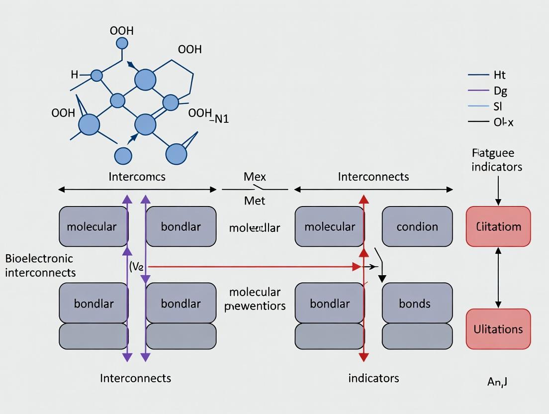
Visualizations
The Scientist's Toolkit: Key Research Reagent Solutions
| Item Name | Function & Relevance to Fatigue Research |
|---|---|
| Polydimethylsiloxane (PDMS), Sylgard 184 | The ubiquitous elastomeric substrate. Its modulus, surface chemistry, and viscoelasticity critically influence stress transfer to thin films. |
| (3-Aminopropyl)triethoxysilane (APTES) | Silane coupling agent. Improves adhesion between inorganic layers (e.g., oxide dielectrics) and polymer substrates, delaying delamination fatigue. |
| Poly(3,4-ethylenedioxythiophene):Poly(styrene sulfonate) (PEDOT:PSS) | Conductive polymer benchmark. Studying its fatigue under strain informs soft, conductive composite design. Often modified with cross-linkers (e.g., GOPS). |
| Ethylene Glycol Dimethyl Acrylate (EGDMA) | Common cross-linker for hydrogels. Increasing its concentration raises elastic modulus and can alter crack propagation behavior under cyclic load. |
| Phosphate Buffered Saline (PBS), pH 7.4 | Standard electrolyte for in-vitro simulated physiological testing. Ionic content drives electrochemical corrosion alongside mechanical stress. |
| Dulbecco's Modified Eagle Medium (DMEM) + 10% FBS | Cell culture media. Provides a biologically active, protein-rich environment for testing biofouling's impact on mechanical integrity. |
| Glycerol or Dimethyl Sulfoxide (DMSO) | Plasticizing additives. Incorporated into hydrogels or polymers to modulate brittleness, reduce stress relaxation, and improve fatigue life. |
| Four-Point Probe Station with Micro-positioners | Essential for accurate, contact-resistance-minimized measurement of sheet resistance changes in fatiguing conductive traces. |
Technical Support Center: Troubleshooting Bioelectronic Interconnect Fatigue
Frequently Asked Questions (FAQs)
Q1: Our thin-film gold interconnects are cracking after 100,000 cycles of 15% uniaxial strain. What material or design factors should we investigate first? A1: This is a classic fatigue failure. Focus on the interplay between substrate modulus and metal film thickness. Cracking often initiates at grain boundaries. Consider implementing a serpentine mesh design to distribute strain, or explore a conductive composite (e.g., PEDOT:PSS with polyurethane) for higher intrinsic stretchability. Ensure your adhesion promoter (e.g., (3-Aminopropyl)triethoxysilane) is correctly applied.
Q2: We observe delamination of the encapsulating silicone layer from our Pt electrode site during cyclic flexion tests. How can we improve adhesion? A2: Delamination is typically a surface energy/chemistry issue. Implement a rigorous surface pretreatment protocol:
- Oxygen plasma treatment of the silicone for 60 seconds at 100W.
- Immediate application of a silicone-based primer (e.g., MED-1511 primer from NuSil).
- Cure the primer before applying the encapsulating top layer. Ensure both silicone layers are from the same manufacturer for compatibility.
Q3: Electrical noise increases dramatically in our recorded signals during dynamic movement experiments. What are the primary troubleshooting steps? A3: This is likely due to intermittent contact from fatigue damage. Follow this diagnostic tree:
- Check Impedance: Measure electrode impedance before, during, and after movement. A spike indicates cracking.
- Inspect Insulation: Use microscopic inspection (SEM recommended) for micro-cracks in the insulation layer.
- Short-Circuit Test: Check for transient short circuits between adjacent traces during flexion.
- Strain Isolation: Ensure your interconnect is properly strain-isolated from the rigid sensor/amplifier chip.
Q4: What is the expected lifetime (cycle count) for a well-designed stretchable interconnect under physiologic strain ranges? A4: Lifetime is highly dependent on materials and strain magnitude. See Table 1 for current performance data from recent literature.
Q5: How do we accurately simulate complex body movements (e.g., shoulder rotation) in a benchtop test? A5: A multi-axis testing rig is required. A simplified protocol involves decomposing the movement into primary axes and sequencing them:
- Program your biaxial or triaxial tester to apply cyclic flexion (X-axis) at 1 Hz.
- Superimpose a lower frequency (0.1 Hz) torsional strain (Y-axis).
- Use a humidity/temperature chamber to simulate the physiologic environment (37°C, 90% RH).
Troubleshooting Guides
Issue: Sudden Catastrophic Failure of Interconnect
- Symptoms: Complete loss of conductivity, visible macroscopic tear.
- Probable Cause: Stress concentration at a geometric feature (e.g., a sharp corner in the trace, or the junction with a rigid component).
- Solution: Redesign the trace geometry to use gradual, filleted curves. Implement a gradient stiffness adapter between rigid and stretchable zones, using a photopatternable polymer like PPF (polypropylene fumarate) with a graded crosslinking density.
Issue: Gradual Drift in Baseline Impedance Over Cycling
- Symptoms: Impedance increases steadily by >10% over 10,000 cycles.
- Probable Cause: Progressive nanoscale cracking or delamination (ratcheting effect).
- Solution: Verify the elastic recovery of your substrate. A viscoelastic substrate (like some PDMS blends) can cause permanent plastic deformation over time, leading to accumulated strain in the metal film. Switch to a more purely elastic substrate (e.g., Ecoflex) or ensure your testing frequency is low enough for full substrate recovery.
Issue: Failure at the Solder Joint or Anisotropic Conductive Film (ACF) Bond
- Symptoms: Failure localized to the connection point to a rigid PCB.
- Probable Cause: The solder/ACF is too stiff, creating a high strain mismatch.
- Solution: Use a strain-relief loop or "S-bend" in the interconnect design leading into the bond pad. Consider using a low-modulus, conductive epoxy (e.g., silver-loaded epoxy) instead of traditional solder, and pot the joint in a soft silicone dome.
Table 1: Fatigue Performance of Bioelectronic Interconnect Materials & Designs
| Material/Design | Substrate | Max Strain (%) | Cycles to Failure | Failure Mode | Key Reference (Year) |
|---|---|---|---|---|---|
| Sputtered Au (50nm) | PDMS (Sylgard 184) | 15% | ~100,000 | Channeling cracks | Liang et al. (2022) |
| Serpentine Au Mesh | Ecoflex 00-30 | 30% | >1,000,000 | Grain boundary voiding | Zhang et al. (2023) |
| PEDOT:PSS/ PU Composite | Hydrogel | 50% | ~500,000 | Conductivity degradation | Kim et al. (2023) |
| Liquid Metal (EGaIn) Embedded | Silicone Rubber | 100% | >5,000,000 | Leakage/oxidation at breach | Wang & Liu (2024) |
| Buckypaper Nanocomposite | Polyimide | 5% (Flexion) | ~200,000 | Delamination | Sharma et al. (2023) |
Experimental Protocols
Protocol 1: Uniaxial Cyclic Strain Test for Thin-Film Interconnects
- Objective: Determine the fatigue life of a conductive trace under repetitive tensile strain.
- Materials: Electro-mechanical tester, custom grips, data logger, potentiostat for impedance.
- Method:
- Mount the sample (e.g., PDMS with patterned Au) in the tester using custom 3D-printed grips that secure the substrate without damaging the trace.
- Apply a pre-strain of 1% to remove slack.
- Program a sinusoidal strain waveform (e.g., 15% peak strain, 0.5 Hz frequency).
- Simultaneously, use a 4-point probe or integrated potentiostat to measure resistance continuously at 100 Hz sampling rate.
- Cycle until resistance increases by 100% (defining failure) or visual cracking is observed.
- Perform post-mortem analysis using SEM.
Protocol 2: Dynamic Flexion Simulation for Spinal Implant Interconnects
- Objective: Simulate the repetitive flexion/extension of the spine on an encapsulated device.
- Materials: Programmable flexion stage, humidity chamber, microscope with video.
- Method:
- Mount the device on a polycarbonate "vertebra" fixture mimicking spinal segment geometry.
- Submerge in phosphate-buffered saline (PBS) at 37°C.
- Program the stage to apply ±30 degrees of flexion at 1 Hz (approximating human gait).
- Use an in-situ monitoring system (e.g., through a transparent window) to record any visual delamination or buckling.
- Pause at set intervals (e.g., every 10,000 cycles) to perform electrochemical impedance spectroscopy (EIS).
Visualizations
Title: Fatigue Failure Pathway in Stretchable Interconnects
Title: Cyclic Strain Testing Workflow for Fatigue Assessment
The Scientist's Toolkit: Research Reagent Solutions
| Item | Function | Example/Supplier |
|---|---|---|
| Ecoflex 00-30 | Ultra-soft, high-toughness silicone elastomer substrate for high-strain applications. | Smooth-On, Inc. |
| PEDOT:PSS (Clevios PH1000) | Conductive polymer, often blended with plasticizers for stretchable conductive composites. | Heraeus Epurio |
| (3-Aminopropyl)triethoxysilane (APTES) | Adhesion promoter to enhance bonding between inorganic metals and polymeric substrates. | Sigma-Aldrich |
| Galinstan or EGaIn | Liquid metal alloy used for ultra-stretchable, self-healing conductive channels. | Geratherm Medical AG |
| MED-1511 Primer | Primes silicone surfaces for covalent bonding, crucial for multilayer encapsulation. | NuSil Technology |
| Polyurethane (PU) Dispersion | Used as an elastic matrix for conductive composites to improve mechanical robustness. | Lubrizol (Tecophilic) |
| Photopatternable Silicone | Allows for precise micropatterning of elastic insulating layers and structures. | Dow (SI 30) |
| Silver Flake/ Silver Nanowires | Conductive fillers for creating percolation networks in elastic composites. | Sigma-Aldrich, Blue Nano |
Technical Support Center: Troubleshooting Fatigue in Bioelectronic Interconnects
FAQs & Troubleshooting Guides
Q1: During cyclic flex testing, our gold traces on polyimide substrates show erratic increases in electrical resistance after ~10,000 cycles, not the gradual increase predicted. What could cause this?
A: This is a classic sign of localized delamination or crack initiation at interface defect sites, leading to sudden, discontinuous failure. The root cause is often contamination or inadequate surface treatment prior to metal deposition. Ensure polymeric substrates undergo O₂ plasma treatment (50-100 W, 30-60 seconds) immediately before deposition to maximize adhesion. Monitor process chamber humidity; keep below 30% RH. Implement in-situ resistance monitoring during cycling to pinpoint the exact cycle of failure.
Q2: Our Parylene-C encapsulation layer is developing micro-cracks after implantation in a simulated physiological environment, leading to device failure. How can we improve barrier integrity?
A: Parylene-C's mechanical performance under hydrational stress is limited. The issue is likely stress corrosion cracking. Two primary solutions:
- Adhesion Promoter: Apply a silane-based adhesion promoter (e.g., A-174 Silane) to the device surface before Parylene deposition. This improves interfacial bonding.
- Multi-Layer Encapsulation: Switch to a multi-layer stack. A common, robust protocol is: Al₂O₃ (30 nm via ALD) / Parylene-C (3-5 µm) / Al₂O₃ (30 nm). The inorganic layers block moisture diffusion, while the Parylene provides mechanical compliance and pin-hole coverage.
Q3: We observe delamination of platinum interconnects from polydimethylsiloxane (PDMS) substrates under minimal strain. What surface modification is most effective?
A: PDMS presents a low-surface-energy challenge. A reliable method is to use an intermediary tie-layer. The following protocol has shown a 300% improvement in adhesion energy:
- Treat PDMS with oxygen plasma (100 W, 1 min).
- Immediately immerse in a 1% (v/v) solution of (3-Mercaptopropyl)trimethoxysilane (MPTMS) in toluene for 60 minutes.
- Rinse with toluene and iso-propanol, then cure at 110°C for 15 min.
- Proceed with metal deposition. The thiol (-SH) group binds strongly to Pt, creating a robust interface.
Q4: How do we accurately measure the fatigue life (Nf) of a thin-film metal trace on a polymer in a simulated bio-environment?
A: Use a custom-built or commercial micro-tensile/flexural tester inside an environmental chamber. Key parameters and a typical result summary are below.
Table 1: Fatigue Test Parameters & Results for Au on Polyimide
| Parameter | Value | Notes |
|---|---|---|
| Substrate | Polyimide (PI-2611), 25 µm thick | Pre-cleaned & plasma treated |
| Metal Trace | Au (300 nm) with Cr adhesion layer (10 nm) | E-beam evaporated |
| Cyclic Strain (ε) | 0.5%, 1.0%, 1.5% | Calculated via beam bending theory |
| Frequency | 1 Hz | Avoids hysteretic heating |
| Environment | PBS, 37°C | Per ASTM F2121 |
| Failure Criteria (Nf) | 20% resistance increase | |
| Avg. Cycles to Failure (Nf) at ε=1.0% | 45,750 ± 2,150 cycles | Mean ± Std Dev, n=10 samples |
Experimental Protocol: Evaluating Interfacial Adhesion via Peel Test
Objective: Quantify the adhesion energy of a metal film on a polymeric substrate before/after environmental aging.
Materials:
- Device sample with metal trace.
- Polyimide or acrylic tape (3M VHB or similar).
- Epoxy resin (e.g., Loctite EA 9466).
- Micro-tensile tester with 10N load cell.
- Environmental chamber (optional for aged samples).
Procedure:
- Sample Preparation: Bond a rigid backing (e.g., glass slide) to the top of the metal trace using a slow-cure, high-strength epoxy. This ensures force is applied to the metal-polymer interface.
- Tape Application: Firmly apply a high-tack tape to the exposed metal trace area.
- Mounting: Clamp the sample backing and the free end of the tape in the tensile tester.
- Testing: Perform a 90-degree or 180-degree peel test at a constant crosshead speed of 10 mm/min.
- Calculation: Adhesion energy (G, in J/m²) is calculated from the average peel force (F, in N) over a stable region: G = (2F / w) for 90° peel, where w is the width of the peeled trace.
- Aging: Repeat on samples aged in Phosphate-Buffered Saline (PBS) at 37°C for 1-4 weeks to assess degradation.
The Scientist's Toolkit: Key Research Reagent Solutions
Table 2: Essential Materials for Bioelectronic Interface Research
| Item | Function | Example/Product Code |
|---|---|---|
| Oxygen Plasma Cleaner | Increases surface energy of polymers for enhanced metal adhesion. | Nordson MARCH, Harrick Plasma |
| (3-Aminopropyl)triethoxysilane (APTES) | Silane coupling agent to create amine-terminated surfaces on oxides for bonding. | Sigma-Aldrich 440140 |
| Polyimide Precursor (PI-2611) | High-performance polymer substrate with excellent thermal and chemical stability. | HD MicroSystems |
| Parylene-C Dimer | For conformal, pinhole-free chemical vapor deposition (CVD) of a bio-inert encapsulation layer. | Specialty Coating Systems |
| Atomic Layer Deposition (ALD) Al₂O₃ Precursors | Trimethylaluminum (TMA) and H₂O for depositing ultra-thin, conformal moisture barriers. | Sigma-Aldrich (for TMA) |
| Phosphate Buffered Saline (PBS), pH 7.4 | Standard solution for simulating physiological conditions during aging tests. | Thermo Fisher 10010023 |
| Cyclic Olefin Copolymer (COC) | Alternative polymer substrate with low moisture absorption and high rigidity. | Topas 5013 |
Diagrams
Title: Bioelectronic Interconnect Fatigue Test Workflow
Title: Interconnect Failure Pathways Under Duress
Technical Support Center
Troubleshooting Guide & FAQ
Q1: During long-term in vivo electrophysiology recording, we observe a steady increase in electrode impedance and a decline in signal-to-noise ratio (SNR). Is this fatigue, and what are the immediate steps? A: Yes, this is a classic symptom of mechanical fatigue at the bioelectronic interface. Immediate steps:
- Isolate the Issue: Perform an in vitro impedance test in phosphate-buffered saline (PBS) using the same measurement parameters. This determines if the change is in the electrode or the biological environment.
- Inspect the Interconnect: Under a microscope, examine the lead from the electrode to the connector for kinks, cracks, or delamination.
- Check Cyclic Loading History: Review experiment logs for the number of animal movement cycles or any external bending events.
Q2: Our flexible microelectrode array suddenly failed (catastrophic failure) after 2 weeks of implantation. Visual inspection shows a broken trace. How can we investigate the root cause? A: Catastrophic failure often results from crack propagation. Follow this protocol:
- Failed Device Analysis:
- Use scanning electron microscopy (SEM) on the fracture site to examine the morphology. Look for fatigue striations, indicative of cyclic stress.
- Perform energy-dispersive X-ray spectroscopy (EDX) on and around the fracture to check for corrosion or material degradation.
- Replicate & Monitor:
- Implement a bench-top accelerated fatigue test (see Protocol 1 below) on devices from the same fabrication batch.
- Use real-time impedance spectroscopy during the accelerated test to correlate mechanical cycles with electrical performance decay.
Q3: What are the best practices to monitor for "signal degradation" proactively in a chronic study? A: Implement a routine monitoring protocol:
- Daily/Bi-daily: Measure single-unit yield and SNR from standard neural signals (e.g., resting-state or evoked potentials).
- Weekly: Perform electrochemical impedance spectroscopy (EIS) at 1 kHz (relevant for neural recording) and 1 Hz (relevant for interface stability). Track the phase angle shift.
- Control Measurement: Always include a stable, non-fatiguing reference electrode (e.g., large-surface-area Pt) to differentiate system drift from interface fatigue.
Experimental Protocols
Protocol 1: Accelerated Fatigue Test for Flexible Interconnects Objective: To simulate years of cyclic bending stress in a controlled laboratory setting. Materials: See "Scientist's Toolkit" below. Method:
- Mount the flexible bioelectronic device on a custom or commercial cyclic bending stage.
- Define bending parameters: Radius (e.g., 5 mm, simulating brain surface curvature), frequency (e.g., 1 Hz), and angle (e.g., 90°).
- Connect the device to an impedance analyzer programmed for intermittent measurement.
- Initiate cycling. Pause at predefined intervals (e.g., every 1,000 cycles) to perform a full EIS sweep (from 10 Hz to 100 kHz).
- Continue until impedance at 1 kHz increases by 200% or catastrophic failure occurs.
- Plot impedance vs. cycle count to determine the "cycles-to-failure" for your design.
Protocol 2: In Situ Impedance and Signal Fidelity Correlation Objective: To directly correlate mechanical fatigue with signal quality loss in a live experiment. Method:
- In an anesthetized animal model, implant the device and acquire baseline electrophysiological data (e.g., spontaneous neural activity) and EIS data.
- Allow recovery and begin chronic recordings over days/weeks.
- During each recording session: a. Record 5 minutes of neural activity. Compute the mean spike SNR and number of detectable single units. b. Immediately after, measure the EIS.
- Post-process: Align SNR/unit count data with impedance magnitude and phase data at the relevant frequency. Use statistical correlation (e.g., Pearson coefficient) to establish the relationship.
Data Presentation
Table 1: Quantitative Progression of Fatigue-Related Failures
| Failure Stage | Typical Impedance Change at 1 kHz | Signal SNR Change | Observable Physical Change | Common Cycle Count (in vivo, approx.) |
|---|---|---|---|---|
| Initial Degradation | +20% to +50% | -10% to -30% | Micro-cracks initiation (not visible) | 1,000 - 10,000 |
| Progressive Fatigue | +50% to +200% | -30% to -70% | Visible trace buckling, delamination onset | 10,000 - 100,000 |
| Catastrophic Failure | >+1000% (Open Circuit) | No Signal | Complete trace fracture, insulation breach | 100,000+ |
Table 2: Key Material Properties Impacting Fatigue Resistance
| Material/Coating | Function | Young's Modulus (GPa) | Typical Fatigue Limit (Cycles, 5mm radius) | Key Advantage for Interconnects |
|---|---|---|---|---|
| Gold (Thin Film) | Conductive Trace | ~79 | 50,000 - 200,000 | High conductivity, standard process |
| PEDOT:PSS | Conductive Polymer Coating | ~2-3 | 100,000 - 500,000* | Lower impedance, more compliant |
| Polyimide | Substrate/Insulator | ~2.5 | 1,000,000+ | Flexible, biocompatible, insulative |
| Silicon Elastomer | Encapsulation | 0.001-0.01 | 500,000+ | Stretchable, moisture barrier |
| Platinum-Iridium | Alloy Electrode | ~200 | 200,000 - 1,000,000 | Corrosion resistant, stable interface |
*Highly dependent on formulation and adhesion.
Visualizations
Title: The Fatigue Failure Cascade in Bioelectronic Interconnects
Title: Technical Support: Fatigue Diagnosis Workflow
The Scientist's Toolkit: Research Reagent Solutions
| Item | Function in Fatigue Research |
|---|---|
| Phosphate-Buffered Saline (PBS), 1X, pH 7.4 | Standard electrolyte for in vitro impedance testing, simulating physiological ionic environment. |
| PEDOT:PSS Dispersion (e.g., Clevios PH1000) | Conductive polymer used to coat electrodes, lowering interfacial impedance and improving mechanical compliance. |
| Polyimide Precursor (e.g., PI-2545) | For spin-coating flexible, robust substrate and insulation layers critical for interconnect longevity. |
| Sylgard 184 PDMS | Silicone elastomer used for encapsulating devices, providing a soft, protective barrier against biological fluids. |
| Artificial Cerebrospinal Fluid (aCSF) | More biologically relevant than PBS for pre-implantation testing, matching ion concentrations of the target tissue. |
| Cyanoacrylate or Epoxy (Medical Grade) | For quick, stable attachment of connectors to skull or casing, relieving strain on the fragile interconnect. |
| Electrochemical Impedance Analyzer | Key instrument for monitoring impedance magnitude and phase, the primary electrical indicator of fatigue. |
Technical Support Center: Troubleshooting & FAQs
FAQ 1: What are the primary failure modes observed in chronically implanted neural probe interconnects?
Answer: The dominant failure modes are mechanical fatigue and electrochemical corrosion at the interconnect sites. Quantitative data from recent studies is summarized below.
| Failure Mode | Location | Typical Time to Failure | Key Stressors | Common Materials Affected |
|---|---|---|---|---|
| Flexural Fatigue Cracking | Thin-film metal trace at strain concentration (e.g., bond pad, sharp bend). | 3-12 months in vivo. | Cyclic micromotion from breathing/pulsation, device tethering. | Gold, Platinum, Iridium Oxide on Polyimide/Parylene C. |
| Delamination & Moisture Ingress | Metal-polymer dielectric interface. | 6-24 months. | Hydrolytic swelling of polymer, poor adhesion, biological fluid exposure. | All polymer-metal laminates (e.g., SiO2/PI/Au). |
| Corrosion & Insulation Failure | Pinholes in insulation or at electrode sites. | 1-9 months. | Applied potential, inflammatory oxidative species (H2O2, NO). | Iridium, Tungsten, Silicon Oxide insulation. |
| Stress-Corrosion Cracking | Grain boundaries of polycrystalline metal traces. | 6-18 months. | Combined mechanical strain and electrochemical potential. | Platinum, Stainless Steel alloys. |
Experimental Protocol for Ex Vivo Accelerated Fatigue Testing:
- Sample Preparation: Fabricate interconnects on flexible substrates (e.g., 25µm polyimide with 200nm Au traces). Potentiostatically deposit PEDOT:PSS or Iridium Oxide on electrode sites.
- Setup: Mount sample on a precision motorized flex stage. Submerge in phosphate-buffered saline (PBS) at 37°C, pH 7.4.
- Cycling Parameters: Apply a sinusoidal or sawtooth bending profile with a radius of curvature of 5mm (mimicking brain tissue movement). Frequency: 2 Hz.
- In-Situ Monitoring: Perform electrochemical impedance spectroscopy (EIS) every 10,000 cycles (1-10 kHz range). Measure DC resistance of the trace continuously.
- Failure Criterion: Define failure as a 50% increase in baseline impedance at 1 kHz OR an open circuit (resistance > 1 MΩ).
- Post-Mortem Analysis: Use SEM/EDX to examine crack morphology and corrosion products.
FAQ 2: How can I differentiate between interconnect fatigue and biological encapsulation as the cause of rising impedance in my cardiac stimulation lead?
Answer: A systematic in vivo and ex vivo electrochemical protocol is required to isolate the failure mechanism.
| Diagnostic Test | Procedure | Interpretation: Fatigue/Corrosion | Interpretation: Fibrotic Encapsulation |
|---|---|---|---|
| Pulse Test | Apply a cathodic-first, charge-balanced biphasic pulse. Monitor voltage transient. | Voltage transient shows excessive polarization or open circuit. | Voltage transient shows increased series resistance but normal shape. |
| EIS Spectrum | Measure impedance from 0.1 Hz to 100 kHz. | Sharp increase at all frequencies, indicating break in conductor. | Increase primarily at low frequencies (<100 Hz), due to diffusion barrier. |
| Potential Step Chronoamperometry | Apply a small potential step (+0.5V). Measure current decay. | Current remains near zero (open). | Current decays slowly, following Cottrell behavior (diffusion-limited). |
| Post-Explanation Analysis | SEM inspection of explanted lead. | Visible cracks, pitting, or delamination at stress points. | Uniform fibrous tissue coating without metallic corrosion. |
Title: Diagnostic Flow for Rising Impedance in Chronic Bioelectronic Implants
The Scientist's Toolkit: Key Research Reagent Solutions
| Item | Function | Example Use Case |
|---|---|---|
| PBS (Phosphate Buffered Saline), pH 7.4 | Simulates ionic body fluid for in vitro electrochemical and corrosion testing. | Accelerated lifetime testing in baths. |
| Hydrogen Peroxide (H2O2) Solution (10-100 µM) | Mimics oxidative stress from inflammatory response (reactive oxygen species). | Testing corrosion resistance of electrode materials. |
| Artificial Cerebrospinal Fluid (aCSF) | Ionically accurate medium for neural interface testing, includes Na+, K+, Ca2+, Mg2+, Cl-. | Neural probe soak testing and ex vivo electrophysiology. |
| Parylene C Deposition System | Provides conformal, biocompatible, moisture-resistant insulating coating. | Insulating thin-film metal traces on flexible probes. |
| Polyimide (PI) Precursors (e.g., HD-4110) | Forms flexible, robust, and thermally stable substrate for microfabricated interconnects. | Spin-coating to create flexible substrate layers. |
| Electroplating Solutions (e.g., Iridium Oxide, PEDOT:PSS) | Deposits high-charge-capacity, low-impedance coatings on electrode sites. | Improving charge injection limits and signal quality. |
| Cyclic Flex Tester with Environmental Chamber | Applies programmable mechanical bending cycles in controlled (temp, humidity, liquid) environments. | Accelerated fatigue life testing of flexible interconnects. |
| Potentiostat/Galvanostat with EIS | Measures electrochemical impedance, corrosion potential, and performs controlled potential experiments. | Characterizing electrode health and failure mechanisms. |
Title: Synergistic Mechanical-Electrochemical Fatigue Failure Pathway
Designing for Durability: Advanced Materials and Engineering Solutions to Combat Fatigue
Troubleshooting Guide for Bioelectronic Interconnect Fatigue Research
FAQs & Troubleshooting
Q1: Our thin-film gold conductors on PDMS are cracking at strain cycles far below the predicted value. What could be the cause? A: This is a common issue often related to poor adhesion and stress concentration at the interface. The mismatch in elastic modulus between the stiff metal and compliant substrate creates localized shear stress. First, ensure you are using an oxygen plasma treatment (e.g., 100W for 60 seconds) on the PDMS prior to metal deposition to improve adhesion. Consider introducing an intermediate compliant layer like a poly(3,4-ethylenedioxythiophene) polystyrene sulfonate (PEDOT:PSS) or a thin silicone adhesive. Also, verify your metal film is not overly thick; for sputtered Au, keep thickness below 100 nm to minimize its neutral mechanical plane effect.
Q2: The electrical resistance of our serpentine-structured copper interconnect increases unpredictably during cyclic stretching tests. How can we diagnose this? A: Unpredictable resistance changes typically indicate the onset of micro-fatigue cracks or delamination. Use in-situ optical microscopy during cycling to observe crack initiation. Ensure your test fixture provides pure, uniform uniaxial strain without lateral constraint. A step-by-step protocol:
- Mount the sample on a cyclic stretcher integrated with a 4-point probe resistance meter.
- Perform in-situ imaging at 10x magnification, focusing on the serpentine's inner bend radius.
- Correlate resistance jumps (e.g., >10% increase from baseline) with visual crack events. A gradual increase suggests void coalescence, while a sharp jump indicates a single macroscopic crack.
Q3: When encapsulating our device with silicone elastomer (Ecoflex), we see delamination and water ingress in accelerated aging tests. How do we improve bonding? A: Delamination is a critical failure mode for bioelectronic interfaces. The key is surface chemistry and mechanical interlocking.
- Protocol for Robust Encapsulation:
- Surface Activation: Treat both the device substrate and the uncured Ecoflex surface with a brief atmospheric plasma (30 seconds).
- Primer Application: Apply a thin, uniform layer of a silicone primer (e.g., MED-151) and allow it to become tack-dry (approx. 5 minutes).
- Bonding & Curing: Carefully place the liquid Ecoflex precursor onto the primed device. Cure at 60°C for 2 hours, applying a slight pressure (approx. 0.5 kPa) during the initial 30 minutes to ensure intimate contact.
Q4: Our PEDOT:PSS conductive polymer films lose conductivity and mechanically degrade after repeated sterilization (autoclaving). Are there material alternatives? A: Standard PEDOT:PSS is hygroscopic and thermally sensitive. For autoclave compatibility (121°C, 15 psi steam), consider these alternatives:
- Ionic Liquid-doped PEDOT:PSS: Adding 5-10 wt% of ionic liquids like 1-ethyl-3-methylimidazolium tetrafluoroborate ([EMIM][BF4]) significantly improves thermal stability and retains >85% conductivity post-sterilization.
- Conductive Elastomer Composites: Use a composite of polyurethane or silicone loaded with 15-20 vol% silver flakes or carbon nanotubes. These materials typically withstand 50+ autoclave cycles with <15% resistance change.
Q5: How do we accurately measure the fatigue life (N_f) of a novel stretchable conductor? A: Follow a standardized electromechanical fatigue test protocol.
- Standard Test Protocol:
- Sample Preparation: Fabricate conductors on your substrate with standardized dimensions (e.g., 50mm x 5mm trace).
- Test Setup: Mount on a motorized cyclic tensile stage. Connect to a digital multimeter for continuous resistance monitoring.
- Parameters: Define your maximum applied strain (εmax, e.g., 20%), a strain rate (e.g., 10% per second), and a waveform (e.g., sinusoidal or trapezoidal).
- Failure Criterion: Define failure as the cycle count (Nf) at which resistance increases by 100% (or another defined threshold like electrical open circuit). Run a minimum of n=5 samples per condition.
Table 1: Fatigue Performance of Common Interconnect Materials
| Material & Structure | Typical Substrate | Max Strain Before Failure (%) | Cycles to Failure (N_f) at 10% Strain | Key Failure Mode |
|---|---|---|---|---|
| Gold (Au), Thin Film | PDMS | 2-5% | < 1,000 | Brittle cracking, delamination |
| Gold, Serpentine Mesh | PDMS | 50-70% | > 100,000 | Stress concentration at bends |
| Eutectic Gallium-Indium (eGaIn) | Ecoflex | > 400% (static) | > 10,000 (at 50%) | Oxide skin rupture, leakage |
| PEDOT:PSS, Pure | PET | 10-15% | ~ 5,000 | Crack propagation, dehydration |
| Silver Flake / Silicone Composite | Silicone | 80-120% | > 50,000 | Percolation network disruption |
| Liquid Metal Embedded Elastomer | SEBS | 250% | > 20,000 | Channel rupture, filler separation |
Table 2: Comparison of Encapsulation Materials
| Material | Water Vapor Transmission Rate (WVTR) [g/m²/day] | Elastic Modulus [MPa] | Adhesion Strength to Au [N/cm] | Biocompatibility (ISO 10993) |
|---|---|---|---|---|
| Polydimethylsiloxane (PDMS), Sylgard 184 | ~ 15-20 | 1.5 - 3.0 | 0.8 - 1.2 | Class VI Passed |
| Silicone Elastomer (Ecoflex 00-30) | ~ 25-35 | 0.03 - 0.08 | 1.5 - 2.0* | Class VI Passed |
| Polyimide (PI) | < 5 | 2,500 - 3,000 | 4.0 - 5.0 (with adhesive) | Generally Compliant |
| Parylene-C (Vapor Dep.) | ~ 0.2 - 0.5 | 2,800 | 0.5 (poor, unless primed) | USP Class VI |
| Polyurethane (Medical Grade) | 50 - 500 (varies) | 5 - 50 | 2.0 - 4.0 | Class VI Passed |
*With proper surface treatment (plasma + primer).
The Scientist's Toolkit: Research Reagent Solutions
| Item | Function | Key Consideration for Fatigue Research |
|---|---|---|
| Oxygen Plasma System | Creates hydroxyl groups on polymer surfaces (PDMS, PI) to dramatically improve metal/polymer adhesion. | Critical for reproducible interfacial strength. Power and time must be optimized to avoid a weak oxidized layer. |
| Silicone Primer (e.g., MED-151, AP-133) | Forms a chemical bridge between inorganic (metal, oxide) and organic (silicone) surfaces for robust bonding. | Essential for long-term encapsulation integrity in wet/cyclic environments. |
| Ionic Liquid Additives (e.g., [EMIM][TFSI]) | Plasticizes and stabilizes conductive polymers (PEDOT:PSS), enhancing stretchability and thermal/ambient stability. | Doping ratio (3-10 wt%) is critical; too much can cause phase separation. |
| Strain-Rate Controlled Cyclic Tensile Tester | Applies precise, repeatable cyclic strain to samples while measuring force. | Must be integrated with electrical resistance measurement for in-situ electromechanical characterization. |
| Four-Point Probe Station | Measures sheet resistance of thin films without contact resistance errors. | Use micro-probes for patterned traces and a shielded setup for sensitive measurements. |
| Environmental Chamber | Controls temperature and humidity during mechanical testing. | Fatigue life (N_f) can vary by an order of magnitude between dry and 95% RH conditions. |
Experimental Workflows & Diagrams
Title: Fatigue Test Workflow for Stretchable Conductors
Title: Fatigue Failure Pathway at Bioelectronic Interconnects
Technical Support & Troubleshooting Center
This support center addresses common experimental challenges in developing fatigue-resistant bioelectronic interconnects, framed within a thesis on mitigating mechanical fatigue at the biotic-abiotic interface.
Frequently Asked Questions (FAQs)
Q1: During cyclic stretching tests, my serpentine Au interconnect consistently fractures at the arc-meander junction. What is the likely cause and solution? A: This is a classic stress concentration failure. The failure occurs due to a suboptimal transition radius between the arc and the straight segment.
- Troubleshooting Guide:
- Verify Design Parameters: Calculate the ratio of your arc radius (R) to the trace width (w). Literature indicates R/w should be >5 to minimize stress concentration. Measure your fabricated design via microscopy.
- Inspect Fabrication: Use SEM imaging to check for microfabrication defects like notches or uneven photoresist reflow at the junction, which act as crack initiation sites.
- Solution: Redesign the junction using a continuous, Euler-spiral (clothoid) transition that gradually changes curvature, or implement a wider radius. Increase the metal thickness if electroplating is an option, as this improves fatigue life.
Q2: My origami-inspired, crinkled Ti/Pt interconnect delaminates from the PDMS substrate after a few hydration cycles in phosphate-buffered saline (PBS). How can I improve adhesion? A: This is an adhesion failure exacerbated by hydrolytic attack at the metal-polymer interface.
- Troubleshooting Guide:
- Surface Treatment: Ensure the PDMS is treated with oxygen plasma immediately before metal deposition. Verify plasma parameters (power, time) are consistent and sufficient to create a robust siloxane (Si–O–Si) layer.
- Use an Adhesion Layer: Incorporate a thin (5-10 nm) chromium (Cr) or titanium (Ti) adhesion layer between the PDMS and your primary conductor (Pt).
- Encapsulation Strategy: Apply a thin, conformal layer of Parylene-C as a moisture barrier. Silane-based adhesion promoters (e.g., (3-Aminopropyl)triethoxysilane, APTES) can also be applied to the PDMS before metal deposition.
Q3: The electrical resistance of my fractal (Peano or Hilbert curve) interconnect increases unpredictably during long-term, low-frequency (1 Hz) dynamic loading. What should I check? A: This points to progressive damage accumulation rather than sudden fracture, often due to microcracking or interfacial sliding.
- Troubleshooting Guide:
- In-Situ Monitoring: Set up simultaneous electrical resistance measurement and optical microscopy (or digital image correlation) during cyclic testing. Look for the specific fractal segment where resistance jump correlates with visible deformation.
- Material Selection: The ductility of the metal is critical. Consider switching from a brittle metal like chromium to a more ductile one like gold, or using a metal alloy (e.g., Au-Ag).
- Substrate Modulus: The substrate modulus should be optimized. A very soft substrate may cause excessive local strain; a slightly stiffer silicone (e.g., modulus of 1-2 MPa) can better distribute the strain across the fractal pattern.
Q4: How do I accurately measure the effective stretchability of a completed interconnect? Is it different from the substrate's stretchability? A: Yes, they are distinct. The interconnect's effective stretchability is the strain at which electrical failure (e.g., a 100% resistance increase) occurs.
- Experimental Protocol:
- Setup: Mount the sample on a uniaxial tensile stage integrated with a four-point probe resistance meter.
- Procedure: Apply a constant strain rate (e.g., 0.1% per second). Simultaneously record applied strain (ε) and normalized resistance (R/R₀).
- Endpoint: The strain at which R/R₀ > 2 is typically defined as the effective stretchability. This is often much higher than the substrate's fracture strain due to the interconnect's geometric design.
Experimental Protocols
Protocol 1: Standardized Fatigue Life Testing for Bioelectronic Interconnects Objective: To quantify the number of cycles to failure (Nf) under simulated physiological motion.
- Sample Preparation: Fabricate interconnect on an elastomeric substrate (e.g., PDMS, Ecoflex). Encapsulate if applicable.
- Mounting: Secure sample ends to a cyclic tensile tester. Ensure interconnect is aligned and unbuckled.
- Environmental Control: Immerse in PBS at 37°C using a temperature-controlled bath, or use a humidity chamber.
- Testing Parameters:
- Waveform: Sinusoidal.
- Frequency: 1 Hz (simulates cardiac or respiratory rhythms).
- Strain Amplitude: 10-30% (physiological range for skin/organ surfaces).
- Monitoring: Record resistance continuously. Use a high-speed camera for deformation tracking.
- Failure Criterion: Define failure as a sustained 100% increase in resistance or visible fracture.
Protocol 2: Characterization of Fractal Interconnect Areal Coverage and Electrical Performance Objective: To correlate fractal order (space-filling property) with conductance and stretchability.
- Design: Generate Hilbert curve designs of orders 2, 3, and 4 with identical trace width and spacing.
- Fabrication: Fabricate all designs in a single batch to ensure material consistency.
- Measurement:
- Use image analysis (ImageJ) to calculate the areal coverage (%) of metal for each design.
- Measure DC resistance (Ω) using a precision multimeter.
- Calculate sheet resistance and effective conductivity.
- Analysis: Plot Areal Coverage vs. Fractal Order and Conductance vs. Fractal Order to identify trade-offs.
Table 1: Comparative Performance of Geometric Interconnect Designs Data synthesized from recent literature (2022-2024).
| Design Type | Max. Achievable Strain (%) | Cycles to Failure @ 20% Strain | Relative Conductance (Normalized to Bulk Metal) | Key Failure Mode |
|---|---|---|---|---|
| Serpentine (Simple) | 50-70 | 10,000 - 50,000 | 0.95 - 0.99 | Fracture at arc-meander junction |
| Serpentine (Fractal-hybrid) | >100 | 50,000 - 200,000 | 0.85 - 0.92 | Microcrack coalescence in straight segments |
| Origami/Crinkled | >200 | 5,000 - 20,000* | 0.70 - 0.85 | Delamination from substrate |
| Hilbert Fractal (3rd Order) | 80-100 | 15,000 - 30,000 | 0.75 - 0.82 | Progressive debonding & necking |
Note: Highly dependent on adhesion promotion strategy.
Table 2: Impact of Encapsulation on Interconnect Lifetime in Hydrated Environments
| Encapsulation Material | Thickness (µm) | Time to 50% Resistance Increase in 37°C PBS (Days) | Water Vapor Transmission Rate (WVTR) g/m²/day |
|---|---|---|---|
| None (Bare Au/PDMS) | N/A | 1 - 3 | N/A |
| PDMS (Sylgard 184) | 50 | 7 - 14 | ~400 |
| Parylene-C | 5 | 60 - 90 | ~0.5 |
| SU-8 | 10 | 30 - 45 | ~2.5 |
Visualizations
The Scientist's Toolkit: Research Reagent Solutions
Table 3: Essential Materials for Fabricating Fatigue-Resistant Interconnects
| Item Name | Function/Application | Example Product/Note |
|---|---|---|
| Sylgard 184 PDMS Kit | Primary elastomeric substrate. Tunable modulus by varying base:curing agent ratio. | Dow Silicones |
| Ecoflex 00-30 | Ultra-soft silicone substrate for high-strain applications (>300% strain). | Smooth-On |
| (3-Aminopropyl)triethoxysilane (APTES) | Silane coupling agent. Improves adhesion of metals to oxidized PDMS surfaces. | Use in vapor phase or dilute solution (0.5% v/v). |
| Parylene-C | Conformal, biocompatible moisture barrier coating. Applied via chemical vapor deposition (CVD). | Specialty Coating Systems |
| AZ 5214E Photoresist | Image reversal photoresist. Enables high-resolution, undercut profiles for liftoff of metal traces. | MicroChemicals |
| Ti/Cr Evaporation Pellets | High-purity source for electron-beam evaporation of thin adhesion layers (5-15 nm). | Kurt J. Lesker Company |
| Au Evaporation Pellets | High-purity source for evaporation or sputtering of the primary conductive layer (50-200 nm). | 99.999% purity recommended. |
| Four-Point Probe Station | For accurate measurement of sheet resistance and conductivity of thin metal films. | Signatone or Jandel Engineering |
Technical Support Center
Troubleshooting Guides & FAQs
Q1: During laser patterning of a polyimide substrate for a stretchable interconnect, I observe inconsistent ablation depth and ragged edges. What could be the cause and solution?
A: Inconsistent ablation is often due to contaminant films or uneven focus. First, ensure the substrate is cleaned with sequential acetone, isopropanol, and DI water baths, followed by oxygen plasma treatment (50 W, 2 mins) to ensure uniform surface energy. Second, calibrate the laser's focal plane using a sacrificial sample. Use a pulsed UV laser (e.g., 355 nm) with a pulse duration <20 ps to minimize thermal damage. Implement an optical setup with a beam profiler to confirm a Gaussian intensity profile. For quantitative reference:
| Issue | Potential Cause | Recommended Solution | Key Parameters |
|---|---|---|---|
| Ragged Edges | Thermal diffusion, incorrect pulse energy | Use shorter pulse (femtosecond/picosecond), optimize fluence | Fluence: 0.5-1.5 J/cm², Rep Rate: 50-200 kHz |
| Inconsistent Depth | Unstable pulse energy, debris accumulation | Regularly clean optics, use beam shutter, employ assist gas (N₂) | Assist Gas Pressure: 10-20 psi |
| Poor Feature Definition | Incorrect focus, substrate vibration | Activate autofocus system, use vibration isolation table | Focus Spot Size: 10-20 µm |
Protocol: Laser Patterning for Polyimide Interconnects
- Substrate Prep: Clean 50 µm polyimide film. Mount on vacuum chuck.
- Laser Calibration: Run test pattern on scrap piece. Measure depth with profilometer. Adjust power until target depth (e.g., 25 µm) is achieved.
- Patterning: Load design file (GDSII). Set scan speed to 200 mm/s. Use galvanometer scanner with 5% overlap between pulses.
- Post-Process: Sonicate in DI water for 5 mins to remove debris. Inspect under microscope.
Q2: In transfer printing of ultrathin silicon nanomembranes onto a PDMS stamp, the yield is low due to fracture. How can I improve adhesion and release kinetics?
A: Fracture during pick-up or printing typically indicates incorrect control of the stamp's adhesion energy. This is governed by the velocity-dependent viscoelastic property of the PDMS stamp (typically a soft, ~50 kPa modulus elastomer like Sylgard 527). Use the following protocol:
| Printing Phase | Critical Parameter | Target Value | Function |
|---|---|---|---|
| Pick-Up | Stamp Approach Velocity | 0.1-0.5 mm/s | Ensures conformal contact |
| Pick-Up | Dwell Time | 60-120 s | Allows van der Waals adhesion to dominate |
| Retraction | Peel-off Velocity | 5-10 mm/s | Fast retraction to fracture sacrificial layer |
| Printing | Stamp Contact Velocity | 0.5-1.0 mm/s | Controlled contact with target |
| Printing | Retraction Velocity | 0.1-0.2 mm/s | Slow retraction to reduce adhesion energy for release |
Protocol: Viscoelastic Transfer Printing of Nanomembranes
- Stamp Fabrication: Cast and cure Sylgard 527 PDMS on a silicon wafer (30 µm thick). Treat surface with brief O₂ plasma (10 W, 10 s) to slightly increase tackiness.
- Pick-Up: Align stamp over donor wafer (with etched Si nanomembranes). Approach slowly at 0.2 mm/s. Apply gentle contact force (~5 N/cm²) for 90 seconds. Retract rapidly at 8 mm/s.
- Printing: Align stamp over target substrate (e.g., PEG hydrogel). Approach at 0.8 mm/s. Make contact and apply light pressure. Initiate extremely slow, stage-controlled retraction at 0.15 mm/s. Use a motorized stage for precise velocity control.
Q3: When fabricating 3D helical microcoils via direct laser writing (DLW) for fatigue-resistant interconnects, the structures collapse during development. How do I prevent this?
A: Collapse is a classic issue due to capillary forces during solvent drying. The solution lies in using a supercritical CO₂ drying process and optimizing photoresist support.
Protocol: 3D Helical Coil Fabrication via Two-Photon Polymerization
- Resist & Substrate: Use a biocompatible photoresist like IP-S (Nanoscribe) on an ITO-coated glass substrate. Apply a drop of resist.
- Writing Parameters: Use a 63x objective lens. Set laser power to 25 mW (at sample). Write helical structure with 150 nm slicing distance and 50% hatching distance. Design temporary cylindrical support pillars at 50 µm intervals around the coil.
- Development & Drying: Crucial Step.
- Develop in PGMEA for 45 minutes.
- Rinse in IPA for 5 minutes.
- Transfer sample to a critical point dryer (CPD). Fill chamber with liquid CO₂ at 10°C. Perform 5 flush cycles to replace IPA with liquid CO₂.
- Heat to 40°C, raise pressure to >1073 psi, transitioning CO₂ to supercritical state.
- Vent slowly at 0.7 L/min over 60 mins. Structures will remain intact.
Research Reagent & Material Solutions
| Item | Supplier (Example) | Function in Bioelectronic Interconnect Research |
|---|---|---|
| Sylgard 527 Silicone Elastomer Kit | Dow Chemical | Used for fabricating viscoelastic stamps for transfer printing; its low modulus enables reliable pick-up and release of fragile devices. |
| IP-S Photoresist | Nanoscribe GmbH | A biocompatible photoresist for high-resolution two-photon polymerization (2PP), used to create 3D fatigue-resistant scaffold structures. |
| Piezoelectric Polymer Film (PVDF-TrFE) | PiezoTech | Serves as a stress-sensing layer integrated into interconnects to monitor mechanical fatigue in situ during cyclic loading. |
| Hydrogel (PEGDA, 4-Arm, 10 kDa) | Sigma-Aldrich | Used as a soft, hydrated target substrate mimicking biological tissue for printing and testing bioelectronic interfaces. |
| Liquid Metal (EGaIn: 75% Ga, 25% In) | Strem Chemicals | Injectable conductive filler for self-healing microfluidic channels within 3D-fabricated interconnects to maintain conductivity after crack formation. |
Experimental Workflow Diagram
Laser, Transfer, and 3D Fabrication Workflow
Laser-Tissue Interaction Pathway Diagram
Laser Parameters Dictate Material Interaction
Technical Support Center: Troubleshooting Bioelectronic Interconnect Experiments
This technical support center provides targeted guidance for common experimental challenges in developing fatigue-resistant bioelectronic interconnects, focusing on adhesion promoters, gradient modulus layers, and self-healing materials.
Frequently Asked Questions (FAQs) & Troubleshooting
Q1: During accelerated fatigue testing (e.g., 100,000 cyclic bends), my thin-film gold interconnect on a PDMS substrate delaminates, despite using a (3-Aminopropyl)triethoxysilane (APTES) adhesion promoter. What could be wrong?
A: Delamination under cyclic loading often indicates insufficient covalent bonding or hydrolysis of the silane layer. Key troubleshooting steps:
- Verify Substrate Pre-treatment: Ensure the PDMS surface is thoroughly activated via oxygen plasma (e.g., 100W for 60 seconds) immediately before silanization. A water contact angle of <10° post-activation confirms success.
- Control Humidity During Silanization: APTES coupling requires optimal ambient moisture. Perform the reaction in a sealed chamber with a relative humidity of 40-60%. Too dry limits silanol formation; too wet promotes multilayer, weak polysiloxane networks.
- Post-Apply a Thermal Cure: After APTES application, cure at 110°C for 10-15 minutes to complete condensation and strengthen the siloxane network.
Q2: I designed a gradient modulus epoxy-acrylate interlayer between my stiff electrode (PEDOT:PSS) and soft neural tissue. My impedance measurements show a significant increase (>50%) after 7 days in simulated interstitial fluid. What is the likely failure mode?
A: This points to interfacial degradation and swelling mismatch. The gradient likely lacks sufficient hydrophobicity or crosslink density at the soft end, allowing fluid ingress and plasticization.
- Solution: Incorporate a hydrophobic monomer (e.g., lauryl methacrylate, 5-10% v/v) into the softest layer of the gradient to reduce water uptake. Ensure graded UV-curing doses increase with layer stiffness to properly crosslink the softer, hydrophilic phases.
Q3: The self-healing polydimethylsiloxane (PDMS) elastomer I synthesized, based on imine-bond chemistry, shows poor autonomic healing efficiency (<30%) at physiological temperature (37°C). How can I improve it?
A: Low healing efficiency at 37°C suggests sluggish imine exchange kinetics or insufficient chain mobility.
- Troubleshoot Catalyst & Plasticizer: Ensure you have incorporated a catalyst (e.g., para-toluenesulfonic acid, 0.5-1 mol%) into the matrix. Consider adding a non-volatile biocompatible plasticizer (e.g., triethyl citrate, <5% w/w) to increase polymer chain mobility, facilitating bond exchange at the crack interface.
Q4: When fabricating my multi-layer stack (Metal / Gradient Polymer / Self-Healing Sealant), I get poor interlayer adhesion. Which adhesion promoter is compatible between these diverse materials?
A: This requires a versatile, possibly multi-functional promoter. Consider a two-step or a hybrid solution:
- For the metal/gradient polymer interface, apply a thiol-based silane (e.g., (3-Mercaptopropyl)trimethoxysilane) which bonds well to metals and offers a thiol group for click chemistry with acrylics.
- For the gradient polymer/self-healing sealant interface, a thin, compliant epoxy-based primer (e.g., a low-Tg polyetheramine-cured epoxy) can provide mechanical interlock and covalent bonds to both layers.
Table 1: Performance of Common Adhesion Promoters for Bioelectronic Interconnects
| Promoter | Target Substrate | Target Film | Peel Strength (N/cm) | Key Failure Mode (after 10⁵ cycles) |
|---|---|---|---|---|
| APTES | SiO₂, Plasma-oxidized PDMS | Gold, ITO | 3.5 - 5.2 | Hydrolytic cleavage at siloxane bond |
| MPTES | Gold, Silver | Conductive Polymers (PEDOT) | 4.8 - 6.1 | Oxidation of thiol to sulfonate |
| DOPA-Polymer | Ti, Wet Tissue | Hydrogel, Elastomer | 2.0 - 3.5 (on wet Ti) | Oxidative degradation of catechol |
| UV-Ozone + Acrylic Primer | Various Polymers | Parylene-C | 5.5 - 7.0 | Cohesive failure within primer |
Table 2: Healing Efficiency of Self-Healing Mechanisms for Elastomers
| Healing Chemistry | Trigger Mechanism | Healing Time @ 37°C | Healing Efficiency* | Best Use Case |
|---|---|---|---|---|
| Diels-Alder | Thermal (60-90°C) | 60 min | >95% | Hermetic seals, non-continuous monitoring |
| Imine Exchange | Autonomic (Ambient) | 24 hrs | 70-85% (with catalyst) | Chronic implants, slow crack repair |
| Hydrogen Bonding | Pressure & Time | 12 hrs | 50-70% | Soft, stretchable matrices |
| Metal-Ligand | Autonomic (Ambient) | 6 hrs | >90% | High-toughness, conductive layers |
*Efficiency measured as % recovery of tensile strength.
Experimental Protocols
Protocol 1: Optimized APTES Adhesion Promotion on Plasma-Treated PDMS
- Surface Activation: Place PDMS substrate in oxygen plasma cleaner. Evacuate chamber to <100 mTorr. Apply plasma at 100W for 60 seconds.
- Silanization: Immediately prepare a 2% (v/v) solution of APTES in anhydrous toluene. Immerse the activated PDMS substrate for 60 minutes at room temperature in a sealed container.
- Rinsing & Curing: Rinse the substrate sequentially with fresh toluene, ethanol, and deionized water to remove physisorbed silane. Cure the adherent layer in an oven at 110°C for 15 minutes.
- Metal Deposition: Proceed with metal (e.g., Au) deposition via sputtering or evaporation within 4 hours.
Protocol 2: Fabricating a Gradient Modulus Interlayer via Sequential Spin-Coating
- Solution Preparation: Prepare three solutions of the same polyurethane acrylate oligomer with increasing concentrations of a crosslinking agent (e.g., 1%, 3%, and 8% w/w of trimethylolpropane triacrylate) and a photoinitiator.
- Layered Deposition: Spin-coat the softest formulation (1% crosslinker) onto your substrate at 2000 rpm for 30s. Expose to UV light (365 nm) for 5 seconds (partial cure). Without delay, spin-coat the next formulation (3%) on top and expose for 10 seconds. Repeat for the final, stiffest layer (8%) with a full 60-second UV cure.
- Post-Processing: Anneal the entire gradient stack at 70°C for 2 hours to ensure inter-layer crosslinking and stress relaxation.
Visualizations
Title: Fatigue Failure Pathway & Mitigation Strategies
Title: Bioelectronic Interconnect Fabrication & Test Workflow
The Scientist's Toolkit: Research Reagent Solutions
Table 3: Essential Materials for Interface Engineering Experiments
| Item | Function / Role | Example Supplier / Product Code |
|---|---|---|
| Oxygen Plasma Cleaner | Creates surface silanol (-OH) groups for silane bonding on polymers/oxides. | Nordson MARCH, Harrick Plasma |
| (3-Aminopropyl)triethoxysilane (APTES) | Classic adhesion promoter; forms covalent bonds between oxides and organic layers/metals. | Sigma-Aldrich, 440140 |
| (3-Mercaptopropyl)trimethoxysilane (MPTES) | Adhesion promoter for noble metal surfaces (Au, Ag) via thiol bonding. | Gelest, SIM6475.7 |
| Polyurethane Acrylate Oligomer | Base resin for creating tunable-modulus, biocompatible gradient layers. | Covestro, Desmolux U100 |
| Diels-Alder Telechelic Polymer (Furan/Maleimide) | Provides thermally reversible self-healing via [4+2] cycloaddition. | Sigma-Aldrich, various custom |
| Diethylene Glycol Diacrylate | Crosslinker to modulate modulus in UV-cured polymer networks. | Sigma-Aldrich, 408304 |
| Photoinitiator (Irgacure 2959) | UV initiator for biocompatible, free-radical polymerization of acrylics. | BASF, 410896 |
| Polydimethylsiloxane (PDMS) | Standard elastomeric substrate (Sylgard 184). | Dow, SYLGARD 184 |
| Simulated Interstitial Fluid (SIF) | Electrolyte for in vitro stability testing of bio-interfaces. | Biotium, 30026 |
Technical Support Center
Troubleshooting Guides & FAQs
Q1: My FEA model of a bioelectronic interconnect shows unrealistic stress concentrations at the electrode-polymer interface, leading to premature predicted failure. What could be the cause?
- A: This is a common meshing and material definition issue. The likely causes and solutions are:
- Incorrect Contact Definition: Ensure the contact between the metal electrode and the encapsulating polymer is defined as "bonded" or uses a cohesive zone model (CZM) with accurately measured interfacial strength properties, not "frictionless."
- Overly Stiff Element Transition: Use a refined, gradual mesh at the interface. Avoid sudden jumps in element size. Consider using tetrahedral to hexahedral transition elements or a swept mesh if geometry permits.
- Missing Material Nonlinearity: The polymer (e.g., PDMS, Parylene C) likely exhibits hyperelastic or viscoelastic behavior. Using a simple linear-elastic model will produce inaccurate stresses. Implement a Mooney-Rivlin or Prony series model calibrated from tensile/relaxation tests.
- Protocol for CZM Parameter Extraction: Perform a micro-scale peel test or nano-indentation on a fabricated electrode-polymer laminate. Record the force-displacement curve during delamination. Use an inverse FEA model of the test to calibrate the traction-separation law parameters (normal and shear strength, fracture energy).
Q2: My in silico fatigue life predictions are orders of magnitude different from my in vitro accelerated cyclic bending tests. How can I calibrate my model?
- A: Discrepancy often stems from the fatigue life model (S-N or E-N curve) used in the FEA post-processing. Follow this calibration protocol:
- Isolate the Material Law: Fabricate a uniform dog-bone sample of your key interconnect material (e.g., thin-film gold, composite conductor).
- Generate Baseline S-N Data: Perform controlled cyclic tensile/bending tests on the sample until failure. Record stress/strain amplitude vs. cycles to failure (Nf).
- Simulate the Test: Create an FEA model of the dog-bone test, replicate the loading, and extract the stress/strain amplitude from the simulation.
- Calibrate & Implement: Use the experimental and simulated data to derive a material-specific E-N curve (for ductile metals) or S-N curve (for polymers/composites). Input this curve into your FEA software's fatigue module (e.g., ANSYS nCode, ABAQUS fatigue).
Q3: How do I model the effect of a corrosive biological environment (like saline) on fatigue life within an FEA simulation?
- A: Directly modeling electro-chemical corrosion coupled with mechanical fatigue is computationally intensive. A pragmatic, experimentally-informed approach is recommended:
- Environmental Degradation Factor (EDF): Conduct identical fatigue tests in two environments: ambient air (control) and simulated bodily fluid (e.g., PBS at 37°C).
- Quantify the Reduction: Calculate the ratio of cycles to failure: EDF = Nf, saline / Nf, air. This yields a scaling factor (typically < 1).
- Apply in Silico: Run your FEA fatigue simulation under "inert" conditions. Multiply the predicted cycles to failure for each critical node/element by the EDF for the corresponding material. This provides a first-order approximation of the in-vivo fatigue life.
Q4: I am seeing convergence errors during the nonlinear, cyclic loading step of my analysis. What steps can I take to resolve this?
- A: Nonlinear cyclic analysis is stability-sensitive. Implement this troubleshooting workflow:
- Increase Incrementation: Reduce the initial and minimum time step size in the solver settings to allow smaller load increments.
- Switch Solvers: For problems with severe material nonlinearity or contact, use an implicit dynamic solver (like ANSYS's Transient Structural) instead of a static solver, as it provides better numerical damping.
- Review Contact: Revisit contact settings. Ensure no penetration is occurring initially. Use a "softened" contact formulation for small deformations.
- Simplify Physics: As a diagnostic step, temporarily remove the fatigue post-processing and run only the cyclic loading step to isolate the instability to the core physics vs. the fatigue calculation.
Table 1: Typical Material Properties for Bioelectronic Interconnect FEA
| Material | Young's Modulus (GPa) | Poisson's Ratio | Yield Strength (MPa) | Fatigue Coefficient (b) - Example | Source / Notes |
|---|---|---|---|---|---|
| Thin-Film Gold | 70 - 80 | 0.42 | 100 - 250 | -0.08 to -0.10 | Sputtered/CVD; highly process-dependent. |
| Platinum-Iridium | 170 - 190 | 0.38 | 350 - 550 | -0.05 to -0.07 | Common electrode material. |
| PDMS (Sylgard 184) | 0.001 - 0.003 | 0.49 | N/A | N/A | Hyperelastic (Ogden model). Shore hardness defines modulus. |
| Parylene C | 2.8 - 4.0 | 0.40 | 55 - 70 | -0.15 | Linear-elastic to small strain; brittle. |
| Polyimide | 2.5 - 3.5 | 0.34 | 230+ | -0.12 | High tensile strength, good fatigue resistance. |
| Liquid Crystal Polymer (LCP) | 10 - 12 | 0.30 | 150 - 200 | -0.09 | Excellent moisture barrier. |
Table 2: Calibrated Environmental Degradation Factors (EDF) from Literature
| Interconnect Structure | Test Environment | Cycles to Failure (Nf) | EDF (Nf, env / Nf, air) | Key Observation |
|---|---|---|---|---|
| Au Trace on PI | Air | 1.2 x 10⁵ | 1.0 (Baseline) | Failure at PI crack propagation into Au. |
| Au Trace on PI | 0.9% NaCl, 37°C | 3.5 x 10⁴ | ~0.29 | Accelerated failure due to pitting corrosion at defect sites. |
| Pt-Ir Coiled Wire | PBS, 37°C | 5.0 x 10⁶ | ~0.67 | Corrosion-fatigue synergy reduces life by ~33%. |
Experimental Protocols
Protocol 1: Accelerated Cyclic Bending Test for Model Validation
- Objective: Generate experimental fatigue life data to validate and calibrate the FEA model.
- Materials: Custom-built or commercial cyclic bending tester (e.g., Instron with bending fixture), bioelectronic interconnect sample, optical microscope.
- Method:
- Mounting: Clamp the device substrate at both ends, leaving the interconnect region suspended over a mandrel of defined radius (R).
- Cycling: Apply a controlled, cyclic displacement to bend the sample between a flat state and a bent state around the mandrel. Frequency should be low (0.5-2 Hz) to minimize heating.
- In-situ Monitoring: Use a digital multimeter in a 4-wire configuration to continuously monitor the electrical resistance of the interconnect trace. A predefined increase in resistance (e.g., 20%) indicates failure.
- Post-mortem Analysis: Use SEM/optical imaging to identify failure mode (crack location, delamination).
- FEA Correlation: Model the exact bending geometry and displacement. Extract the maximum principal strain/stress in the trace at peak bending. Correlate this value with the experimental cycles to failure (Nf).
Protocol 2: Cohesive Zone Model (CZM) Parameter Extraction via Peel Test
- Objective: Measure interfacial fracture energy for accurate FEA modeling of delamination.
- Materials: Universal testing machine, fabricated metal-polymer laminate sample, flexible substrate, precision fixtures.
- Method:
- Sample Prep: Fabricate a sample where one end of the metal film is free from the polymer substrate to act as a peel tab.
- Peel Test: Mount the sample and peel the metal film from the polymer at a constant angle (90° or 180°) and a slow, constant rate (e.g., 1 mm/min).
- Data Acquisition: Record the force (F) vs. displacement curve throughout the stable peeling process.
- Calculation: The interfacial fracture energy (Gc) is calculated as Gc = (2F / w) for a 90° peel, where w is the width of the peel arm. This value is used directly in the CZM definition in FEA.
The Scientist's Toolkit: Research Reagent Solutions
Table 3: Essential Materials for Bioelectronic Interconnect Fatigue Research
| Item | Function in Research | Example/Notes |
|---|---|---|
| Polyimide Substrates | Flexible, biocompatible base for thin-film microfabrication. | Kapton HN, UPILEX; defines mechanical backbone. |
| PDMS (Sylgard 184) | Encapsulant/elastomeric substrate; simulates soft tissue. | Two-part silicone; mixing ratio & cure temp control modulus. |
| Parylene C Deposition System | Provides conformal, pinhole-free biocompatible barrier coating. | SCS Labcoater series; critical for insulation & moisture protection. |
| Cyclic Bend Tester | Applies controlled mechanical fatigue loads to devices. | Custom-built or adapted from tensile testers with fixtures. |
| 4-Point Probe Station with SMU | For in-situ electrical integrity monitoring during fatigue tests. | Keysight B1500A; sensitive resistance measurement is failure criterion. |
| Cohesive Zone Model Software Module | Enables simulation of interfacial delamination in FEA. | Built into ANSYS, ABAQUS; requires calibrated fracture energy (Gc). |
| Environmental Chamber | Houses test equipment to simulate body temperature & fluid exposure. | Temperature-controlled bath or chamber for in-vitro fatigue testing. |
Visualizations
Title: FEA-Based Fatigue Life Prediction Workflow
Title: Factors Integrated into Predictive FEA Model
Diagnosing and Mitigating Fatigue Failures: A Practical Troubleshooting Guide
Troubleshooting Guides & FAQs
Q1: During SEM imaging of my polymer-metal bioelectronic interconnect, I observe charging artifacts that obscure surface cracks. What are the primary solutions? A: Charging in non-conductive or semi-conductive polymer substrates is common. Implement these steps:
- Apply a Thin Conductive Coating: Sputter-coat a 5-10 nm layer of gold/palladium or carbon. For EDX analysis later, carbon coating is preferred to avoid interfering spectral peaks.
- Use Low-Vacuum or Environmental SEM Mode: If available, this mode allows imaging of uncoated samples by using gas to dissipate charge.
- Optimize SEM Parameters: Reduce accelerating voltage (e.g., to 5 kV or below) and use a smaller spot size.
Q2: My EDX spectral data from a fatigued interconnect shows unexpected oxygen and carbon peaks at the metal fracture surface. Is this contamination or a real signal? A: This requires systematic analysis to differentiate. Follow this protocol:
- Establish a Baseline: First, take an EDX spectrum from a known, pristine area of the metal trace.
- Compare Fatigued and Pristine Areas: Acquire multiple spectra from the fatigued fracture surface.
- Check for Consistency: If carbon and oxygen are only on the fatigued surface and increase with cycling, it may indicate polymer decomposition products or environmental corrosion. If present uniformly, it's likely surface contamination.
- Mitigation: Use a plasma cleaner on samples prior to insertion to remove ambient hydrocarbons. Ensure tools for handling samples are clean.
Q3: My 4-point probe resistance measurements on thin-film interconnects are unstable and noisy during cyclic fatigue testing. How can I improve signal stability? A: Unstable readings often stem from poor contact or external interference.
- Contact Check: Verify probe tips are clean, sharp, and aligned. Use a microscope to confirm placement within the interconnect trace, avoiding the edges. A probe force of 40-60g is typical for thin films.
- Electrical Shielding: Use shielded cables for the probe and ensure the sample stage is properly grounded. Enclose the setup in a Faraday cage if possible.
- Current Source Stability: Use a low-noise, stable current source. For thin films, a current of 1-10 mA is typical, but verify it does not induce joule heating. Start with 1 mA.
- Data Acquisition: Use a digital multimeter with high input impedance (>10 GΩ) and averaging function (e.g., 10-50 readings per data point).
Q4: How do I correlate nano-scale cracks seen in SEM with macroscopic resistance changes measured by the 4-point probe? A: This is a core challenge. Implement a correlated multi-scale analysis:
- Marker Lithography: Before fatigue testing, deposit microscopic fiducial markers (e.g., via FIB) near the 4-point probe contact points.
- Measure In-Situ or Ex-Situ: Conduct 4-point probe measurement at regular fatigue intervals.
- Locate Exact Measurement Area: After final failure, use the fiducial markers in the SEM to navigate to the exact region probed.
- Image and Quantify: Acquire high-resolution SEM images of that specific region. Quantify crack density, length, and width using image analysis software.
Q5: What are the critical control experiments for a study on early-stage fatigue in bioelectronic interconnects? A: Essential controls include:
- Material Controls: Test pristine interconnects with no applied cyclic strain to establish baseline resistance and SEM morphology.
- Environmental Controls: Perform tests in both ambient and relevant bio-simulant fluid (e.g., PBS at 37°C) to isolate mechanical from electrochemical effects.
- Process Controls: Include interconnects from different fabrication batches to account for process variation.
- Measurement Controls: For 4-point probe, validate setup by measuring a known standard (e.g., a calibrated thin-film resistor).
Experimental Protocols
Protocol 1: Correlated SEM/EDX Analysis of Fatigue Fracture Surface
Objective: To identify micro-crack initiation sites and elemental composition changes at fatigue fractures.
- Sample Preparation: Subject bioelectronic interconnect to predetermined fatigue cycles (e.g., 10%, 50%, 90% of mean time to failure). Coat with 5 nm carbon using a sputter coater.
- SEM Imaging: Load sample. Use an accelerating voltage of 10-15 kV for topographical contrast. Perform initial survey at low mag (500X). Locate fracture site and image at progressively higher magnifications (2000X, 5000X, 10000X) to identify crack initiation points (often at electrode edges or material imperfections).
- EDX Spectroscopy: At key sites (pristine metal, crack initiation, crack propagation zone), perform spot or area analysis. Use a live time of 60-100 seconds to ensure sufficient counts. Ensure detector is optimally positioned.
- Data Analysis: Overlay elemental maps on SEM images. Quantify atomic % of key elements (e.g., Au, Pt, C, O, from substrate/encapsulation) at each site. Tabulate data.
Protocol 2: In-Situ Resistance Monitoring via 4-Point Probe During Cyclic Fatigue
Objective: To quantitatively track the evolution of electrical resistance as a function of mechanical fatigue cycles.
- Setup Calibration: Calibrate the 4-point probe against a standard reference wafer. Mount the bioelectronic device on a cyclic bending stage. Align four micro-manipulated probes onto the metal interconnect line. Ensure probes are in a linear array with equal spacing (s = 1 mm typical).
- Initial Measurement: Measure the initial resistance (R0) using a constant current (I). Calculate sheet resistance (Rs) if needed: Rs = 4.532 * (V/I) for a thin film on an insulating substrate.
- Cyclic Testing: Initiate cyclic bending (e.g., 1 Hz, 1% strain). Program an automated system to pause bending at set intervals (e.g., every 100 cycles), measure resistance (Rn), and resume.
- Data Processing: Normalize resistance: Rnnorm = (Rn / R0). Plot Rnnorm vs. cycle number (N). Define failure threshold (e.g., 20% increase in R).
Data Presentation
Table 1: Typical EDX Elemental Analysis at Different Stages of Interconnect Fatigue
| Sample Condition | Location Analyzed | Atomic % (Mean ± Std Dev) | Key Observation |
|---|---|---|---|
| Pristine | Interconnect Center | Au: 95.2 ± 1.1, C: 4.1 ± 0.8, O: 0.7 ± 0.2 | Baseline composition |
| After 1k Cycles | Crack Initiation Point | Au: 87.5 ± 2.3, C: 8.9 ± 1.5, O: 3.6 ± 0.9 | Increase in C/O suggests local delamination or contamination ingress. |
| After 5k Cycles | Crack Propagation Zone | Au: 82.1 ± 3.5, C: 12.4 ± 2.1, O: 5.5 ± 1.2 | Further increase in C/O, correlating with crack opening. |
| After 10k Cycles (Fail) | Fracture Surface | Au: 76.8 ± 4.2, C: 16.7 ± 2.8, O: 6.5 ± 1.4 | Significant non-metal presence, indicating possible oxidation or polymer residue. |
Table 2: 4-Point Probe Resistance Evolution During Cyclic Bending Fatigue
| Fatigue Cycle Count (N) | Normalized Resistance (Rn/R0) | SEM Observation Correlation |
|---|---|---|
| 0 | 1.00 ± 0.02 | Smooth, featureless film. |
| 500 | 1.05 ± 0.03 | First observable surface roughening. |
| 2,000 | 1.18 ± 0.04 | Isolated nano-voids (<100 nm) at edges. |
| 5,000 | 1.45 ± 0.07 | Formation of micro-cracks (>1 µm) propagating from edges. |
| 7,500 | 2.10 ± 0.15 | Network of interconnected cracks. |
| 10,000 | >5.00 (Open Circuit) | Complete electrical open, physical separation. |
Diagrams
Title: Correlated Microscopy & Electrical Analysis Workflow
Title: Fatigue Analysis Decision Logic & Technique Role
The Scientist's Toolkit: Research Reagent Solutions & Essential Materials
| Item | Function in Experiment |
|---|---|
| Carbon Conductive Tape | Mounts non-conductive samples to SEM stub while providing a grounding path to reduce charging. |
| Carbon Sputter Coater | Applies an ultra-thin, conductive carbon layer to insulating polymer samples for SEM/EDX, minimizing spectral interference vs. metal coatings. |
| Focused Ion Beam (FIB) System | Used to deposit platinum fiducial markers for site-specific correlation and to create cross-sections of cracks for subsurface analysis. |
| Micro-manipulated 4-Point Probe Head | Allows precise, aligned placement of four independent probe tips onto microscopic interconnect lines for reliable resistance measurement. |
| Cyclic Mechanical Testing Stage | A miniaturized bend/flex stage compatible with SEM or probe stations to apply controlled, cyclic strain to devices. |
| Bio-simulant Fluid (e.g., PBS) | Provides a physiologically relevant environment for in-situ or post-test corrosion and fatigue studies. |
| Plasma Cleaner | Removes organic contamination from sample surfaces prior to SEM/EDX analysis, ensuring accurate compositional data. |
| Image Analysis Software (e.g., ImageJ, DigitalMicrograph) | Quantifies crack density, length, and width from SEM micrographs for statistical correlation with electrical data. |
Troubleshooting Guides & FAQs
FAQ 1: During cyclic bend testing of my thin-film polymer/metal bioelectronic interconnect, I observe fine line cracks in the metal trace after 10,000 cycles. What is the most likely cause and how can I mitigate it?
- Answer: This is a classic case of fatigue-induced crack initiation due to strain mismatch. The primary cause is the difference in elastic moduli and strain tolerance between the rigid metal conductor (e.g., Au, Pt) and the flexible polymer substrate (e.g., polyimide, Parylene C). Repeated bending creates shear stress at the interface, leading to crack nucleation, typically at feature edges or micro-defects.
- Mitigation Protocol:
- Material Selection: Use more ductile metals (e.g., gold over chromium) or composite nanowires.
- Geometry Optimization: Design traces with wavy ("serpentine") or fractal geometries to localize strain.
- Interface Engineering: Apply a thin, compliant adhesion promoter (e.g., a silane-based primer) between layers to distribute stress.
- Encapsulation: Use a neutral mechanical plane design by embedding the trace within the polymer substrate.
FAQ 2: My implanted bioelectrode shows significant signal degradation after 4 weeks. Visual inspection under a microscope reveals peeling of the electrode layer from the substrate. Is this delamination, and how can I test for it?
- Answer: Yes, this is delamination – the separation of bonded layers. In bioelectronics, it's often accelerated by hydrated in-vivo environments which weaken interfacial bonds and promote hygroscopic stress.
- Diagnostic & Testing Protocol (Adhesion Test):
- Tape Test (ASTM D3359): Apply and remove a standardized pressure-sensitive tape over a cross-hatched pattern cut into the film. Compare the amount of removed material to adhesion classification scales.
- Peel Test (ASTM D6862): Use a mechanical tester to measure the force required to peel a 1 cm wide strip of the film from the substrate at a 90-degree or 180-degree angle. This provides quantitative adhesion energy data (J/m²).
- Soak Test: Immerse devices in phosphate-buffered saline (PBS) at 37°C and perform periodic peel tests to quantify adhesion degradation over time.
FAQ 3: I notice that the resistance of a critical micron-scale interconnect in my chronic monitoring device is steadily increasing during accelerated life testing. What failure mode should I suspect?
- Answer: Suspect electromigration. At high current densities (>10⁵ A/cm² for Au), electron wind can cause the directional diffusion of metal atoms, leading to voids (open circuits) at the cathode and hillocks/hydrothermal blistering (short circuits) at the anode.
- Troubleshooting Steps:
- Measure Current Density: Calculate J = I / (trace cross-sectional area). Ensure it is below the threshold for your material.
- Temperature Control: Electromigration is thermally activated. Use micro-thermocouples or IR imaging to check for localized Joule heating. Improve heat dissipation.
- Material/Design Fix: Increase trace width/thickness (reducing J), use alloys (e.g., Au-Pd) which are more resistant than pure metals, or implement redundant parallel traces.
Table 1: Characteristic Parameters for Common Failure Modes in Bioelectronic Interconnects
| Failure Mode | Typical Location | Key Driving Force | Accelerating Factors | Quantitative Metric (Example Range) |
|---|---|---|---|---|
| Crack Initiation | Metal trace, edge of features | Cyclic Strain (ε) | High strain amplitude (>0.5%), brittle materials, poor adhesion | Fatigue Life (Nf): 10³ - 10⁷ cycles |
| Delamination | Polymer/Metal Interface | Interfacial Shear Stress (τ) | Moisture, poor surface energy match, thermal cycling | Adhesion Energy (Gc): 1 - 50 J/m² |
| Electromigration | Grain boundaries/via holes | Current Density (J), Temperature (T) | J > 1 MA/cm², T > 150°C, high temperature gradient | Mean Time to Failure (MTTF): 10 - 10,000 hrs |
Table 2: Standard Test Protocols for Failure Analysis
| Test | Standard | Measured Output | Relevance to Thesis |
|---|---|---|---|
| Cyclic Bend Test | ASTM F2871 | Resistance change (ΔR/R₀) vs. bend cycles | Simulates mechanical fatigue from body movement. |
| Accelerated Aging | ISO 10993 | Adhesion strength post-soak | Tests interfacial stability in simulated biofluids. |
| Electromigration Test | JEDEC JEP154 | Median time to failure (t₅₀) at stress current | Assesses long-term electrical reliability under bias. |
Experimental Protocols
Protocol 1: In-Situ Resistance Monitoring During Cyclic Bending
- Objective: Quantify crack initiation and growth via electrical continuity.
- Methodology:
- Fabricate a test device with a meandering metal trace on a flexible substrate.
- Mount the device on a motorized bend tester with a defined radius (e.g., 5 mm).
- Connect the trace to a digital multimeter or source measurement unit using a low-force probe station.
- Program the bend tester for cyclic flexion (e.g., 0.5 Hz frequency).
- Continuously log resistance every 100 cycles. A sharp or gradual increase indicates crack formation/propagation.
- Correlate resistance jumps with post-mortem imaging (SEM) of the trace.
Protocol 2: Electrothermal Analysis for Electromigration Risk Assessment
- Objective: Identify hotspots and quantify current density thresholds.
- Methodology:
- Using a micro-fabricated test structure, apply a series of constant current steps.
- Simultaneously, measure the voltage drop across a known segment of the trace to calculate its temperature rise using the temperature coefficient of resistance (TCR) of the metal.
- Alternatively, use infrared thermal microscopy to map 2D temperature profiles.
- Plot current density (J) vs. temperature rise (ΔT). The point where ΔT becomes nonlinear indicates significant Joule heating, defining a safe operating limit.
- Perform long-term stress tests at 80% of this limit to determine MTTF.
Diagrams
Diagram 1: Bioelectronic Interconnect Fatigue Analysis Workflow
Diagram 2: Primary Failure Mode Pathways & Interactions
The Scientist's Toolkit: Research Reagent Solutions
Table 3: Essential Materials for Bioelectronic Interconnect Reliability Research
| Item | Function | Example Product/ Specification |
|---|---|---|
| Flexible Substrate | Base material providing mechanical support and flexibility. | Polyimide film (e.g., Kapton HN, 25-125 µm thick), Parylene C coating. |
| Conductive Trace Material | Forms the electrical interconnect. | Evaporated/Sputtered Gold (≥200 nm), with adhesion layer (Cr, Ti, 10-20 nm). |
| Adhesion Promoter | Enhances bonding between dissimilar layers, combating delamination. | (3-Aminopropyl)triethoxysilane (APTES) for metal/polymer adhesion. |
| Encapsulation Layer | Provides environmental barrier and mechanical stability. | Polydimethylsiloxane (PDMS, Sylgard 184), Epoxy (SU-8), Parylene C. |
| Simulated Biofluid | For accelerated aging tests in realistic chemical environment. | Phosphate-Buffered Saline (PBS), pH 7.4, 0.01M. |
| Conductive Polymer Coating | Can improve interfacial compliance and electro-mechanical performance. | Poly(3,4-ethylenedioxythiophene) polystyrene sulfonate (PEDOT:PSS). |
| Failure Analysis Dyes | Visually identify crack paths and delamination areas. | Non-conductive fluorescent penetrant liquid for optical inspection. |
Accelerated Life Testing Protocols for Simulating Years of Implantation
Troubleshooting Guides & FAQs
FAQ 1: My accelerated life test samples show fatigue cracks earlier than predicted by the model. What are the most likely causes? Answer: Premature cracking typically stems from discrepancies between the accelerated test environment and actual physiological conditions. Key factors to investigate include: (1) Inaccurate Load Profile: The simulated physiological loading (e.g., from breathing, heart pulsation) may have higher amplitude or different frequency content than in vivo. (2) Aggressive Electrolyte: The test solution (e.g., phosphate-buffered saline at 37°C) may have a different pH or specific ion concentration than the target tissue, accelerating corrosion-fatigue. (3) Stress Concentrations: Microscopic flaws introduced during device fabrication or test fixture assembly can act as crack initiation sites. Verify sample preparation and clamping mechanism.
FAQ 2: How do I correlate accelerated test cycles to real-time implantation years?
Answer: Correlation is based on a damage accumulation model. The standard approach uses the Miner's Rule (Palmgren-Miner linear damage hypothesis) for mechanical fatigue, coupled with Arrhenius-based acceleration for chemical processes like corrosion. The foundational equation is:
D_accelerated = Σ (n_i / N_i) = D_real-time
Where n_i is the number of cycles applied at a specific stress level in the test, and N_i is the number of cycles to failure at that stress level derived from in vivo or benchmark data. The acceleration factor (AF) is calculated as:
AF = (Time_in_vivo / Time_test) = (Failure Cycle Rate_in_vivo) / (Failure Cycle Rate_test)
Validation through comparison with real-time aged samples is critical.
FAQ 3: What is the recommended control for isolating mechanical fatigue from general corrosion in my test setup? Answer: Implement a three-pronged control strategy:
- Static Soak Control: Identical samples are immersed in the same electrolyte at the same temperature but without applied mechanical cycling. This isolates pure corrosion effects.
- Inert Environment Control: Test samples in a dry nitrogen atmosphere or with a protective coating to prevent corrosion, while applying mechanical cycles. This isolates pure mechanical fatigue.
- Open Circuit Potential (OCP) Monitoring: Continuously monitor OCP of cycling samples. A sudden shift in OCP during cycling often indicates a breach of the insulation layer or encapsulation, signaling the onset of a new failure mode.
FAQ 4: My potentiostat records noisy electrochemical impedance spectroscopy (EIS) data during mechanical cycling. How can I improve signal quality? Answer: Noise is common due to motion-induced changes in the electrical double layer and solution resistance. Mitigation steps include:
- Synchronization: Trigger EIS measurements at the same phase point of each mechanical cycle (e.g., at peak strain) using a function generator synced to both the mechanical tester and potentiostat.
- Filtering: Apply a low-pass filter in your potentiostat's software settings, set to a frequency just above the maximum frequency of your EIS sweep.
- Electrode Stabilization: Ensure working and counter electrodes are physically secured and positioned to minimize relative movement. Use a pseudo-reference electrode (e.g., Pt wire) placed closer to the working electrode to reduce solution resistance noise.
- Averaging: Increase the number of measurements per frequency point.
Key Experimental Protocols
Protocol 1: Cyclic Bend Testing for Interconnect Fatigue Assessment Objective: To simulate repetitive flexing of a bioelectronic interconnect in a subcutaneous or epicardial environment. Methodology:
- Fixture Setup: Mount the interconnect sample on a motorized bend fixture with a defined radius of curvature (e.g., 5 mm, 10 mm).
- Environmental Control: Submerge the fixture in a temperature-regulated bath (37°C ± 0.5°C) filled with simulated body fluid (SBF) per ISO 23317.
- Cycling Parameters: Apply a sinusoidal bending profile at a frequency of 1-5 Hz. The strain amplitude (ε) is calculated by
ε = t / (2R + t), wheretis substrate thickness andRis bend radius. - In-situ Monitoring: Use a 4-wire resistance measurement system to record electrical continuity of the interconnect trace every 1000 cycles. Perform periodic EIS (e.g., every 10,000 cycles) to monitor insulation integrity.
- Failure Criterion: Define failure as a 20% increase in resistance or a complete open circuit.
Protocol 2: Accelerated Corrosion-Fatigue of Encapsulation Edges Objective: To evaluate the synergistic effect of mechanical stress and corrosion on the metal/polymer encapsulation interface. Methodology:
- Sample Preparation: Fabricate test structures with a thin-film metal trace (e.g., Pt, Au) terminating at an edge sealed by a polymer (e.g., polyimide, Parylene C).
- Applied Stress: Use a uniaxial tensile tester with an environmental chamber to apply a constant mean stress (e.g., 50% of yield strength) with a small superimposed cyclic component (R-ratio = 0.8-0.9).
- Electrochemical Cell: In the chamber, contain the sample edge within a custom electrochemical cell filled with deaerated PBS at 80°C (accelerated via Arrhenius equation).
- Potentiodynamic Polarization: Periodically interrupt cycling to perform a potentiodynamic polarization scan around the OCP to measure corrosion current density.
- Post-Mortem Analysis: Use scanning electron microscopy (SEM) and energy-dispersive X-ray spectroscopy (EDX) to characterize crack propagation paths and corrosion products.
Data Presentation
Table 1: Common Acceleration Factors for Implant Simulation Tests
| Test Parameter | Typical In Vivo Condition | Accelerated Test Condition | Basis for Acceleration | Approximate Acceleration Factor (AF)* |
|---|---|---|---|---|
| Temperature | 37°C (310 K) | 87°C (360 K) | Arrhenius Model (Ea~0.7eV for hydrolysis) | ~12x (for chemical degradation) |
| Mechanical Frequency | 1 Hz (Heartbeat) | 10-50 Hz | Increased cycles per unit time (fatigue) | 10x - 50x |
| Solution Aggressiveness | Interstitial Fluid | 0.1M HCl or High [Cl⁻] | Increased corrosion rate | 2x - 20x (material dependent) |
| Strain/Stress Amplitude | 1-3% strain | 5-10% strain | Coffin-Manson relationship | 5x - 100x |
Note: AFs are multiplicative. A combined AF can be in the 100s-1000s range. Actual AF must be validated for specific material system.
Table 2: Common Failure Modes & Diagnostic Techniques in ALT
| Failure Mode | Primary ALT Simulation Method | Key Diagnostic Technique | Measurable Metric for Failure | ||
|---|---|---|---|---|---|
| Metal Trace Fatigue Crack | High-Cycle Bend/Flex Test | 4-Point Probe Resistance, Optical/SEM Imaging | R > 120% of initial, visible crack | ||
| Polymer Encapsulation Delamination | Thermal-Humidity Cycling + Mechanical Stress | Electrochemical Impedance Spectroscopy (EIS) | Drop in | Z | at low frequency (0.1 Hz) |
| Corrosion at Interface | Applied Potential/Stress in Electrolyte | Potentiodynamic Polarization, EDX | Increased corrosion current (i_corr), Chloride detection | ||
| Insulation Water Uptake | 85°C/85%RH Soak | EIS, Gravimetric Analysis | Shift in EIS time constant, % weight gain |
Diagrams
Diagram 1: Accelerated Life Testing Workflow (81 characters)
Diagram 2: Stress-Corrosion Synergy at Interconnects (77 characters)
The Scientist's Toolkit: Research Reagent Solutions
| Item | Function in ALT for Bioelectronics |
|---|---|
| Simulated Body Fluid (SBF) | Aqueous solution with ion concentrations (Na⁺, K⁺, Ca²⁺, Mg²⁺, Cl⁻, HCO₃⁻, HPO₄²⁻, SO₄²⁻) similar to human blood plasma. Used as the primary corrosive electrolyte for immersion tests. |
| Phosphate-Buffered Saline (PBS) | A simpler, more stable electrolyte than SBF, primarily containing NaCl and phosphate buffer. Standard for initial screening of corrosion and insulation integrity. |
| Lactated Ringer's Solution | Isotonic solution that mimics the ionic balance of interstitial fluid. Useful for testing devices intended for subcutaneous or tissue-integrated applications. |
| Polymethylmethacrylate (PMMA) Spacer | Used to create crevice corrosion cells on test fixtures, simulating the confined, oxygen-depleted environment at the device-tissue interface. |
| Silicone Oil (Temperature Bath) | High-temperature immersion fluid for dry thermal cycling tests, where ionic corrosion is not a factor and pure thermal-mechanical fatigue is being studied. |
| Adhesion Promoter (e.g., Silane A-174) | Applied to substrate surfaces prior to polymer encapsulation in test samples to standardize and maximize initial adhesion, ensuring tests measure degradation, not poor fabrication. |
| Fluorescent Dye (e.g., Rhodamine B) | Added to test electrolyte to visually track fluid ingress into micro-cracks or delaminations under a fluorescence microscope during or after testing. |
| Reference Electrode (e.g., Ag/AgCl, Saturated Calomel) | Essential for all electrochemical measurements (EIS, Potentiodynamic) to provide a stable, known potential against which the working electrode (the device) is measured. |
Technical Support Center
Troubleshooting Guides & FAQs
Q1: During in vivo testing, my thin-film encapsulation develops micro-cracks after 5000 bending cycles, leading to a rapid increase in water vapor transmission rate (WVTR). How can I improve the mechanical compliance without severely compromising the barrier?
- A: This is a classic fatigue failure at the encapsulation-interconnect interface. We recommend a multi-layer approach.
- Diagnose the Failure Mode: Use SEM imaging on the failed area to confirm if cracks are in the barrier layer itself or at the interface with the underlying polymer substrate.
- Solution Protocol: Implement a graded, hybrid layer stack.
- Base Layer: Apply a soft, compliant silicone (e.g., PDMS, modulus ~1-2 MPa) as a stress-absorbing buffer.
- Intermediate Layers: Alternate thin, inorganic (e.g., 50 nm SiON) and organic (e.g., Parylene C) layers. This decouples defects and creates a tortuous path for moisture.
- Top Layer: A final thin PDMS layer for biocompatibility and strain isolation.
- Verification Test: Perform a cyclic bending test (e.g., 1% strain, 1 Hz) on the new stack while monitoring electrical impedance of an encapsulated test trace. Target >1 million cycles without failure or significant impedance change.
Q2: My atomic layer deposition (ALD) alumina barrier is exhibiting excellent initial WVTR (<10⁻⁴ g/m²/day) but delaminates from the polyimide substrate upon dynamic flexing. What surface treatment or adhesion promoter should I use?
- A: Delamination indicates poor interfacial adhesion under strain.
- Root Cause: ALD films are highly conformal but can have limited chemical bonding to polymer surfaces.
- Experimental Protocol:
- Surface Activation: Treat the polyimide substrate with a brief O₂ plasma (50 W, 30 sec). This creates hydroxyl and carboxyl groups.
- Primer Application: Immediately apply a single molecular layer of (3-aminopropyl)triethoxysilane (APTES) via vapor phase deposition. This silane couples with the activated surface and provides a reactive -NH₂ group for the ALD process.
- ALD Process: Proceed with TMA and H₂O pulses for Al₂O₃ deposition at 80°C. The initial ALD cycles will react with the amine groups, ensuring strong chemical bonding.
- Quantitative Adhesion Test: Perform a tape test (ASTM D3359) and a quantitative microscratch test post-flexing to measure adhesion strength improvement.
Q3: How do I accurately measure the water vapor transmission rate (WVTR) for ultra-barrier films intended for chronic implants? Our calcium test seems unreliable at very low WVTRs.
- A: The optical calcium test has limits near 10⁻⁵ g/m²/day. For implant-grade barriers (<10⁻⁶ g/m²/day), use a MOCON-based coulometric sensor.
- Detailed Protocol:
- Sample Preparation: Encapsulate your device or a dummy substrate with the barrier. Ensure edges are hermetically sealed in a test fixture.
- Test Standard: Follow ASTM F1249. The test chamber is divided into two sides: one with dry carrier gas (N₂), the other with humidified gas (100% RH at 37°C).
- Measurement: The water vapor permeating through the film is carried by dry N₂ to a coulometric sensor, which electrolyzes the water and measures the current required, directly correlating to WVTR.
- Data: Run the test until a stable steady-state reading is achieved (may take several days for ultra-barriers).
- Detailed Protocol:
Q4: We observe that adding more barrier layers eventually causes the entire encapsulated interconnect to stiffen, inducing strain concentration at the edges. Is there a quantitative model to optimize the number of layers?
- A: Yes, a mechanical model balancing composite flexural rigidity versus defect density is key.
- Modeling Approach: Use classical laminate theory (CLT) to calculate the effective bending stiffness (EI) of your multi-layer stack.
- Key Variables: Layer thickness (t), Young's modulus (E), and Poisson's ratio (ν) for each material.
- Trade-off Table: The goal is to minimize EI while achieving the target WVTR.
| Number of Dyad Layers (SiNx/Parylene) | Total Thickness (µm) | Calculated Bending Stiffness (EI, N·m²) x10⁻¹⁰ | Measured WVTR (g/m²/day) | Lifetime in PBS at 37°C (Days to Failure) |
|---|---|---|---|---|
| 1 | 2.5 | 1.2 | 5.0 x 10⁻² | <7 |
| 3 | 7.5 | 32.5 | 1.2 x 10⁻³ | ~30 |
| 5 | 12.5 | 150.8 | 2.5 x 10⁻⁵ | >180 |
| 7 | 17.5 | 405.9 | <10⁻⁶ | >365 (predicted) |
| 10 | 25.0 | 1182.0 | <10⁻⁶ | Mechanical Failure at 50k cycles |
- Recommendation: For a dynamic implant, 5-7 dyads may offer the optimal trade-off. Validate with in situ electrochemical impedance spectroscopy (EIS) during cyclic bending.
The Scientist's Toolkit: Key Research Reagent Solutions
| Item Name & Supplier Example | Function in Encapsulation Research |
|---|---|
| Parylene C (Specialty Coating Systems) | A vapor-deposited, conformal, biocompatible polymer layer. Provides a hydrophobic barrier and excellent dielectric properties. |
| ALD Precursors (TMA, H₂O) | Trimethylaluminum (TMA) and water are used to deposit ultra-thin, pinhole-free alumina (Al₂O₃) barrier films at low temperatures. |
| Polyimide Substrate (e.g., Kapton HN) | A high-temperature, chemically stable polymer film used as a flexible substrate for bioelectronic interconnects. |
| PDMS (Sylgard 184, Dow) | Soft silicone elastomer used as a compliant, stress-relieving top coat or interlayer to enhance mechanical compliance. |
| APTES (Sigma-Aldrich) | Silane adhesion promoter that forms a molecular bridge between hydroxylated polymer surfaces and inorganic ALD or evaporated layers. |
| Conductive Epoxy (EPO-TEK H20E) | Used for making reliable, encapsulated electrical connections that can withstand some flexing. |
| PBS Buffer (pH 7.4, Thermo Fisher) | Standard physiological saline solution for in vitro accelerated aging and soak testing of encapsulation integrity. |
Diagrams
DOT Code for Encapsulation Optimization Workflow
DOT Code for Multi-layer Barrier Structure
Troubleshooting Guides & FAQs
Q1: During cyclic stretch testing of my thin-film gold interconnect, I observe a sudden, permanent increase in resistance (>50%) after a certain number of cycles. What is the likely cause and how can I mitigate it? A: This typically indicates the initiation and propagation of a fatigue crack through the conductive layer. Mitigation strategies include: 1) Interface Modification: Apply an adhesion promoter (e.g., (3-Aminopropyl)triethoxysilane) to the substrate before metal deposition to improve metal-polymer adhesion. 2) Geometry Optimization: Redesign the interconnect into a "horseshoe" or serpentine shape to localize strain away from the electrical path. 3) Layer Integration: Introduce a thin, conductive compliant interlayer (e.g., PEDOT:PSS or a silver nanowire mesh) between the rigid metal and elastomer.
Q2: My encapsulated bioelectronic device fails at the wire-to-pad solder joint during in vivo mobility studies. How can I improve joint robustness without compromising signal integrity? A: This is a classic mechanical-electrical trade-off. Implement the following protocol:
- Replace Solder with Isotropic Conductive Adhesive (ICA): Use a silver-epoxy ICA. It provides a more flexible, strain-relieving bond.
- Employ a "Strain Relief" Loop: Design the wire to have a small, loose loop before the joint to absorb macro-movements.
- Potting with Soft Silicone: Apply a local dab of medical-grade silicone elastomer (e.g., NuSil MED-6217) over the joint to distribute stress.
Q3: When testing my interconnect under simultaneous electrical bias and mechanical strain, I notice intermittent signal dropout. How should I diagnose this? A: This suggests the formation of micro-cracks that temporarily lose contact. Follow this diagnostic workflow:
- In-Situ Monitoring: Use a high-speed data logger to correlate resistance spikes (R > 1 MΩ) with specific phases of the strain cycle.
- Post-Mortem Analysis: Perform Scanning Electron Microscopy (SEM) on the failed device. Look for "crazing" patterns in the metal or delamination at the interface.
- Check Substrate Creep: Ensure your polymeric substrate (e.g., PDMS) has fully cured and is not exhibiting permanent deformation under load, which strains the metal film.
Q4: I am seeing a baseline drift in impedance measurements from my cortical surface electrode array after repeated flexing. What could be causing this? A: Gradual delamination or water ingress are likely culprits. To diagnose and address:
- Perform Electrochemical Impedance Spectroscopy (EIS): A low-frequency (e.g., 1-10 Hz) impedance decrease often indicates crack formation or delamination, increasing the electroactive area. An increase can suggest non-conductive oxide formation.
- Enhance Barrier Layer: Add a thin, conformal parylene-C coating (2-5 µm) via chemical vapor deposition. This improves moisture resistance without significantly affecting flexibility.
- Validate with Accelerated Aging: Soak devices in phosphate-buffered saline at 37°C while applying cyclic bend (e.g., 1 Hz, 5% strain) and track impedance daily.
Q5: How do I quantitatively choose the thickness of a conductive layer to optimize for both conductance and flex endurance? A: This requires a specific experiment. Use the protocol below to generate data for a trade-off curve.
Experimental Protocol: Determining Optimal Metal Film Thickness
Objective: To find the metal (e.g., Au) film thickness that maximizes conductivity before mechanical failure under cyclic strain. Materials: Polyimide or PDMS substrates, E-beam evaporation system, profilometer, 4-point probe, custom-built stretch/flex tester. Method:
- Sample Fabrication: Deposit gold films with thicknesses of 50 nm, 100 nm, 200 nm, and 500 nm on identically prepared substrates. Use a 10 nm Cr or Ti adhesion layer.
- Baseline Measurement: Measure sheet resistance (Rs) for each sample using a 4-point probe. Calculate conductivity (σ).
- Mechanical Testing: Subject samples to 10,000 cycles of 2% uniaxial tensile strain at 1 Hz.
- In-Situ Monitoring: Record resistance every 100 cycles.
- Failure Criterion: Define failure as a >20% permanent increase in resistance from the initial value.
- Post-Test Analysis: Use SEM to characterize crack density and morphology for each thickness.
Data Presentation: Thickness Trade-off Analysis
Table 1: Electrical vs. Mechanical Performance of Au Films under Cyclic Strain
| Film Thickness (nm) | Initial Sheet Resistance (Ω/sq) | Conductivity (S/m) | Cycles to Failure (N_f) | Observed Primary Failure Mode |
|---|---|---|---|---|
| 50 | 1.5 | 4.5e7 | 3,200 | Island formation, complete cracking |
| 100 | 0.8 | 7.8e7 | 8,500 | Dense network of micro-cracks |
| 200 | 0.4 | 1.2e8 | 12,100 | Widely spaced macro-cracks |
| 500 | 0.15 | 2.7e8 | 4,700 | Delamination from substrate |
Key Takeaway: The 200 nm film offers the best trade-off, balancing high conductivity with superior fatigue life, as it is thick enough to bridge micro-cracks but not so thick as to induce high bending stress leading to delamination.
The Scientist's Toolkit: Key Research Reagent Solutions
| Item & Supplier/Example | Function in Research |
|---|---|
| Polyimide Substrate (e.g., Kapton HN) | High-temperature, chemically stable film providing a robust, flexible base for metallization. |
| PDMS Kit (e.g., Sylgard 184) | Silicone elastomer for creating stretchable substrates and encapsulation layers. |
| Parylene-C Deposition System | Provides a conformal, bio-inert, and moisture-resistant barrier coating. |
| (3-Aminopropyl)triethoxysilane | Silane adhesion promoter to create strong covalent bonds between inorganic and organic layers. |
| Silver Epoxy (e.g., Epo-Tek H20E) | Isotropic conductive adhesive for creating robust, flexible electrical joints. |
| PEDOT:PSS Dispersion (e.g., Clevios) | Conductive polymer used as a compliant interfacial layer to improve strain tolerance. |
Visualization: Experimental & Conceptual Diagrams
Title: Fatigue Analysis Workflow for Bioelectronic Interconnects
Title: Stress-Induced Signal Degradation Pathways
Benchmarking Performance: Validating and Comparing Interconnect Technologies for Longevity
Technical Support Center: Troubleshooting & FAQs
Q1: During cyclic bend testing per ASTM F1980, our thin-film metallic interconnect exhibits erratic resistance changes rather than a smooth increase. What could cause this? A: Erratic resistance changes typically indicate intermittent contact rather than uniform material fatigue. Follow this troubleshooting protocol:
- Check Fixturing: Ensure the device is securely clamped without slippage. Re-verify the bend radius using a radius gauge per clause 7.2 of ASTM F1980.
- Inspect for Delamination: Use a high-magnification optical microscope (50-100X) to examine the interconnect/polymer substrate interface for signs of localized buckling or peel-off.
- Clean Contact Points: Clean the four-point probe contact pads with isopropyl alcohol and ensure consistent probe pressure. A micromanipulator with gold-plated probes is recommended.
- Protocol for Diagnosis: Perform a static bend hold test. Hold the device at the maximum bend radius for 24 hours while monitoring resistance. A stable reading suggests a dynamic fixturing issue; a drifting reading suggests slow crack propagation.
Q2: When following ISO 19291 for fatigue life (S-N curve) testing, our results show extremely high scatter. How can we improve consistency? A: High scatter in S-N data is often due to uncontrolled variables in sample preparation or the test environment. Implement these steps:
- Standardize Sample Preparation: Create a strict protocol for device fabrication, including substrate cleaning (e.g., O₂ plasma treatment for 2 minutes at 100W), metal deposition rate (e.g., 1 Å/s for gold), and photolithography developer time.
- Control Environmental Conditions: Conduct all tests in a controlled atmosphere (23±1°C, 50±5% RH) as specified in ISO 19291:2018, clause 5. Use an environmental chamber if necessary.
- Validate Strain Calculation: Recalculate the applied strain (ε) using the formula ε = t/(2Rₙ) , where t is total sample thickness and Rₙ is the neutral bend radius. Ensure your tester's reported radius aligns with this calculation.
- Increase Sample Size: For initial characterization, increase the sample size (n) per stress level to at least 8-10 devices to obtain statistically meaningful data.
Q3: Our accelerated aging tests (per ASTM F1980) in PBS solution at 37°C cause corrosion, confounding the pure mechanical fatigue signal. How do we isolate the mechanical effect? A: You must decouple electrochemical corrosion from mechanical fatigue. Use this experimental methodology:
- Apply a Protective Barrier: Deposit a thin, conformal, inert layer (e.g., 200 nm of Parylene C or silicon nitride) via chemical vapor deposition over the interconnect, leaving only the probe contact pads exposed.
- Implement a Control Group: Test three parallel sample sets:
- Set A: Bare interconnect, tested in air (mechanical only).
- Set B: Bare interconnect, tested in PBS (combined mechanical & corrosion).
- Set C: Coated interconnect, tested in PBS (mechanical dominant).
- Post-Test Analysis: Use Scanning Electron Microscopy (SEM) with Energy Dispersive X-ray Spectroscopy (EDS) on failed segments to identify pitting (corrosion) versus cleavage fracture (fatigue).
Q4: Which specific ISO standard is relevant for testing the dynamic fatigue of stretchable, screen-printed silver-polymer interconnects? A: While no standard is exclusively for printed stretchable electronics, ISO 19291:2018 (Fracture toughness testing of metallic biomaterials) provides the foundational framework for cyclic loading. For stretchable substrates, you must adapt the gripping and strain calculation. Reference ISO 527-3:2018 (Plastics — Determination of tensile properties — Part 3: Test conditions for films and sheets) for substrate characterization. The most critical adaptation is the use of a video extensometer or digital image correlation (DIC) system to measure true local strain on the printed trace, as the substrate strain may not equal the trace strain.
Table 1: Comparison of Relevant ASTM/ISO Standards for Fatigue Testing
| Standard Designation | Title | Key Scope for Bioelectronic Interconnects | Typical Test Parameters (Example) |
|---|---|---|---|
| ASTM F1980-21 | Standard Guide for Accelerated Aging of Sterile Barrier Systems for Medical Devices | Adapted for accelerated mechanical fatigue via cyclic bending/stretching. | Bend Radius: 1-5 mm, Cycles: 10,000 - 1,000,000, Frequency: 0.5-2 Hz. |
| ISO 19291:2018 | Metallic biomaterials — Determination of fatigue crack growth rate using compact tension (CT) and single-edge tension (SE(T)) specimens | Fracture mechanics approach to characterize crack propagation in thin films. | Stress Intensity Factor Range (ΔK): 1-10 MPa√m, R-ratio: 0.1, Frequency: 5-50 Hz. |
| ASTM E2948-16(2021) | Standard Test Method for Conducting Rotating Bending Fatigue Tests of Solid Round Fine Wire | Applicable to fine wire conductors used in neurostimulation leads. | Wire Diameter: 25-250 µm, Rotation Speed: 3000-5000 rpm, Max Surface Strain: 0.1-0.5%. |
| ISO 12106:2017 | Metallic materials — Fatigue testing — Axial-strain-controlled method | High-cycle fatigue testing under strain control for ductile interconnect materials. | Strain Amplitude (εₐ): 0.001-0.01, Waveform: Sine, Temperature: 23°C or 37°C. |
Table 2: Research Reagent Solutions & Essential Materials
| Item Name | Function & Specification |
|---|---|
| Polydimethylsiloxane (PDMS) | Elastic substrate (Sylgard 184, 10:1 base:curing agent ratio). Simulates soft tissue modulus. |
| Phosphate Buffered Saline (PBS), pH 7.4 | Ionic testing medium for simulated physiological environment. Must be 0.01M for corrosion studies. |
| Parylene C Conformal Coating | Vapor-deposited, pinhole-free barrier layer (~1-10 µm thick) for environmental isolation. |
| Four-Point Probe Station | For sheet resistance measurement. Requires micromanipulated probes (tungsten or gold-plated). |
| Digital Image Correlation (DIC) System | Non-contact optical method to map full-field strain on deformed interconnects. |
| Cyclic Test Fixture (Custom) | Precise radius-controlled bending/stretching fixture compatible with an electrodynamic tester. |
Experimental Protocol: Isolating Fatigue Crack Initiation
Objective: To determine the number of cycles to crack initiation (Nᵢ) in a gold thin-film interconnect on a polyimide substrate under cyclic bending.
Methodology:
- Sample Preparation:
- Sputter deposit 500 nm of gold on a 50 µm thick polyimide sheet.
- Pattern interconnects (10mm x 0.5mm) via photolithography and wet etching.
- Encapsulate the top surface with 5 µm of epoxy, leaving ends for electrical probing.
- Test Setup:
- Mount sample on a motorized bend fixture with a controlled radius (R=2mm).
- Connect to a digital multimeter for continuous 4-wire resistance monitoring.
- Set cyclic bending frequency to 1 Hz.
- Data Acquisition:
- Record resistance (R) every 100 cycles.
- Calculate the normalized resistance change: ΔR/R₀ = (Rₙ - R₀)/R₀.
- Failure Criterion:
- Define crack initiation (Nᵢ) as the cycle count at which ΔR/R₀ shows a sustained increase of 5% over baseline noise.
- Define final failure (N_f) as an open circuit (ΔR/R₀ > 1000%).
Visualization of Workflows
Title: Standardized Fatigue Test Workflow for Bioelectronics
Title: Mechanical Fatigue Failure Pathway in Bioelectronic Interconnects
Troubleshooting & FAQs for Bioelectronic Interconnect Fatigue Studies
Q1: During cyclic bending tests, my gold film interconnects show premature cracking. What are the primary causes and solutions? A: This is a classic mechanical fatigue failure. Gold (Au), while highly conductive and biocompatible, has a relatively high modulus and can work-harden.
- Cause: Stress concentration at the film-substrate interface or at microstructural defects. Inadequate adhesion under cyclic strain leads to crack initiation and propagation.
- Solution:
- Interface Engineering: Implement a chromium (Cr) or titanium (Ti) adhesion layer (5-10 nm) between the Au and the substrate (e.g., polyimide).
- Geometry Optimization: Design the interconnect in a "horseshoe" or serpentine shape to distribute strain more evenly.
- Deposition Parameter Tuning: Use magnetron sputtering at a higher pressure to create a less dense, more compliant Au film, or anneal the film to reduce residual stress.
Q2: My platinum (Pt) electrodes exhibit a significant increase in electrochemical impedance after 10,000 stimulation cycles. Is this due to mechanical or electrochemical degradation? A: It is likely a combination of both, but the primary culprit is often electrochemical dissolution.
- Cause: Platinum dissolves during the anodic phase of charge-balanced stimulation, especially when driven outside its water window or with asymmetric waveforms.
- Solution:
- Stimulation Protocol: Ensure perfectly charge-balanced, biphasic pulses with a cathodic-first phase. Keep the potential within safe limits (-0.6V to +0.8V vs. Ag/AgCl).
- Surface Area: Increase the real surface area by electrodepositing Pt Black. This lowers the charge density per unit area, reducing dissolution.
- Material Combination: Use Pt as a thin layer over a more durable mechanical base (e.g., a tough hydrogel or a PEDOT:PSS composite).
Q3: My PEDOT:PSS films delaminate or lose conductivity when subjected to prolonged wet cycling. How can I improve their adhesion and hydration stability? A: PEDOT:PSS is a hydrogel-like organic conductor susceptible to swelling and mechanical weakening in aqueous environments.
- Cause: Differential swelling between the PEDOT:PSS layer and the substrate, combined with potentially weak physical adhesion.
- Solution:
- Cross-linking: Add cross-linkers like (3-glycidyloxypropyl)trimethoxysilane (GOPS) at 1-3% v/v to the PEDOT:PSS solution before deposition. This creates a more robust network.
- Secondary Doping: Post-treatment with ethylene glycol or dimethyl sulfoxide (DMSO), followed by mild baking (60-80°C), enhances conductivity and film cohesion.
- Adhesion Promoters: Use oxygen plasma treatment on the substrate immediately before spin-coating to increase surface energy and bonding sites.
Q4: When comparing materials, what are the key quantitative metrics I should track for a durability study? A: You should measure a combination of electrical, electrochemical, and mechanical metrics before, during, and after cyclic testing.
Table 1: Key Metrics for Cyclic Durability Analysis
| Metric Category | Specific Measurement | Tool/Method | Significance for Durability |
|---|---|---|---|
| Electrical | Sheet Resistance (Ω/sq) | 4-point probe | Tracks crack formation or material degradation. |
| Electrochemical | Electrochemical Impedance Spectroscopy (EIS) at 1 kHz | Potentiostat | Monitors changes in charge transfer capability at the electrode-electrolyte interface. |
| Electrochemical | Charge Storage Capacity (C/cm²) | Cyclic Voltammetry (CV) | Indicates loss of active surface area. |
| Mechanical | Crack Onset Strain (%) | In-situ microscopy during bending/straining | Fundamental measure of film flexibility. |
| Functional | Signal-to-Noise Ratio (SNR) Decay | Recording setup in vitro | Overall functional performance indicator. |
Experimental Protocol: Standardized Cyclic Bending Test for Interconnect Durability
- Fabrication: Deposit test material (Au, Pt, PEDOT:PSS) on a flexible substrate (e.g., 50μm thick polyimide) using standardized parameters (sputtering for metals, spin-coating for PEDOT:PSS). Pattern into identical, simple serpentine traces.
- Initial Characterization: Measure baseline sheet resistance (R₀) and impedance (Z₀ at 1kHz in PBS).
- Cycling Setup: Mount sample on a custom or commercial cyclic bending stage. Define a fixed bending radius (e.g., 5mm) to impose a known strain. Submerge in phosphate-buffered saline (PBS) at 37°C.
- Testing: Cycle at a physiologically relevant frequency (e.g., 1 Hz). Pause testing at defined intervals (e.g., 100, 1k, 10k, 100k cycles).
- Interval Measurement: Blot sample dry. Measure resistance (Rₙ) and perform EIS. Document surface morphology with optical or scanning electron microscopy.
- Analysis: Calculate normalized resistance (Rₙ/R₀). Plot Rₙ/R₀ and |Z| at 1kHz vs. cycle number. Determine cycle count to failure (e.g., when Rₙ/R₀ > 2).
Diagram: Experimental Workflow for Interconnect Fatigue Testing
Title: Durability Test Workflow for Bioelectronic Interconnects
The Scientist's Toolkit: Research Reagent Solutions
Table 2: Essential Materials for Interconnect Fatigue Experiments
| Item | Function & Role in Durability Research |
|---|---|
| Polyimide Substrate (e.g., Kapton) | Industry-standard flexible substrate with high thermal stability and chemical resistance. Provides a mechanically robust base for thin-film deposition. |
| Chromium (Cr) or Titanium (Ti) Pellets (99.99+%) | Source material for sputtering thin (5-10 nm) adhesion layers beneath Au or Pt films to prevent delamination. |
| PEDOT:PSS Aqueous Dispersion (e.g., Clevios PH1000) | High-conductivity polymer dispersion. The base material for soft, conductive coatings. Requires additives (GOPS, DMSO) for stability. |
| (3-Glycidyloxypropyl)trimethoxysilane (GOPS) | Cross-linking agent for PEDOT:PSS. Forms covalent bonds within the film and with OH-rich substrates, dramatically improving adhesion and wet stability. |
| Dimethyl Sulfoxide (DMSO), Anhydrous | Secondary dopant for PEDOT:PSS. Enhances conductivity and promotes morphological rearrangement for a more stable film. |
| Phosphate Buffered Saline (PBS), 1X, pH 7.4 | Standard physiological electrolyte for in-vitro soaking and electrochemical testing. Provides a controlled ionic environment to simulate body fluid. |
| Flexible Encapsulant (e.g., Polydimethylsiloxane - PDMS) | Used to encapsulate finished devices, leaving only electrode sites exposed. Protects interconnects from environmental factors and mechanical abrasion. |
Technical Support Center: Troubleshooting Bioelectronic Interconnect Fatigue Experiments
FAQs & Troubleshooting Guides
Q1: Our in vitro cyclic bending tests show excellent interconnect durability, but the same construct fails prematurely in a murine model. What are the primary factors for this discrepancy? A: This common issue highlights the "in vitro-in vivo gap." In vitro tests often fail to replicate the complex, dynamic biological environment. Key factors include:
- Dynamic Mechanical Loads: In vivo, movements are multi-axial (bending, twisting, stretching) and irregular, not the simplified uniaxial cycling typical in lab tests.
- Biofouling and Inflammation: Protein adsorption, cellular encapsulation, and the foreign body response create additional mechanical stress and can alter the local chemical environment, accelerating corrosion.
- Hydration & Ionic Content: Continuous exposure to saline biological fluids promotes electrochemical degradation not present in dry-air lab tests.
Protocol Adjustment: Implement an accelerated aging in vitro protocol that better simulates in vivo conditions:
- Submerge the interconnect in phosphate-buffered saline (PBS) or simulated body fluid (SBF) at 37°C.
- Use a multi-axis fatigue tester to apply combined bending and torsion cycles.
- Introduce controlled, transient voltage/current pulses across the interconnect during mechanical cycling to simulate active use.
- Periodically perform electrochemical impedance spectroscopy (EIS) to monitor for insulation breakdown or increased resistance.
Q2: How can we effectively monitor the onset of mechanical fatigue and interconnect failure in vivo? A: Direct, real-time monitoring is challenging. We recommend a combination of pre-implant characterization and indirect in vivo signaling.
Experimental Protocol for Pre-Implant Characterization:
- Instrument the Interconnect: Serially measure and record the baseline electrical resistance (R0) and impedance spectrum (via EIS) of each interconnect channel.
- Create a Failure Correlation Table: Perform in vitro tests to failure. Correlate specific changes in electrical signatures (e.g., a 20% increase in resistance, a shift in impedance phase angle) with microscopic evidence of crack initiation in the conductor or insulation.
In Vivo Monitoring Workflow:
- Baseline Measurement: Record R and EIS immediately post-implantation (Day 0).
- Chronic Tracking: At regular intervals (e.g., weekly), remotely or percutaneously measure the same parameters.
- Data Triangulation: Correlate electrical changes with behavioral outputs (e.g., signal fidelity loss in a neuromodulation device) and terminal histology.
Q3: What are the best material and design strategies to enhance the in vivo fatigue resistance of flexible bioelectronic interconnects? A: The strategy must address both mechanical and biological interfaces.
| Strategy | In Vitro Advantage | In Vivo Consideration | Recommended Validation Test |
|---|---|---|---|
| Substrate: Polyimide vs. Parylene C | Polyimide has superior tensile strength. | Parylene C has lower water vapor transmission and better bio-inertness, reducing inflammatory stress. | Soak samples in 37°C PBS for 4 weeks, then perform peel adhesion and flex tests. |
| Conductor: Thin-Film Gold vs. Composite | Sputtered gold has stable, predictable resistivity. | Gold is prone to cracking. Graphene-PDMS or silver flake-polymer composites can withstand higher strain. | Perform >1,000,000 cycles of stretching at 10-15% strain while monitoring resistance in real-time. |
| Encapsulation: Silicone vs. Hydrogel | Silicone rubber provides robust mechanical protection. | Stiff silicone can cause tissue irritation. Soft, hydrophilic hydrogel coatings improve biocompatibility. | Measure the foreign body response (capsule thickness) in a rodent subcutaneous model after 4 weeks. |
| Geometric Design: Straight Trace vs. Serpentine | Straight traces are simpler to model and fabricate. | Serpentine ("horse") designs localize strain, preventing propagation of cracks. | Use finite element analysis (FEA) to model strain distribution, then validate with digital image correlation (DIC) during bending. |
The Scientist's Toolkit: Research Reagent Solutions for Interconnect Fatigue Studies
| Item | Function & Rationale |
|---|---|
| Simulated Body Fluid (SBF), pH 7.4 | Provides an ionic solution mimicking blood plasma for in vitro corrosion and aging studies. |
| Polydimethylsiloxane (PDMS) Elastomer Kit | For creating realistic tissue-mimicking substrates or soft encapsulation layers for mechanical testing. |
| Electrochemical Impedance Spectroscope | Critical for non-destructively tracking insulation integrity and interfacial degradation of interconnects. |
| Fluorescent Microspheres (1µm) | Mixed into encapsulation materials to visually track crack initiation and propagation under microscopy. |
| Cyanoacrylate Fibrin Adhesive | Used in terminal studies to carefully explant devices without damaging fragile, fatigued interconnects. |
| Micro-CT Contrast Agent (e.g., Iodine) | For non-destructive 3D imaging of implanted interconnects to identify gross physical deformations. |
Diagrams
Title: Primary In Vivo Factors Leading to Interconnect Failure
Title: Bridging In Vitro and In Vivo Validation Workflow
Technical Support Center
Troubleshooting Guides & FAQs
Q1: Our chronically implanted bioelectronic device shows a gradual decline in signal amplitude over 6 months. What are the primary failure modes to investigate? A: A gradual signal decline is strongly indicative of mechanical fatigue at the interconnect. The primary correlated failure modes are:
- Interconnect Crack Propagation: Cyclic strain from physiological movement (breathing, muscle flexion) leads to microcracks in metallic traces (e.g., Au, Pt). This increases electrical resistance.
- Delamination at the Bio-Interface: Loss of adhesion between the polymer substrate (e.g., polyimide, parylene-C) and the conductor or encapsulation layer, allowing fluid ingress.
- Insulation Degradation: Hydrolysis or swelling of the polymeric insulation, reducing impedance and causing current leakage.
Recommended Protocol: Failure Analysis
- Step 1: Perform electrochemical impedance spectroscopy (EIS) across relevant frequencies (e.g., 1 Hz to 1 MHz). A drop in impedance magnitude at low frequencies suggests insulation failure.
- Step 2: Use micro-CT scanning or post-explant optical microscopy with dye penetrants to visualize crack networks and delamination.
- Step 3: Conduct four-point probe resistance measurements along the interconnect length to localize high-resistance segments.
Q2: How can we experimentally simulate and accelerate mechanical fatigue in vitro to predict long-term interconnect performance? A: Use a customized bioreactor system for accelerated mechanical testing under physiologically relevant conditions.
Experimental Protocol: Accelerated Fatigue Testing
- Sample Preparation: Fabricate interconnects on flexible substrates with standardized geometry.
- Setup: Mount samples in a tensile testing system or a custom flexion jigger integrated into a 37°C PBS bath.
- Conditions: Apply cyclic tensile strain or bending (e.g., 1-10% strain, 1 Hz frequency) to simulate months/years of implantation in weeks.
- Monitoring: Perform in-situ or intermittent measurements of resistance and insulation impedance. Use the data to construct a strain-cycle (S-N) curve for your material stack.
Q3: What are the key material and design parameters we should log to correlate with eventual functional failure? A: Capture these quantitative parameters at implant (T=0) and monitor changes during explant analysis.
Table 1: Key Parameters for Correlation Analysis
| Parameter Category | Specific Metric | Measurement Technique | Correlates With |
|---|---|---|---|
| Electrical | DC Resistance (Ω) | 4-point probe | Conductor cracking, delamination |
| Insulation Impedance at 1 kHz (Ω) | EIS | Insulation degradation, fluid ingress | |
| Charge Storage Capacity (C/cm²) | Cyclic Voltammetry | Electrode site integrity | |
| Mechanical | Crack Density (cracks/mm) | SEM/AFM post-explant | Applied cyclic strain history |
| Adhesion Strength (J/m²) | Peel test | Delamination risk | |
| Biological | Fibrous Capsule Thickness (µm) | Histology | Chronic inflammatory response |
| Immunohistochemistry (CD68+) | Histology | Macrophage-driven degradation |
Q4: Our data shows intermittent signal dropouts. Could this be related to interconnect issues rather than biological noise? A: Yes. Intermittent dropouts are a classic symptom of a failing mechanical connection, such as a crack that momentarily opens under specific strain. This is distinct from biological noise (e.g., biofouling, gliosis). To diagnose:
- Synchronize high-speed electrical recording with video monitoring of the implant site or strain gauges.
- A strong temporal correlation between movement/breathing and signal dropout points to a fatiguing interconnect.
- Post-explant analysis using focused ion beam (FIB)-SEM can reveal nano-scale cracks responsible for intermittent opens.
The Scientist's Toolkit: Research Reagent Solutions
Table 2: Essential Materials for Interconnect Integrity Research
| Item | Function & Rationale |
|---|---|
| Polyimide Substrate (e.g., Kapton) | Industry-standard flexible film providing mechanical support and insulation. High thermal stability allows for processing. |
| Parylene-C Conformal Coating | USP Class VI biocompatible polymer deposited via chemical vapor deposition (CVD). Provides excellent moisture barrier and insulation. |
| Epoxy Silane (e.g., (3-Glycidyloxypropyl)trimethoxysilane) | Adhesion promoter. Forms covalent bonds between inorganic (metal, SiO2) and organic (polymer) layers, reducing delamination. |
| Phosphate Buffered Saline (PBS) with 0.1% H2O2 | In vitro aging solution. Simulates inflammatory environment through reactive oxygen species (ROS) generation, accelerating oxidative material degradation. |
| Fluorescein Dye | Penetrant for optical microscopy. Visualizes microcracks and pores in encapsulation layers post-explant or after in vitro testing. |
| Conductive Adhesive (e.g., Silver Epoxy) | For reliable ex-vivo electrical connections to fragile explanted devices during failure analysis. |
Experimental Workflow & Analysis Pathways
Diagram 1: Long-Term Interconnect Study Workflow
Diagram 2: Failure Mode Correlation Logic
Technical Support Center: Troubleshooting Bioelectronic Interfacing Experiments
This technical support center provides targeted guidance for common experimental challenges in the development of fatigue-resistant bioelectronic interconnects, framed within the context of addressing mechanical fatigue.
Frequently Asked Questions (FAQs)
Q1: My thin-film gold interconnect on PDSUnder cyclic strain (15%), electrical resistance increases sharply after ~10,000 cycles, contradicting literature claims of stability up to 100,000 cycles. What could be the cause? A: This premature failure is often due to interfacial adhesion issues or film quality. First, ensure the PDMS substrate is properly plasma-treated (O₂ plasma, 50W, 60 seconds) to increase surface energy. Second, verify the metal deposition parameters. For e-beam evaporation, a slow deposition rate (0.3-0.5 Å/s) with a thin chromium or titanium adhesion layer (5 nm) is crucial for a dense, low-defect film. Rapid deposition leads to porous films prone to crack propagation.
Q2: When testing a hydrogel-elastomer hybrid interconnect, the measured impedance is highly variable and noisy. How can I improve signal fidelity? A: This typically indicates poor ionic/electronic interfacial stability or dehydration. Ensure the hydrogel is uniformly doped with conducting polymers (e.g., PEDOT:PSS) and is sufficiently ionically conductive (>10 S/m). Seal the edges of the hydrogel with a thin, impermeable silicone barrier (e.g., PDMS, 50 µm) to prevent dehydration during testing. Apply a consistent, gentle pressure at the interface during measurement to ensure stable contact.
Q3: Cracks are visibly propagating from the edges of my serpentine interconnect design during mechanical testing. How can I mitigate this? A: Edge-initiated cracking suggests stress concentration. Redesign the serpentine geometry to utilize "self-similar" fractal curves or horseshoe shapes with larger radii at the turning points. Literature shows increasing the arc radius from 50 µm to 200 µm can improve fatigue life by 300%. Also, consider applying a thin, strain-isolating encapsulation layer (e.g., 20 µm of polyimide) over the high-strain regions.
Q4: My liquid metal (EGaIn) traces encapsulated in a microchannel frequently rupture upon stretching, leading to open circuits. A: Rupture is often due to poor wetting of the channel walls or excessive oxidation. Pre-treat the microchannel walls with a monolayer of mercaptosilane to improve wetting. Ensure the EGaIn alloy is fresh and minimally oxidized. Applying a gentle vacuum to fill the channel completely, removing all air bubbles, is critical. Design channels with a cross-sectional aspect ratio close to 1 (e.g., 100 µm x 100 µm) to prevent bead separation.
Quantitative Performance Data from Recent Literature
Table 1: Comparative Performance of State-of-the-Art Interconnect Materials & Designs
| Material/Design | Substrate/Matrix | Max Strain (%) | Cycles to Failure (Key Strain%) | Conductivity (S/cm) | Key Fatigue-Resistance Mechanism | Ref. (Year) |
|---|---|---|---|---|---|---|
| Buckled Au Nanoribbon | Pre-strained PDMS | 50% | >50,000 (30%) | ~4.1 x 10⁵ | Compressive buckling, out-of-plane wrinkles | (Nat. Commun. 2023) |
| PEDOT:PSS-Hydrogel Hybrid | Polyurethane-PEG Hydrogel | 100% | >10,000 (50%) | ~40 | Dynamic hydrogen bonds, energy dissipation | (Science 2024) |
| Fractal Serpentine Au | Silicone Elastomer | 60% | >200,000 (20%) | ~2.2 x 10⁵ | Geometry-driven strain distribution | (Adv. Mater. 2023) |
| Liquid Metal (EGaIn) Embedded | SEBS Copolymer | 500% | >5,000 (200%) | ~3.4 x 10⁴ | Liquid phase, no solid-state cracking | (PNAS 2023) |
| Carbon Nanotube Yarn Coil | Ecoflex | 200% | >15,000 (100%) | ~1.2 x 10³ | Helical coil spring structure | (Nat. Electron. 2024) |
Detailed Experimental Protocols
Protocol 1: Fabrication and Fatigue Testing of Buckled Gold Nanoribbon Interconnects Objective: Create and characterize stretchable interconnects via the buckling instability method.
- Substrate Preparation: Stretch a PDMS slab (Sylgard 184, 20:1 base:curing agent) uniaxially to 25-30% strain. Mount on a rigid holder.
- Film Deposition: Deposit a 5 nm Cr adhesion layer, followed by a 50 nm Au layer via e-beam evaporation at 0.3 Å/s.
- Pattern Transfer: Use standard photolithography and wet etching (KI/I₂ solution for Au, CR-7 for Cr) to define nanoribbon patterns (e.g., 100 µm wide, 5 mm long).
- Release and Buckle Formation: Carefully release the pre-strain. The metal film forms controlled, wavy buckles perpendicular to the release direction.
- Fatigue Testing: Mount the sample on a cyclic stretcher. Apply sinusoidal strain (e.g., 15% amplitude, 0.5 Hz). Monitor resistance in-situ with a digital multimeter. Failure is defined as a >100% increase in baseline resistance.
Protocol 2: Electro-Mechanical Characterization of Hydrogel-Based Electrodes Objective: Measure impedance stability under cyclic deformation.
- Hydrogel Synthesis: Prepare a precursor solution: 15% w/w acrylamide, 0.1% w/w MBAA crosslinker, 0.5% w/w LiTFSI salt, and 0.3% w/v PEDOT:PSS dispersion. Degas with N₂ for 15 min.
- Polymerization: Add APS (0.1% w/w) and TEMED (0.05% v/v) to initiate free-radical polymerization. Pour into a mold with an embedded Pt wire and cure at 60°C for 1 hour.
- Encapsulation: Bond a laser-cut, oxygen-inhibited silicone membrane (e.g., Dragon Skin) around the hydrogel to create a hydration seal, leaving a contact window.
- Testing Setup: Connect to a potentiostat (e.g., Ganny Reference 600+). Immerse in PBS at 37°C.
- Cyclic Impedance: Apply a 10 mV RMS sinusoidal perturbation from 100 Hz to 100 kHz (EIS). While measuring EIS at a set interval (e.g., every 100 cycles), subject the sample to controlled uniaxial strain cycles (e.g., 0-30%) using a motorized stage.
Visualizations
The Scientist's Toolkit: Research Reagent Solutions
Table 2: Essential Materials for Fatigue-Resistant Interconnect Research
| Item | Function/Justification | Example Product/Supplier |
|---|---|---|
| Sylgard 184 Silicone Kit | Standard, tunable modulus elastomer for substrates/encapsulation. | Dow Chemical |
| Ecoflex 00-30 | Ultra-soft silicone (modulus ~30 kPa) for high-strain applications. | Smooth-On |
| PEDOT:PSS Dispersion (Clevios PH1000) | Conductive polymer for transparent, flexible electrodes/hydrogel doping. | Heraeus |
| Eutectic Gallium-Indium (EGaIn) | Liquid metal for ultra-stretchable, self-healing traces. | Sigma-Aldrich |
| SU-8 Photoresist Series | High-aspect-ratio photoresist for creating microfluidic channel molds. | Kayaku Advanced Materials |
| Polyimide Tape (Kapton) | Thin, flexible, and thermally stable substrate or strain-isolating layer. | DuPont |
| Chromium or Titanium Pellets (4N-5N) | High-purity source for e-beam deposition of adhesion layers. | Kurt J. Lesker |
| (3-Aminopropyl)triethoxysilane (APTES) | Silane coupling agent for improving adhesion to oxide surfaces. | Sigma-Aldrich |
| Lithium Bis(trifluoromethanesulfonyl)imide (LiTFSI) | Hygroscopic salt for enhancing ionic conductivity in hydrogels. | TCI Chemicals |
| Polyurethane Acrylate (PU) Pre-polymer | For synthesizing tough, elastomeric hydrogels. | Sigma-Aldrich (e.g., CN9021) |
Conclusion
Addressing mechanical fatigue is paramount for the transition of bioelectronic devices from acute research tools to chronic clinical solutions. Foundational understanding reveals a complex interplay of materials and biological motion. Methodological advances in compliant design and novel materials offer powerful solutions, while rigorous troubleshooting and optimization are essential for refinement. Finally, robust validation and comparative analysis provide the critical evidence needed to select and trust a technology for long-term implantation. The future lies in integrated, multi-scale approaches—combining predictive modeling, smart materials with sensing capabilities, and bio-integrative designs—to create interconnects that not only withstand fatigue but also adapt and report on their own mechanical health, unlocking a new era of reliable, lifelong bioelectronic therapies.