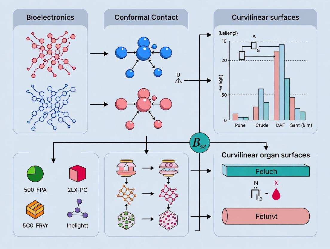
Conformal Contact Technologies: Overcoming Topographic Challenges for Next-Generation Biomedical Devices on Curvilinear Organ Surfaces
This article provides a comprehensive technical review for researchers and biomedical engineers on achieving robust conformal contact between bioelectronic devices and the complex, dynamic surfaces of internal organs.
Conformal Contact Technologies: Overcoming Topographic Challenges for Next-Generation Biomedical Devices on Curvilinear Organ Surfaces
Abstract
This article provides a comprehensive technical review for researchers and biomedical engineers on achieving robust conformal contact between bioelectronic devices and the complex, dynamic surfaces of internal organs. We first explore the fundamental principles and bio-mechanical challenges posed by organ topography. Next, we detail current methodological approaches, including material innovations and fabrication techniques, with specific applications in cardiac, neural, and epidermal interfaces. We then address critical troubleshooting, optimization strategies, and stability considerations for *in vivo* environments. Finally, we discuss validation frameworks, performance benchmarking, and comparative analysis of leading technologies. The synthesis aims to guide the development of more reliable, high-fidelity diagnostic and therapeutic interfaces.
The Conformal Challenge: Fundamental Principles and Biomechanical Barriers on Dynamic Organ Surfaces
Technical Support Center: Troubleshooting Conformal Contact
Frequently Asked Questions (FAQs)
Q1: My bioelectronic device does not adhere uniformly to the wet, curved surface of a beating heart. What are the primary factors to check? A: Achieving conformal contact on dynamic, curvilinear organs requires balancing multiple factors. First, verify the mechanical modulus of your device substrate; it should typically be in the low MPa to kPa range (e.g., PDMS ~1-2 MPa, silicone elastomers ~100 kPa) to match soft tissue. Second, ensure your adhesive strategy accounts for surface energy and hydration. Simple hydrophobic adhesion often fails. Consider bioadhesives (e.g., gelatin-methacryloyl, dopamine-based polymers) or suction-based mechanical fixation. Third, device thickness is critical; aim for <100 µm to ensure flexibility and minimize sheer stress.
Q2: I am experiencing signal drift and high impedance in my conformal electrode recordings. What could be the cause? A: Signal drift and high impedance often indicate poor interfacial contact, despite apparent physical adhesion. This is a classic failure of simple adhesion versus true conformal contact. Key troubleshooting steps:
- Measure Contact Impedance In-situ: Use electrochemical impedance spectroscopy (EIS) to verify the electrode-electrolyte-tissue interface. A stable, low impedance (<10 kΩ at 1 kHz) is a good indicator.
- Check for Microscopic Gaps: Conformal contact requires nanoscale intimacy. Even a micron-scale gap filled with interstitial fluid can drastically increase impedance. Ensure your device material can flow into tissue microtopography.
- Interface Material: Standard metals (Pt, Au) may require conductive hydrogel or porous conductive polymer (e.g., PEDOT:PSS) coatings to stabilize the electrochemical interface on dynamic tissue.
Q3: How do I quantitatively assess whether I have achieved true conformal contact vs. macroscopic adhesion? A: Researchers use a combination of quantitative metrics, summarized in the table below.
Table 1: Quantitative Metrics for Assessing Conformal Contact
| Metric | Measurement Technique | Target Value for Conformal Contact | Indicates |
|---|---|---|---|
| Contact Angle | Goniometry on tissue/organ mimic | < 30° (high wettability) | Intimate molecular-level interaction |
| Peel Adhesion Strength | 90° or 180° peel test | 0.1 - 10 N/m (context-dependent) | Mechanical bonding strength |
| Interfacial Toughness | Shear-lag or blister test | > 10 J/m² for dynamic organs | Energy to propagate delamination |
| Effective Contact Strain | Digital Image Correlation (DIC) | > 99% surface area contact | Percentage of surface in atomic proximity |
| Electrical Contact Impedance | Electrochemical Impedance Spectroscopy (EIS) | Stable, low value (e.g., < 5 kΩ at 1 kHz) | Quality of electronic interface |
Troubleshooting Guides
Issue: Device Delamination Under Cyclic Mechanical Strain (e.g., on lung or heart) Root Cause: The adhesion energy is insufficient to overcome repeated strain energy release at the interface. Solution Protocol:
- Design: Implement a strain-isolating, "filamentary serpentine" mesh design for the device interconnects.
- Interface Functionalization: Apply a bioadhesive layer. A common protocol:
- Material: Oxidized dextran (Dextran-Aldehyde) or dopamine-modified hyaluronic acid.
- Application: Spin-coat a 2-5 µm layer onto the device substrate.
- Bonding: Press device onto the tissue surface with light pressure (5-10 kPa) for 60-90 seconds. The aldehyde or catechol groups form covalent bonds with tissue amines.
- Validation: Perform cyclic stretch testing (e.g., 10,000 cycles at 10-15% strain) while monitoring electrical continuity or direct observation under microscopy.
Issue: Inconsistent Signal-to-Noise Ratio (SNR) Across Electrode Array on Curved Surface Root Cause: Variable contact pressure and intimacy across the array due to non-conformal wrapping. Solution Protocol:
- Device Fabrication: Ensure ultra-thin, substrate-less electrode arrays. Use parylene-C (< 10 µm) or polyimide (< 5 µm) as a carrier.
- Conformal Transfer: Use a water-soluble tape (e.g., polyvinyl alcohol) or a sacrificial layer to transfer the device onto the organ. This allows the device to drape naturally without internal stress.
- Contact Force Mapping: Use a thin, flexible pressure sensor film placed between the device and a calibration surface to map contact force distribution. Optimize device geometry and application method to achieve uniform pressure (±10% variation).
- Post-Hoc Validation: After experiment, use confocal microscopy or optical coherence tomography (OCT) on a tissue phantom to image the cross-sectional interface for gaps.
The Scientist's Toolkit: Research Reagent Solutions
Table 2: Essential Materials for Conformal Biointerface Research
| Item | Function & Rationale | Example Product/Chemical |
|---|---|---|
| Soft Elastomer Substrate | Provides mechanical compliance to match tissue modulus and enable bending without delamination. | PDMS (Sylgard 184), Ecoflex (00-30), Hydrogels (PAAm, PEGDA) |
| Conductive Polymer Coating | Reduces electrochemical impedance, improves charge injection limit, and adds mechanical flexibility vs. bare metal. | Poly(3,4-ethylenedioxythiophene):Polystyrene sulfonate (PEDOT:PSS) |
| Tissue-Adhesive Polymer | Forms covalent/non-covalent bonds with tissue surface proteins, enhancing wet adhesion. | Gelatin-Methacryloyl (GelMA), Dopamine-Methacrylate, Oxidized Dextran |
| Sacrificial Release Layer | Enables fabrication and handling of ultra-thin devices; dissolves to release device for transfer. | Poly(vinyl alcohol) (PVA), Poly(methyl methacrylate) (PMMA), Sugar (Sucrose) |
| Hydration & Interface Control Gel | Maintains a stable ionic interface, prevents drying, and can act as an adhesive electrolyte. | Agarose gel (0.5-2%), Photo-crosslinkable hydrogel (e.g., PEG-NHS) |
Experimental Workflow for Validating Conformal Contact
Diagram Title: Conformal Contact Validation Workflow
Key Signaling Pathways in Mechanotransduction at Biointerface
Diagram Title: Impact of Contact Quality on Tissue Signaling
Technical Support Center
Troubleshooting Guides & FAQs
Q1: Our 3D-printed phantom organ model does not achieve stable conformal contact with our sensing array. The contact seems patchy and inconsistent. What could be the issue? A: This is a classic issue of mismatched effective modulus. The phantom's bulk compliance may be correct, but its surface topography (micro-scale roughness) or the viscoelastic properties of its coating material may prevent intimate contact.
- Solution: First, quantify your phantom's surface roughness using profilometry (see Protocol 1). Compare this to literature values for the target organ. If roughness is >10% of your sensor's feature size, consider using a thinner, softer silicone elastomer (e.g., Dow Sylgard 527) dip-coating to fill micro-asperities. Ensure your testing apparatus applies a gentle, uniform preload (0.5-2 kPa) to initiate contact without excessive deformation.
Q2: When measuring dynamic motion (e.g., simulated heartbeat), our optical coherence tomography (OCT) data shows motion artifacts, blurring the surface strain map. How can we improve data fidelity? A: This is typically a result of low temporal resolution relative to the motion speed.
- Solution: Implement or switch to a high-speed OCT system (>100 frames/sec). Synchronize your image acquisition trigger with the motion generator's (e.g., pulsatile pump) cycle using a hardware trigger. Apply a post-processing digital image correlation (DIC) algorithm specifically designed for cyclical motion to track surface points across frames. Ensure your sample has a fine, stochastic speckle pattern (e.g., applied titanium dioxide powder) for the DIC algorithm to track effectively.
Q3: The compliance values we measure via Atomic Force Microscopy (AFM) indentation on ex vivo liver tissue vary by over 200% across samples from the same source. Are we doing something wrong? A: Significant variation often stems from uncontrolled hydration state and testing environment, which drastically affects soft tissue mechanics.
- Solution: Strictly control the physiological buffer environment. Use a fluid cell and maintain the sample fully submerged in phosphate-buffered saline (PBS) at 37°C throughout preparation and testing (see Protocol 2). Limit total ex vivo time to <4 hours. Implement a standardized pre-conditioning protocol (5-10 indentation cycles at the test location before data collection) to achieve a repeatable mechanical state.
Q4: Our finite element analysis (FEA) model of a device conforming to a lung surface fails to converge when we incorporate measured surface topography data. The mesh becomes too complex. A: Direct incorporation of high-resolution topography into a global FEA model is computationally prohibitive.
- Solution: Use a multi-scale modeling approach. Create a global macro-scale model with smooth curvature. For critical contact regions, develop a separate, localized micro-scale sub-model. Apply the displacement boundary conditions from the macro-model to the sub-model, which incorporates the detailed topography. This isolates the contact complexity and makes the problem tractable.
Q5: How do we validate that our "conformal contact" is sufficient for effective drug delivery patch adhesion and release? A: Conformal contact is a means to an end. Validation must be functional.
- Solution: Perform an integrated experiment. First, map the actual contact area using a pressure-sensitive film (e.g., Fujifilm Prescale) placed between your patch and a curved phantom. Then, in a separate setup using the same preload, measure the shear adhesion force required to detach the patch. Finally, use a Franz diffusion cell with a curved receiver compartment to quantify the drug flux through a tissue-mimicking hydrogel membrane. Sufficient contact should yield >90% of the theoretical maximum flux.
Experimental Protocols
Protocol 1: Surface Topography Mapping of Soft Biomaterials using Optical Profilometry Objective: To non-destructively quantify the surface roughness (Sa, Sz) and waviness of soft organ phantoms or ex vivo tissue samples.
- Sample Mounting: Secure the sample on a stable stage using a non-invasive adhesive (e.g., cyanoacrylate on its back side only). Ensure the region of interest is horizontal.
- System Setup: Use a white-light interferometer or confocal profilometer. Select a 10X or 20X objective. Set the vertical scanning range to fully capture the surface height variation.
- Acquisition: Acquire data from at least three distinct, representative areas (e.g., 1 mm² each). For tissues, keep the surface moist with a fine mist of PBS during scanning (if not using an environmental chamber).
- Analysis: Use instrument software to level and flatten the data. Apply a Gaussian filter (cut-off wavelength λc = 80 μm) to separate roughness from form and waviness. Extract areal roughness parameters: Sa (arithmetical mean height), Sz (maximum height), and Sdr (developed interfacial area ratio).
Protocol 2: Nanoindentation for Local Compliance Mapping of Hydrated Tissue Objective: To measure the elastic modulus (E) of soft tissue at micro-scale resolution under physiologically relevant conditions.
- Sample Preparation: Section fresh or properly thawed tissue to ~3mm thickness using a vibratome. Immerse in PBS. Mount in a custom fluid cell filled with PBS.
- AFM Configuration: Use a colloidal probe (5-10μm diameter silica sphere) attached to a cantilever with a known spring constant (0.1-1 N/m). Calibrate in fluid using the thermal tune method.
- Environment Control: Maintain fluid cell temperature at 37°C ± 0.5°C using an in-line heater or stage-top incubator.
- Indentation Grid: Program a 10x10 grid indentation map over a 100x100 μm area. Set approach velocity to 2 μm/s, indentation depth to 1-2 μm (≤10% sample height), and a 1-second hold at peak load.
- Data Processing: Fit the retract curve of each force-displacement measurement with the Hertz contact model for a spherical indenter to extract the reduced modulus (Er), then calculate the sample modulus (E_sample).
Data Presentation
Table 1: Representative Biomechanical Properties of Human Organ Surfaces Data synthesized from recent AFM and suction cup studies (2020-2023).
| Organ | Approx. Elastic Modulus (E) | Characteristic Roughness (Sa) | Key Dynamic Motion (Frequency) | Citation Context |
|---|---|---|---|---|
| Brain (Cortex) | 0.5 - 1.5 kPa | 0.2 - 0.5 μm | Pulsatile (Cardiac, 1-2 Hz) | Intracortical probe integration |
| Liver (Glisson's Capsule) | 5 - 15 kPa | 1 - 3 μm | Respiratory (~0.2 Hz) | Laparoscopic sensor adhesion |
| Heart (Epicardium) | 20 - 50 kPa | 5 - 20 μm | Contractile (1-2 Hz) | Epicardial pacing/patch deployment |
| Lung (Visceral Pleura) | 10 - 25 kPa | 10 - 50 μm | Respiratory (~0.2 Hz) | Pleural pressure sensing |
| Kidney (Capsule) | 25 - 75 kPa | 2 - 5 μm | Pulsatile/Respiratory | Perirenal device anchoring |
Table 2: Common Coating Materials for Improving Conformal Contact
| Material | Typical Formulation/Name | Function | Effective Modulus | Best For |
|---|---|---|---|---|
| Silk Fibroin | Aqueous solution, layer-by-layer deposition | Biodegradable adhesive interface; reduces impedance | 100-500 MPa (film) | Neural, cardiac interfaces |
| Polydimethylsiloxane (PDMS) | Sylgard 527 (1:1 mix) | Ultra-soft, curable elastomer filler | 3 - 50 kPa | Filling macro-scale curvature |
| Hyaluronic Acid (HA) Hydrogel | Methacrylated HA, photo-crosslinked | Hydrated, lubricating, drug-eluting layer | 1 - 10 kPa | Lung, gastrointestinal surfaces |
| Polyethylene Glycol (PEG) | Star-PEG with adhesive peptides | Non-fouling, tethers bioactive molecules | 10 - 100 kPa (gel) | Vascular, ocular surfaces |
The Scientist's Toolkit: Research Reagent Solutions
| Item | Function in Conformal Contact Research |
|---|---|
| Sylgard 527 Silicone Dielectric Gel | Two-part, mixable elastomer for creating ultra-soft (kPa range) organ phantoms or device coatings. |
| Fujifilm Prescale Pressure Film | Color-changing film to visually map and quantify pressure distribution and actual contact area between two surfaces. |
| Matrigel Basement Membrane Matrix | Biologically-derived hydrogel for coating surfaces to mimic the extracellular matrix and improve biocompatibility/adhesion. |
| Fluorescein Isothiocyanate (FITC)-Dextran | Fluorescent tracer of varying molecular weights to quantify drug/permeant diffusion in curved Franz cell assays. |
| Titanium Dioxide (TiO2) Powder | Applied as a fine, white speckle pattern on sample surfaces for optical strain mapping via Digital Image Correlation (DIC). |
| Polybead Microspheres (10µm) | Used as colloidal probes for AFM cantilevers to perform nanoindentation on soft, hydrated tissues. |
| Dulbecco's Phosphate Buffered Saline (DPBS) | Standard isotonic buffer for maintaining hydration and ionic balance in ex vivo tissue during biomechanical testing. |
Visualizations
Diagram 1: Workflow for Conformal Contact Experiment Design
Diagram 2: Key Factors in Conformal Contact Mechanics
Troubleshooting Guides & FAQs
Q1: My ultrathin electronic film is cracking during transfer to the target organ surface. What could be wrong? A: Cracking during transfer is often due to inadequate support during the release from the fabrication substrate. Ensure you are using a water-soluble tape (like polyvinyl alcohol) or a thermal release tape as a temporary handle. The transfer process should be performed on a droplet of deionized water or in a humidity-controlled chamber (>80% RH) to minimize surface tension stresses. If using a sacrificial layer (e.g., poly(methyl methacrylate) - PMMA), verify it is fully dissolved with a fresh solvent bath.
Q2: The measured stretchability of my polymer substrate is 50% lower than the literature value for the same material. How can I troubleshoot this? A: Discrepancies in stretchability often stem from fabrication or testing variables.
- Curing/Annealing: Verify the precise temperature, time, and environment (e.g., nitrogen glovebox) for polymer curing. Incomplete crosslinking reduces elasticity.
- Sample Geometry: Ensure your dog-bone tensile specimen conforms to ASTM D412 or ISO 37 standards to prevent premature failure at the grips.
- Strain Rate: Conduct tensile tests at multiple strain rates (e.g., 1 mm/min, 10 mm/min, 100 mm/min). Some viscoelastic materials are highly rate-sensitive.
- Defect Inspection: Use optical microscopy to check for micro-tears, dust inclusions, or uneven thickness prior to testing.
Q3: How do I quantitatively evaluate the conformal contact quality of my device on a curved biological surface? A: Conformality can be quantified via the Contact Adhesion Index (CAI). The protocol involves:
- Spin-coat a fluorescent dye (e.g., Rhodamine B) onto your device surface.
- Gently apply the device to the target curvilinear surface (e.g., an explanted heart or a 3D-printed phantom with matching topography).
- Apply a uniform, low pressure (e.g., 1 kPa for 60 seconds).
- Carefully remove the device and image the amount of dye transferred onto the target surface using fluorescence microscopy.
- Calculate: CAI = (Area of Fluorescent Transfer / Total Device Area) x 100%. A CAI > 90% indicates excellent conformal contact.
Q4: My conformal sensor’s electrical performance degrades after 100 bending cycles. What are the primary failure modes to investigate? A: Focus on the interfaces and neutral mechanical plane design.
- Metal Trace Fracture: Use scanning electron microscopy (SEM) to check for microcracks in the conductive traces, especially at the edge of the substrate. Consider switching to a wavy or serpentine trace geometry.
- Delamination: Check adhesion between the metal layer and the polymer substrate. Implement oxygen plasma treatment or an adhesion promoter (like APTES or Ti/Au underlayer) before metallization.
- Neutral Plane Mismatch: Ensure the active layer is positioned near the structure's neutral mechanical plane. Encapsulate the device with a top layer of the same substrate material to shift the neutral plane.
Q5: What are the critical parameters for achieving reliable, long-term adhesion of an ultrathin device to a wet, dynamic organ surface? A: Long-term bioadhesion requires a multi-faceted strategy:
- Surface Energy: Treat the device surface with oxygen plasma to increase hydrophilicity and temporarily enhance wettability.
- Mechanical Interlocking: Use a substrate with a nano- or micro-structured surface (e.g., molded micropillars from PDMS) to increase contact area.
- Chemical Bonding: Employ a bioadhesive interfacial layer. A common protocol: apply a thin (<5 µm) layer of a dopamine-modified methacrylated hyaluronic acid hydrogel. Crosslink via brief UV exposure (365 nm, 20 mW/cm² for 30 sec) in situ.
- Handling Force: Pre-apply the adhesive layer and use a soft, porous elastomeric stamp for placement to avoid squeezing the adhesive away from the interface.
Experimental Protocols
Protocol 1: Fabrication of a Stretchable, Ultrathin Polyimide Substrate
- Spin-coat Sacrificial Layer: Spin-coat 5% (w/v) PMMA in anisole on a clean silicon wafer at 3000 rpm for 60 sec. Bake at 180°C for 2 min.
- Spin-coat Polyimide: Spin-coat the polyimide precursor (e.g., PI-2545) at 4000 rpm for 60 sec.
- Soft Bake: Bake on a hotplate at 120°C for 2 min.
- Cure: Fully imidize in a nitrogen oven using a stepped cure: 150°C for 15 min, 250°C for 15 min, 350°C for 60 min. Allow to cool slowly.
- Release: Immerse the wafer in acetone to dissolve the PMMA layer. Transfer the free-floating polyimide film to a Petri dish with fresh acetone, then to isopropanol, and finally deionized water before pickup with a custom frame or transfer tool.
Protocol 2: In-Vitro Bendability Testing on a Cylindrical Mandrel
- Setup: Acquire a set of precision stainless-steel mandrels with diameters from 10 mm down to 1 mm.
- Mounting: Gently wrap your thin-film device around a mandrel, ensuring no air gaps. Secure the ends with biocompatible Kapton tape.
- Electrical Monitoring: Connect the device to a source meter (e.g., Keithley 2450) while mounted. Measure baseline resistance (R0).
- Cycling: Manually (or using a motorized stage) unwrap and re-wrap the device around the mandrel for a set number of cycles (e.g., 1000).
- Measurement: After cycling, measure the final resistance (R). Calculate the normalized change in resistance: ΔR/R0 = (R - R0)/R0. Plot ΔR/R0 vs. bending radius and cycle count.
Data Presentation
Table 1: Comparison of Key Material Properties for Conformal Electronics
| Material | Typical Thickness | Effective Modulus | Fracture Strain | Water Vapor Permeability | Best Use Case |
|---|---|---|---|---|---|
| Poly(dimethylsiloxane) (PDMS) | 50-500 µm | 0.5 - 2 MPa | >100% | High (>1000 g/m²/day) | Stretchable substrates, epidermal patches |
| Polyimide (PI) | 5-25 µm | 2.5 - 8 GPa | < 3% | Very Low (<10 g/m²/day) | Ultrathin, flexible neural implants |
| Parylene-C | 5-30 µm | 2.8 - 4 GPa | 2 - 3% | Low (~5 g/m²/day) | Conformal barrier/encapsulation layer |
| Silk Fibroin | 1-10 µm | 5 - 10 GPa (dry) | 4 - 30% (wet) | Tunable | Bioresorbable, ultrathin substrates |
| Hydrogel (PAAm-Alginate) | 100-1000 µm | 1 - 100 kPa | >500% | Very High | Soft, wet adhesives for dynamic organs |
Table 2: Quantitative Conformality Metrics on Phantom Heart Surface
| Device Architecture | Thickness | Bending Stiffness (EI, nN·m²) | Contact Adhesion Index (CAI) | Minimum Stable Bending Radius |
|---|---|---|---|---|
| Bulk Silicone Sheet | 1 mm | ~1.2 x 10⁶ | 42% | 5.0 mm |
| Structured PDMS (Micropillars) | 100 µm | ~1.5 x 10³ | 78% | 1.5 mm |
| Ultrathin Polyimide + Serpentine Au | 8 µm | ~0.9 x 10¹ | 96% | 0.2 mm |
| Nanomesh PEDOT:PSS | 800 nm | ~0.5 x 10⁻¹ | 99% | 0.05 mm |
Mandatory Visualization
Diagram 1: Conformal Device Development Workflow
Diagram 2: Signal Pathway from Conformal Contact to Data
The Scientist's Toolkit
Research Reagent Solutions for Conformality Experiments
| Item | Function & Rationale |
|---|---|
| Poly(dimethylsiloxane) (PDMS), Sylgard 184 | The quintessential elastomer for stretchable substrates and stamps. Tunable modulus (by base:curing agent ratio). Provides flexibility and gas permeability. |
| Polyimide Precursor (PI-2545 or similar) | For fabricating robust, biocompatible, ultrathin (<10 µm) substrates. Essential for minimizing bending stiffness (EI). |
| Poly(methyl methacrylate) (PMMA) | A common sacrificial layer material. Dissolved in acetone or anisole to release free-standing thin films from rigid carrier wafers. |
| (3-Aminopropyl)triethoxysilane (APTES) | Adhesion promoter. Used as a molecular glue to improve bonding between inorganic layers (e.g., metals, oxides) and organic polymer substrates. |
| Dopamine Hydrochloride | Key component for creating versatile, water-resistant bioadhesive coatings via self-polymerization into polydopamine, which sticks to virtually all surfaces. |
| Hyaluronic Acid (Methacrylated) | Formulated into UV-crosslinkable hydrogels for soft, wet bioadhesives that interface with dynamic organ surfaces without causing damage. |
| Polystyrene Sulfonate (PEDOT:PSS) | Conductive polymer mixture. Can be processed into highly conformable, stretchable conductive traces or transparent electrodes for sensing. |
| Water-Soluble Tape (e.g., PVA Tape) | Provides temporary mechanical support for handling ultrathin, fragile devices during transfer to target surfaces. Dissolves upon contact with water. |
Technical Support & Troubleshooting Center
Frequently Asked Questions (FAQs)
Q1: My bio-interface film delaminates from the curvilinear organ surface during in vivo implantation. What are the primary causes and solutions?
A: Delamination typically results from insufficient conformal contact or mismatched mechanical properties. Ensure surface energy modification (e.g., plasma treatment) matches the target tissue's wettability. Utilize shear-thinning hydrogels or in-situ polymerizing adhesives like gelatin-methacryloyl (GelMA) to improve adhesion. Monitor elastic modulus; it should be within 10-20% of the target tissue's modulus to prevent stress-induced peeling.
Q2: How do I diagnose a foreign body reaction (FBR) to my implant, and what material modifications can mitigate it?
A: Key indicators of FBR are a thickened fibrotic capsule (>50 µm), persistent inflammation (e.g., elevated TNF-α, IL-1β), and macrophage fusion into foreign body giant cells. To mitigate:
- Surface Topography: Implement micro-scale (<5 µm) patterning to disrupt macrophage fusion.
- Surface Chemistry: Integrate anti-inflammatory moieties (e.g., covalently bonded CD200 peptide).
- Drug Release: Incorporate localized, sustained release of dexamethasone (0.1-1 µg/day) from the interface coating.
Q3: My permeable membrane for drug elution shows significantly reduced flux (J) after 30 days. How can I restore or predict permeability?
A: Flux reduction is often due to protein fouling or pore collapse. Perform SEM imaging to check pore integrity. Use table data (see Table 1) to select materials with higher inherent stability. Pre-coat membranes with non-fouling polymers like poly(ethylene glycol) (PEG) or zwitterionic poly(sulfobetaine methacrylate). Model long-term flux using the following equation, where P is permeability, ΔC is concentration gradient, and t is time, accounting for a fouling factor (α):
J(t) = (P * ΔC) / (1 + α*t)
Q4: What are the best practices for accelerating long-term stability testing of bio-interfaces under physiological conditions?
A: Use an accelerated aging protocol:
- Environmental Stress: Incubate in PBS at 45°C (following Arrhenius kinetics, this accelerates degradation ~3x compared to 37°C).
- Mechanical Stress: Subject to cyclic strain (e.g., 10% strain at 1 Hz) in a bioreactor simulating peristalsis or pulsation.
- Biological Stress: Incubate with concentrated collagenase (for protein-based materials) or reactive oxygen species (H₂O₂ solution) to simulate inflammatory oxidative stress.
- Key Metrics: Monitor mass loss, elastic modulus change, and permeability weekly. Failure is defined as >20% change from baseline.
Experimental Protocols
Protocol 1: Assessing Conformal Contact on Ex Vivo Curvilinear Surfaces
- Objective: Quantify the contact adhesion energy (γ) of a bio-interface film on a tissue surface.
- Materials: PDMS stamp, bio-interface film, fresh ex vivo organ (e.g., rodent heart), microbalance, vertical stage.
- Procedure:
- Laminate the bio-interface film onto a flexible PDMS stamp.
- Bring the stamp into gentle contact with the rinsed, moist organ surface for 60 seconds.
- Attach the organ to a microbalance and the stamp to a motorized vertical stage.
- Retract the stage at a constant speed (e.g., 10 µm/s) while recording the force from the microbalance.
- Calculate work of adhesion (W = Force/Width). Repeat over 5 organ samples (n=5).
Protocol 2: In Vitro Permeability and Fouling Test
- Objective: Measure the solute permeability coefficient (P) of a membrane and its change after protein exposure.
- Materials: Side-by-side diffusion cells, magnetic stirrers, test membrane, PBS, analyte (e.g., 100 µM fluorescein isothiocyanate-dextran, 70 kDa), BSA solution (40 mg/mL), spectrophotometer.
- Procedure:
- Mount the membrane between donor and receptor compartments filled with PBS. Stir at 600 rpm, 37°C.
- Add analyte to the donor compartment. Take 100 µL samples from the receptor compartment every 15 min for 2 hours. Measure concentration (C) via absorbance/fluorescence.
- Calculate initial permeability:
P_initial = (dC/dt * V) / (A * ΔC), where V is receptor volume, A is membrane area. - Flush system. Circulate BSA solution in donor compartment for 24 hrs to simulate fouling.
- Repeat step 2-3 to calculate P_fouled. Report % retention:
(P_fouled / P_initial)*100.
Data Presentation
Table 1: Comparative Properties of Common Bio-Interface Materials
| Material | Elastic Modulus (kPa) | Water Permeability (10^-12 m^2) | Stable In Vivo Period (Weeks) | Primary Degradation Mode |
|---|---|---|---|---|
| Poly(dimethylsiloxane) (PDMS) | 1500-2000 | 0.001 | >52 (Inert) | Hydrolytic (slow) |
| Poly(lactic-co-glycolic acid) (PLGA) 85:15 | 1000-1500 | 0.05 | 8-12 | Hydrolytic bulk erosion |
| Gelatin-Methacryloyl (GelMA) 5% | 10-50 | 2.1 | 2-4 | Enzymatic (collagenase) |
| Poly(ethylene glycol) Diacrylate (PEGDA) | 100-500 | 1.5 | 4-8 | Oxidative cleavage |
| Silk Fibroin | 5000-10000 | 0.1 | >52 (Slow proteolysis) | Proteolytic surface erosion |
Table 2: Troubleshooting Summary: Symptoms & Actions
| Observed Problem | Potential Root Cause | Recommended Diagnostic Test | Corrective Action |
|---|---|---|---|
| Film Cracking | Mismatched modulus, brittle material | Tensile test to failure | Plasticize material (e.g., add glycerol), reduce crosslink density. |
| Excessive Fibrosis (>100 µm capsule) | High surface roughness, pro-inflammatory chemistry | Histology (H&E, Masson's Trichrome) | Polish to Ra < 100 nm; graft anti-fouling polymers (PEG). |
| Unpredictable Drug Release | Pore clogging, bulk degradation | HPLC of release medium, SEM | Switch to surface-eroding polymer; add porogen (salt leaching). |
| Loss of Electrical Signal (for sensors) | Delamination, protein adsorption | Electrochemical Impedance Spectroscopy (EIS) | Improve adhesion; coat with conducting polymer (PEDOT:PSS). |
Visualizations
Title: Immune Response Pathway & Mitigation for Bio-Interfaces
Title: Bio-Interface Development & Validation Workflow
The Scientist's Toolkit: Research Reagent Solutions
| Item | Function in Bio-Interface Research |
|---|---|
| Poly(dimethylsiloxane) (PDMS), Sylgard 184 | Gold-standard elastomer for flexible substrates and stamps; tunable modulus by base:curing agent ratio. |
| Gelatin-Methacryloyl (GelMA) | Photocrosslinkable hydrogel that promotes cell adhesion; key for soft, conformal interfaces. |
| Poly(ethylene glycol) Diacrylate (PEGDA) | Biocompatible, hydrophilic crosslinker; forms non-fouling hydrogels to control permeability. |
| Dexamethasone | Synthetic glucocorticoid; incorporated for localized, sustained anti-inflammatory release. |
| Sulfo-SANPAH Crosslinker | Heterobifunctional crosslinker (NHS-ester and photoactive group) for covalent bonding of biomolecules to surfaces under UV light. |
| Fluorescein Isothiocyanate (FITC)-Dextran (various MW) | Fluorescent tracer molecules used to quantitatively measure membrane permeability and integrity over time. |
| Plasma Cleaner (O₂ or Ar Plasma) | Critical for modifying surface energy of polymers (e.g., PDMS) to increase hydrophilicity and improve bonding/laminating. |
| Electrospinning Apparatus | Used to fabricate nano-/micro-fibrous membranes with high porosity for controlled permeability in barrier layers. |
Fabrication Strategies and In Vivo Applications: From Material Design to Functional Interfaces
Technical Support & Troubleshooting Center
This support center addresses common experimental challenges in fabricating and applying soft electronic materials for achieving conformal contact on curvilinear organ surfaces. The information is synthesized from current literature and best practices in the field.
FAQs & Troubleshooting Guides
Q1: My intrinsically stretchable polymer (e.g., PDMS-SBS composite) cracks upon cyclic stretching beyond 50% strain. What could be the cause? A: This is typically due to insufficient dynamic cross-linking or phase separation. Ensure your polymer composite is mixed uniformly using a speed mixer (e.g., at 2000 rpm for 2 minutes) and cured at the recommended temperature gradient (e.g., 65°C for 2 hrs, then 85°C for 1 hr). If using a double-network strategy, verify the stoichiometric ratio of your covalent and reversible (e.g., hydrogen) bonds.
Q2: The conductivity of my silver flake/elastomer composite degrades significantly after 1000 stretch-release cycles. How can I improve durability? A: This is a common issue related to microcrack propagation and filler dislocation. Solutions include:
- Pre-stretching the elastomer substrate during composite application to create aligned conductive pathways.
- Incorporating a secondary nanofiller (e.g., 0.5 wt% carbon nanotubes) to bridge microgaps between silver flakes.
- Using a softer elastomer matrix (e.g., Ecoflex with modulus ~60 kPa) to reduce stress on the percolation network.
Q3: My transferred nanomembrane (e.g., Parylene-C) wrinkles or delaminates from the curvilinear organ model surface. How do I ensure conformal adhesion? A: Wrinkling indicates compressive stress; delamination indicates poor adhesion. Follow this protocol:
- Surface Energy Matching: Treat the organ model surface with oxygen plasma (50 W, 30 sec) to increase its hydrophilicity.
- Wet Transfer: Use a water-soluble sacrificial layer (e.g., Polyvinyl alcohol) and perform a wet transfer, allowing the membrane to float and conform before gently draping.
- Mechanical Confinement: Apply a thin, conformal coating of a hydrogel (e.g., Polyacrylamide, 2% w/v) over the adhered membrane to provide pressure and maintain contact.
Q4: How do I quantify the level of conformal contact achieved between my device and a biological surface?
A: The standard metric is the Contact Adhesion Efficiency (CAE). Calculate using the formula:
CAE (%) = [1 - (A_gap / A_total)] * 100
Where A_gap is the non-contact area imaged via optical coherence tomography or confocal microscopy, and A_total is the total device area. A CAE > 95% is typically required for reliable bio-interfacing.
Table 1: Performance Comparison of Conductive Composites for Stretchable Electrodes
| Composite Material | Filler Loading (wt%) | Initial Conductivity (S/cm) | Conductivity at 50% Strain (S/cm) | Max Tolerable Strain | Key Application |
|---|---|---|---|---|---|
| Silver Flakes / SEBS | 65 | 4,200 | 850 | 180% | Epicardial sensing |
| Liquid Metal / Ecoflex | 75 | 3.6 x 10⁴ | 2.1 x 10⁴ | 400% | Peripheral nerve cuff |
| PEDOT:PSS / PUA | 1 (PEDOT) | 0.8 | 0.75 | 100% | Cortical surface mapping |
| Carbon Nanotube / PDMS | 3 | 120 | 15 | 150% | Strain sensing |
Table 2: Nanomembrane Substrates for Bio-Integration
| Membrane Material | Thickness (nm) | Effective Modulus (MPa) | Water Vapor Transmission Rate (g/m²/day) | Biodegradation Time | Best For |
|---|---|---|---|---|---|
| Poly(lactic-co-glycolic) | 500 | 2.1 | 245 | 21-42 days | Transient implants |
| Parylene-C | 1,000 | 3.2 | 0.2 | Non-degradable | Chronic interfaces |
| Silk Fibroin | 400 | 5.0 | 310 | Tunable (hrs-yrs) | Drug-delivery wraps |
| Silicon Nitride | 100 | 270 | 0 | Non-degradable | Ultrathin barriers |
Experimental Protocols
Protocol 1: Fabrication of an Intrinsically Stretchable Conducting Composite Electrode
- Solution Preparation: Dissolve 1g of styrene-ethylene-butylene-styrene (SEBS) in 20ml of toluene by magnetic stirring for 4 hours.
- Filler Integration: Add 65% by weight of silver flakes (avg. diameter 5µm) to the solution.
- Homogenization: Mix the slurry using a dual-asymmetric centrifugal speed mixer at 2200 rpm for 3 minutes, followed by 30 minutes of bath sonication to break agglomerates.
- Film Casting: Pour the mixture into a PTFE mold and doctor-blade to a 300µm wet thickness.
- Curing: Let the film dry at room temperature for 12 hours, then vacuum-anneal at 80°C for 2 hours to remove residual solvent and enhance filler contact.
Protocol 2: Deterministic Transfer of a Nanomembrane to a Curved Surface
- Sacrificial Layer Spin-Coat: Spin-coat a 2µm layer of poly(methyl methacrylate) (PMMA) on a silicon handling wafer.
- Membrane Deposition: Deposit your target nanomaterial (e.g., 500 nm Parylene-C via chemical vapor deposition).
- Pattern & Release: Pattern the membrane via photolithography and etching. Immerse the wafer in acetone to dissolve the PMMA, releasing the membrane onto the solvent surface.
- Conformal Pick-Up: Lower a pre-shaped, adhesive-coated elastomer stamp (e.g., PDMS) to pick up the floating membrane.
- Stamp & Apply: Gently press the stamp with the adhered membrane onto the target organomorphic surface. Apply uniform pressure (5 kPa) for 60 seconds, then peel back the stamp at a slow, consistent angle (< 10°/sec).
Visualization Diagrams
Title: Nanomembrane Transfer to Curved Surface Workflow
Title: Troubleshooting Conductive Composite Durability
The Scientist's Toolkit: Research Reagent Solutions
Table 3: Essential Materials for Conformal Bio-Interface Research
| Item | Function & Rationale |
|---|---|
| SEBS (Styrene-Ethylene-Butylene-Styrene) | A thermoplastic elastomer providing intrinsic stretchability and a robust matrix for conductive fillers. |
| Ecoflex 00-30 | A very soft, platinum-catalyzed silicone elastomer (modulus ~30 kPa) for ultra-conformal substrates. |
| Galinstan | A low-toxicity liquid metal alloy (Ga-In-Sn) for ultra-stretchable, self-healing conductive traces. |
| Parylene-C | A vapor-deposited, biocompatible polymer that forms uniform, pinhole-free nanomembranes for insulation. |
| Polyvinyl Alcohol (PVA) | A water-soluble sacrificial layer for releasing fabricated devices from handling wafers. |
| (3-Aminopropyl)triethoxysilane (APTES) | A silane coupling agent to improve adhesion between inorganic/organic layers and tissue surfaces. |
| Polyacrylamide Hydrogel | A tunable, conformal coating to provide mechanical confinement and hydration at the bio-interface. |
| 4',6-Diamidino-2-Phenylindole (DAPI) | A fluorescent nuclear stain used in ex-vivo validation of device-tissue integration without damage. |
Technical Support Center: Conformal Contact for Curvilinear Organ Surfaces
Troubleshooting Guide & FAQs
Q1: During transfer printing of a polymer mesh, my stamp fails to release the mesh onto the curved biological surface. What could be wrong?
A: This is often an issue of stamp surface energy mismatch or contact mechanics.
- Check Stamp Material: PDMS stamps are common. Ensure the stamp's surface energy is lower than that of the receiving surface. If using a flat stamp on a curve, the contact area may be insufficient.
- Troubleshooting Steps:
- Surface Treatment: Treat the target organ surface with oxygen plasma (low power, 10-30 seconds) to temporarily increase its hydrophilicity and improve adhesion.
- Stamp Modification: Silanize the PDMS stamp with (tridecafluoro-1,1,2,2-tetrahydrooctyl)trichlorosilane to lower its surface energy further, promoting release.
- Apply Mechanical Assistance: Use a soft, compliant secondary stamp or a rolling applicator to apply gradual, conformal pressure.
- Protocol - Plasma Activation for Improved Adhesion:
- Place the target tissue or tissue-mimetic hydrogel substrate in a plasma cleaner chamber.
- Evacuate the chamber to a base pressure of ~0.2 mbar.
- Introduce oxygen gas at a flow rate of 20 sccm.
- Apply RF power at 30 W for 15 seconds.
- Perform the transfer printing within 5 minutes of treatment.
Q2: My implanted mesh electronics are not achieving stable, conformal contact and are dislodging from the beating heart surface. How can I improve integration?
A: This indicates insufficient mechanical coupling. Relying solely on van der Waals forces is often inadequate for dynamic organs.
- Primary Cause: Mismatch in the effective modulus between the mesh and the dynamic, soft tissue.
- Solution: Employ Bioadhesives.
- Select a hydrogel-based bioadhesive (e.g., gelatin methacryloyl (GelMA), dopamine-modified hyaluronic acid) that can form covalent or strong physical bonds with both the mesh polymer and the tissue epi-layer.
- Application Method: Apply a thin, uniform layer of adhesive precursor onto the mesh before placement. Gently position the mesh on the organ. Crosslink the adhesive in situ using light (for GelMA) or pH/ionic change.
Q3: During deterministic assembly of micro-LEDs onto a soft, curved substrate, my placement accuracy exceeds 50 µm. What parameters should I optimize?
A: Placement accuracy in pick-and-place assembly is sensitive to viscoelastic relaxation and adhesion control.
- Key Parameters to Optimize:
- Dwell Time: The time the elastomeric stamp contacts the micro-device. Too short (<100 ms) results in weak pickup; too long (>1 s) causes excessive deformation and misplacement upon release.
- Retraction Speed: A fast retraction speed (>10 mm/s) is critical for clean release from the stamp onto the target.
- Stamp Geometry: The stamp tip's shape and size must match the device. For curved surfaces, use a stamp with a crowned tip profile.
Q4: How do I quantify the degree of conformal contact achieved by my implanted device?
A: Conformal contact is assessed by the contact angle and the effective contact area ratio.
- Method 1: Ex Vivo/In Situ Microscopy.
- Protocol: After implantation, fix the tissue-device construct (e.g., with 4% PFA). Section transversely. Use confocal microscopy to image the interface. The angle (θ) between the device tangent and the tissue tangent at the interface is the contact angle. θ < 15° indicates excellent conformity.
- Method 2: Impedance Spectroscopy.
- Protocol: Use the implanted device as an electrode. Measure electrochemical impedance (EIS) at 1 kHz. A lower impedance indicates a larger, more intimate contact area. Track impedance over time to monitor stability.
Quantitative Data Summary
Table 1: Performance Comparison of Fabrication Techniques for Conformal Contact
| Technique | Typical Resolution | Conformal Contact Metric (θ) | Best For Surfaces With: | Key Limitation |
|---|---|---|---|---|
| Transfer Printing | 1 µm - 500 µm | 10° - 30° | Moderate curvature (Radius > 1 mm) | Stamp design complexity for high curvature |
| Mesh Electronics | Sub-µm - 100 µm | < 10° | High, dynamic curvature (Beating heart, brain) | Requires injection/placement surgery |
| Deterministic Assembly | 10 nm - 100 µm | 5° - 20° | Predefined, heterogeneous layouts | Sequential process, slower for large arrays |
Table 2: Troubleshooting Common Conformal Contact Failures
| Symptom | Likely Cause | Diagnostic Test | Corrective Action |
|---|---|---|---|
| Device delamination | Weak interfacial adhesion | Measure peel force (< 0.1 N/m) | Apply bioadhesive; nano-scale surface patterning |
| Device cracking | Mechanical modulus mismatch | Measure device & tissue modulus | Use ultra-low modulus polymers (e.g., PGS, < 1 MPa) |
| Poor electrical signal | High interfacial impedance | EIS at 1 kHz (> 1 MΩ) | Improve contact via conductive hydrogel coating |
| Uncontrolled release | Stamp kinetics mismatch | High-speed video of release | Tune retraction speed and dwell time |
Experimental Protocol: Assessing Conformal Contact via Confocal Microscopy Title: Ex Vivo Conformal Contact Angle Measurement
- Implantation: Implant your device (mesh or printed array) onto the target curvilinear organ (e.g., rodent kidney).
- Fixation: Perfuse the animal with 4% paraformaldehyde (PFA) in PBS to fix the device-tissue interface in situ.
- Excision & Sectioning: Carefully excise the organ with the device attached. Embed in OCT compound. Cryo-section 20 µm thick slices transverse to the device-tissue interface.
- Staining: Stain tissue with phalloidin (actin) and DAPI (nuclei). If device is polymer-based, stain with a compatible dye (e.g., DiO).
- Imaging: Use a confocal microscope with a 40x water-immersion objective to obtain high-resolution z-stacks of the interface.
- Analysis: Use image analysis software (e.g., FIJI/ImageJ) to trace the device profile and the tissue surface profile. Calculate the tangent angles at points of contact. Report the average contact angle (θ) and standard deviation.
Visualizations
Title: Conformal Contact Failure Diagnosis Path
Title: Deterministic Assembly & Transfer Printing Workflow
The Scientist's Toolkit: Essential Research Reagents & Materials
Table 3: Key Reagents for Conformal Contact Research
| Item | Function/Benefit | Example Use Case |
|---|---|---|
| Polydimethylsiloxane (PDMS) | Elastomeric stamp material; tunable modulus. | Transfer printing stamp for micro-LEDs. |
| Gelatin Methacryloyl (GelMA) | Photocrosslinkable bioadhesive hydrogel. | Bonding mesh electronics to heart surface. |
| Poly(glycerol sebacate) (PGS) | Ultra-soft, biodegradable elastomer. | Substrate for compliant mesh electronics. |
| (Tridecafluoro-1,1,2,2-tetrahydrooctyl)trichlorosilane | Low-surface-energy release coating. | Treating PDMS stamps for reliable release. |
| Poly(3,4-ethylenedioxythiophene):Polystyrene sulfonate (PEDOT:PSS) | Conductive polymer coating. | Improving electrode-tissue interface impedance. |
| Oxygen Plasma | Increases surface hydrophilicity/reactivity. | Activating tissue surface before device bonding. |
| Dulbecco's Phosphate Buffered Saline (DPBS) | Physiological buffer for ex vivo work. | Hydrating tissues during implantation steps. |
| 4% Paraformaldehyde (PFA) | Tissue fixative. | Fixing device-tissue interfaces for imaging. |
Technical Support Center
Troubleshooting Guide: Common Experimental Issues
Issue 1: Poor Signal-to-Noise Ratio (SNR) in Recorded Electrophysiological Data
- Possible Cause: Incomplete or non-conformal contact between the patch electrode array and the epicardial surface.
- Solution: Verify tissue surface preparation. Ensure thorough removal of epicardial fat and moisture. Re-apply the patch using the calibrated gentle vacuum suction protocol (see Experimental Protocol 1). Check impedance values across all channels; channels with impedance >1.5 MΩ typically indicate poor contact.
- Thesis Context: This directly relates to the core challenge of maintaining stable, uniform interfacial contact on the dynamic, curvilinear heart surface, which is critical for signal fidelity.
Issue 2: Patch Delamination During Cardiac Contraction
- Possible Cause: Insufficient adhesive bonding or mismatch in the mechanical compliance of the patch substrate relative to the myocardial tissue.
- Solution: Confirm the use of the recommended silicone-based bio-adhesive layer. Pre-warm the patch to 37°C before application to improve material pliability. Ensure the patch design (e.g., mesh, filament serpentine) is aligned with the major strain axis of the heart.
- Thesis Context: Addresses the sub-hypothesis that material compliance and anisotropic design are prerequisites for chronic conformality on mechanically active organs.
Issue 3: Inconsistent Activation Map Acquisition Across Trials
- Possible Cause: Variability in patch placement anatomical registration or drift in the reference electrode.
- Solution: Implement the fiducial marker protocol (3 markers on the right atrial appendage, left ventricular apex, and right ventricle). Use the provided software script to digitally align maps based on fiducials before comparison.
- Thesis Context: Highlights the importance of reproducible spatial registration in validating the functional performance of conformal biosensor arrays.
Frequently Asked Questions (FAQs)
Q1: What is the recommended sterilization method for the conformal epicardial patch? A: Low-temperature hydrogen peroxide gas plasma sterilization (e.g., STERRAD) is required. Do not use autoclaving, gamma irradiation, or ethylene oxide, as these methods degrade the electronic components and polymer substrates.
Q2: What is the maximum recommended duration for continuous mapping using the patch in an acute porcine model? A: Under approved IACUC protocols, stable recording with high SNR can be maintained for up to 6 hours. Performance degradation often occurs after 8 hours due to protein fouling and localized edema, which breaks conformal contact.
Q3: How do I process the raw voltage data to construct an activation map? A: Use the custom MATLAB toolbox provided. The standard workflow is: (1) Apply a 1-500 Hz bandpass filter. (2) Identify local activation times (LATs) using the -dV/dt max algorithm. (3) Interpolate LATs across the electrode grid using cubic spline interpolation. (4) Plot isochrones.
Q4: Our lab is studying drug-induced arrhythmogenesis. Can these patches detect early afterdepolarizations (EADs) or delayed afterdepolarizations (DADs)? A: Yes, the high spatial density (electrode spacing ≤ 2 mm) and high sampling rate (≥ 2 kHz) are specifically designed to capture such localized pro-arrhythmic events. The "Alternans & EAD Detection" module in the analysis software can automate this.
Q5: How does the conformal contact research translate to other organ surfaces? A: The core thesis principles—engineering substrate modulus to match tissue, using geometric designs (fractals, meshes) to accommodate strain, and developing tissue-specific adhesives—are directly applicable to cortical, pleural, or gastric surface mapping.
Data Presentation
Table 1: Performance Comparison of Patch Configurations
| Configuration | Electrode Density (el/cm²) | Mean Contact Impedance (kΩ) | SNR (dB) | Stable Conformal Contact Duration (min) |
|---|---|---|---|---|
| Rigid PCB Array | 4 | 850 ± 120 | 18 ± 3 | 15 ± 5 |
| Flexible Polyimide Array | 16 | 450 ± 80 | 25 ± 4 | 45 ± 10 |
| Conformal Silicone Mesh (Current Study) | 36 | 220 ± 30 | 41 ± 5 | 360 ± 45 |
Table 2: Impact of Conformal Contact on Arrhythmia Mapping Accuracy
| Metric | Non-Conformal Array | Conformal Epicardial Patch | Improvement |
|---|---|---|---|
| Activation Wavefront Velocity Error (%) | 22.5 | 6.8 | 69.8% |
| Latent PVC Foci Detection Rate | 3/10 | 10/10 | 233% |
| Circuit Isthmus Localization Precision (mm) | 5.2 | 1.5 | 71% |
Experimental Protocols
Protocol 1: AcuteIn VivoPlacement and Mapping
- Animal Preparation: Establish general anesthesia in a large animal (porcine) model. Perform median sternotomy to expose the heart.
- Epicardial Preparation: Gently clear superficial fat and blot surface moisture with lint-free gauze.
- Patch Application: Position the sterilized patch on the target ventricular region. Apply gentle, uniform manual pressure for 30 seconds. Activate the integrated low-vacuum suction (-25 kPa) via the connected control unit.
- Validation: Monitor real-time impedance across all channels. Accept if >85% of channels show impedance <300 kΩ.
- Pacing & Recording: Use a bipolar probe for pacing induction. Record unipolar electrograms at 2 kHz sampling rate. Perform programmed electrical stimulation (S1-S2) protocols.
- Termination: Release vacuum, gently irrigate patch-tissue interface with warm saline for removal.
Protocol 2: Quantitative Conformality Assessment (Ex Vivo)
- Interface Pressure Mapping: Place a thin-film tactile pressure sensor (e.g., Tekscan I-Scan) on a silicone heart phantom.
- Testing: Apply the epicardial patch according to Protocol 1, Step 3.
- Data Acquisition: Record the pressure distribution map from the sensor at 10 Hz for 60 seconds.
- Analysis: Calculate the Conformality Index (CI) = (Area of contact with pressure > 0.5 kPa) / (Total patch area). A CI > 0.9 is considered excellent conformal contact.
Visualizations
Title: Workflow for Acute EP Mapping with Conformal Patch
Title: Key Layers for Stable Bio-Interface
The Scientist's Toolkit: Research Reagent Solutions
Table 3: Essential Materials for Conformal Epicardial Patch Experiments
| Item | Function | Example/Specification |
|---|---|---|
| Conformal Epicardial Patch | High-density electrode array for signal acquisition. | Custom, 256-channel, silicone-mesh embedded. |
| Low-Vacuum Control Unit | Applies calibrated suction for patch adhesion. | Programmable, range: -10 to -40 kPa. |
| Tissue-Compatible Bio-Adhesive | Enhances interfacial contact and stability. | Silicone-based, cytocompatible hydrogel (e.g., Biolnky). |
| Multi-Channel Amplifier/DAQ | Conditions and digitizes electrophysiological signals. | 512 channels, 0.05–5000 Hz bandwidth, 16-bit resolution. |
| Programmable Electrical Stimulator | Induces paced rhythms and arrhythmias for testing. | Iso-flex isolated stimulator with A-M systems amplifier. |
| Silicone Heart Phantom | Ex vivo platform for testing conformality and placement. | 3D-printed, tissue-mimetic mechanical properties. |
| Thin-Film Pressure Sensor | Quantitatively measures patch-tissue contact pressure. | Tekscan I-Scan system, <0.1 mm thickness. |
| Data Analysis Suite | Processes signals and generates activation/voltage maps. | Custom MATLAB toolbox with LAT detection algorithms. |
Troubleshooting & FAQs: Conformal Contact for Curvilinear Organ Surfaces
FAQ 1: Why is my implanted cortical surface electrode array failing to maintain stable electrophysiological recordings over time?
Answer: This is commonly due to a loss of conformal contact caused by fibrotic encapsulation or mechanical mismatch. The foreign body response creates an insulating layer, increasing impedance and signal-to-noise ratio. Ensure your device uses ultra-soft materials (e.g., elastomers like PDMS with a Young's modulus <100 kPa) and consider surface coatings like PEDOT:PSS or anti-inflammatory drug elution to mitigate fibrosis.
FAQ 2: How can I improve the adhesion of my peripheral nerve cuff interface on a small, moving nerve without causing compression injury?
Answer: Achieving stable, non-sliding contact on small-diameter nerves requires a balance of adhesive force and compliance. Utilize a shape-memory polymer (SMP) cuff designed to be implanted in a temporary, expanded state. Upon gentle heating to body temperature, it contracts to a pre-programmed diameter, achieving a snug, conformal fit without excessive pressure. Monitor the Nerve Conduction Velocity (NCV) post-implantation; a drop >15% indicates potential over-compression.
FAQ 3: What are the primary failure modes for thin-film, conformal electrode arrays during chronic implantation?
Answer: The main failure modes are:
- Delamination: Separation of conductive metal traces from the polymer substrate.
- Fatigue Fracture: Cracking of traces at strain concentration points due to cyclic loading (e.g., from pulsating brain or moving limb).
- Hydrolysis: Water ingress degrading insulation or adhesive layers. Solution: Implement robust encapsulation (e.g., alternating Parylene C and silicone oxide layers), use serpentine or fractal trace designs to distribute strain, and conduct accelerated aging tests in PBS at 37°C.
Table 1: Quantitative Comparison of Conformal Interface Materials
| Material | Young's Modulus | Typical Application | Key Advantage | Chronic Issue (>4 weeks) |
|---|---|---|---|---|
| Polyimide | 2.5 - 8.5 GPa | Cortical Surface Array | Excellent photolithographic patterning | High stiffness leads to gliosis |
| PDMS (Sylgard 184) | 360 kPa - 2 MPa | Nerve Cuff, Enclosure | Biocompatible, oxygen permeable | Can absorb small molecules, hydrophobic |
| Parylene C | 2.8 - 4.0 GPa | Conformal Insulating Coating | USP Class VI, excellent barrier | Can develop micro-cracks under strain |
| Hydrogel (PEG/Peptide) | 0.5 - 50 kPa | Adhesive Interfacial Layer | Modulus matches neural tissue | Swelling/degradation rate control |
Detailed Experimental Protocols
Protocol 1: Assessing Conformal Contact via Impedance Spectroscopy and Histology Objective: Quantify the bio-integration and electrical stability of a cortical surface electrode. Methodology:
- Implant a flexible electrode array onto the rat primary somatosensory cortex.
- Measure electrochemical impedance spectroscopy (EIS) at 1 kHz daily for 4 weeks. Record the magnitude (|Z|) and phase.
- At endpoint, perfuse-fix the animal and extract the brain.
- Section the tissue at the implant site (30 µm thick) and stain with H&E for general histology and anti-GFAP for astrocytes.
- Quantify the glial scar thickness (µm) and correlate with the rate of impedance increase over time.
Protocol 2: Validating Nerve Cuff Conformality via Finite Element Analysis (FEA) and In Vivo Validation Objective: Ensure nerve cuff design exerts minimal pressure (<20 mmHg) on a sciatic nerve. Methodology:
- Modeling: Create a 3D FEA model of the nerve cuff (in ABAQUS or COMSOL) simulating its closure around a cylindrical nerve model. Apply material properties from Table 1.
- Simulate the contact pressure distribution. Iterate design (cuff thickness, radius of curvature) until max pressure < 20 mmHg.
- Fabricate the optimized cuff and implant on rat sciatic nerve (n=6).
- Measure compound muscle action potential (CMAP) amplitude and latency intraoperatively and at 2-week post-op. A significant change indicates non-conformality or injury.
- Correlate the in vivo results with the FEA-predicted pressure map.
Visualizations
Diagram 1: Chronic Implant Failure Pathway
Diagram 2: Conformal Cuff Development Workflow
The Scientist's Toolkit: Research Reagent Solutions
Table 2: Essential Materials for Conformal Neural Interface Research
| Item | Function/Application | Example Product/Model |
|---|---|---|
| Soft Lithography Kit | Fabrication of PDMS-based microelectrode arrays. Provides molds and elastomer. | SU-8 Master Mold & Sylgard 184 (Dow) |
| Conductive Polymer Ink | Creating soft, compliant electrode sites. Lower impedance than pure metals. | PH1000 PEDOT:PSS (Heraeus) |
| Biocompatible Adhesive | Bonding layers of flexible implants without cytotoxic effects. | MED-1000LV (NuSil) |
| Shape-Memory Polymer | For self-fitting nerve cuffs that deploy in situ. | DiAPLEX MM Series (Mitsubishi) |
| Anti-Fibrotic Agent | Coatings to suppress glial scar/fibrosis formation. | Dexamethasone, Losartan |
| Impedance Spectrometer | Critical for monitoring electrode-tissue interface stability in vivo. | Spectrum Analyzer MFIA (Zurich Instruments) |
| Finite Element Analysis Software | Modeling mechanical interaction between device and curvilinear organ surface. | COMSOL Multiphysics, ABAQUS |
Technical Support Center: Troubleshooting & FAQs
Frequently Asked Questions
Q1: How can I improve the consistency of cell seeding and monolayer formation on the curved membrane of my epidermal-on-a-chip device? A1: Inconsistent seeding is often due to improper surface treatment and fluid dynamics. First, ensure the PDMS membrane is treated with O2 plasma (50-100 W for 45-60 seconds) and immediately coated with 50 µg/mL collagen IV for 1 hour at 37°C. Use a low-flow seeding protocol: inject cell suspension (1.5-2.0 x 10^6 cells/mL) at 5 µL/min for 10 minutes, then let the chip static for 20 minutes before initiating perfusion at 10 µL/min. Pre-wetting all channels with PBS prior to seeding is critical.
Q2: My organ-on-a-chip model fails to establish a stable endothelial-epithelial barrier. What are the key parameters to check? A2: Barrier failure typically relates to shear stress and differentiation timing. Refer to the quantitative parameters in Table 1. Ensure you apply physiological shear stress (0.5-2.0 dyn/cm² for epidermal, 5-20 dyn/cm² for endothelium) only after the cells have adhered for 24 hours in static conditions. Measure TEER daily; a value below 200 Ω·cm² for skin models indicates poor barrier formation. Check your medium formulation for appropriate differentiation factors (e.g., high calcium >1.2 mM for keratinocytes).
Q3: I am observing high rates of cell death in my multi-organ chip during a 7-day drug exposure experiment. How can I improve viability? A3: Sustained viability requires optimizing the recirculating medium volume and conditioning. Increase the reservoir medium volume to at least 1 mL per million cells. Implement a medium conditioning phase: circulate the medium through each tissue compartment separately for 24 hours before connecting them for cross-talk. This allows each tissue to secrete necessary trophic factors. Monitor lactate levels; a concentration >15 mM is indicative of metabolic stress and requires medium refreshment.
Q4: How do I achieve reliable conformal contact between a curved epidermal layer and a sensor array for transepithelial electrical resistance (TEER) measurement? A4: Conformal contact for TEER on curvilinear surfaces requires a custom flexible electrode array. Use photolithography to pattern gold microelectrodes (100 nm thickness) on a flexible polyimide substrate (25 µm thick). Apply a thin, uniform layer of conductive hydrogel (e.g., PEG-DOPA) as an interface between the electrode and the tissue. Apply gentle, uniform pressure (0.5-1.0 kPa) using a pneumatic bladder system. Calibration with known resistivity solutions (e.g., 0.1 M KCl) is essential before each experiment.
Q5: Air bubbles frequently form in the microfluidic channels, disrupting the tissue. How can I prevent and remove them? A5: Bubbles often form due to temperature changes and priming errors. Prime all channels from outlet to inlet with degassed PBS + 0.1% pluronic F-127 using a syringe pump at 2 µL/min. Keep the chip and all media at 37°C in an incubator for 1 hour before use to eliminate temperature-driven gas solubility changes. If a bubble forms, stop perfusion, tilt the chip so the bubble moves to a dedicated "bubble trap" chamber, and carefully withdraw it with a syringe via a side port.
Troubleshooting Guides
Issue: Poor Differentiation of Epidermal Layer in Skin-on-a-Chip
- Symptoms: Thin epithelium, lack of stratified layers, low expression of differentiation markers (involucrin, filaggrin).
- Potential Causes & Solutions:
- Cause: Inadequate air-liquid interface (ALI) establishment. Solution: Confirm the apical chamber is completely drained and exposed to humidified air (95% humidity, 5% CO2). Ensure basal medium level in the basal channel is precisely maintained at the membrane level.
- Cause: Incorrect medium composition for differentiation. Solution: Switch to differentiation medium (e.g., DMEM with 1.2 mM Ca2+, 10 ng/mL EGF, 1 µM hydrocortisone) precisely 24 hours after reaching 100% confluence. Add 50 µg/mL L-ascorbic acid fresh daily.
- Cause: Excessive shear stress from basal perfusion. Solution: Reduce basal flow rate to 1-5 µL/min during the differentiation phase (days 1-7 post-ALI).
Issue: Unphysiological Crosstalk in Linked Multi-Organ Chip
- Symptoms: One tissue type overwhelms the shared medium, causing toxicity or starvation in another tissue.
- Diagnosis & Calibration Protocol:
- Quantify Secretion/Bioabsorption Rates: Culture each tissue unit independently for 24 hours in the planned shared medium volume. Use ELISA/MS to measure key metabolites (e.g., albumin, urea, cytokines) and the drug compound of interest.
- Calculate Scaling Factors: Use the data to calculate the appropriate tissue size/scaling ratio (e.g., Liver:Skin often targeted at ~1:100 in cellularity). See Table 2 for example scaling data.
- Implement a Proportional-Integrative-Derivative (PID) Controller: In automated systems, use the measured metabolite levels (e.g., albumin as a liver health marker) in the shared medium to dynamically adjust the flow rate between compartments via a software-controlled PID loop to maintain homeostasis.
Data Presentation
Table 1: Critical Quantitative Parameters for Epidermal-on-a-Chip Viability and Barrier Function
| Parameter | Target Range | Measurement Method | Impact on Conformal Contact Research |
|---|---|---|---|
| Transepithelial Electrical Resistance (TEER) | 200 - 1000 Ω·cm² | Flexible electrode array, daily measurement | Primary metric for barrier integrity; low TEER invalidates drug permeability data. |
| Shear Stress (Basal Channel) | 0.5 - 2.0 dyn/cm² | Calculated from Q=µ, validated with particle image velocimetry | Higher stress thins epithelium, affecting curvature and sensor contact. |
| Medium Recirculation Rate | 0.1 - 0.5 mL/hr | Syringe pump calibration | Dictates nutrient/waste turnover; critical for maintaining tissue health during long-term contact studies. |
| Differentiation Marker Expression (qPCR Fold Change) | Involucrin: >50x, Filaggrin: >20x | RT-qPCR normalized to Day 0 | Confirms model physiological relevance for compound metabolism studies. |
| Cell Seeding Density | 1.5 - 2.0 x 10^6 cells/mL | Hemocytometer/automated counter | Optimal for achieving confluent, uniform monolayers on curved surfaces. |
Table 2: Example Tissue Scaling for a 4-Organ Chip (Liver/Gut/Skin/Kidney)
| Tissue Compartment | Cell Number (approximate) | Surface Area (mm²) | Relative Scaling Factor | Primary Function in Screen |
|---|---|---|---|---|
| Liver (Hepatocytes) | 50,000 | 10 | 1.0 (Reference) | Metabolism, Toxicity |
| Gut (Caco-2) | 100,000 | 5 | 2.0 | Absorption, Metabolism |
| Skin (Epidermal) | 500,000 | 50 | 10.0 | Barrier, Absorption |
| Kidney (Proximal Tubule) | 25,000 | 15 | 0.5 | Excretion, Toxicity |
Experimental Protocols
Protocol 1: Establishing a Differentiated Epidermal Layer on a Curvilinear PDMS Membrane Objective: To create a stratified, keratinized epidermal equivalent for drug penetration studies under dynamic flow. Materials: See "The Scientist's Toolkit" below. Method:
- Chip Preparation & Coating: Sterilize the PDMS-chip (with curved membrane) in 70% ethanol for 20 min, UV expose for 30 min. Activate the membrane surface with oxygen plasma (80 W, 60 sec). Immediately pipette 50 µL of collagen IV solution (50 µg/mL in 0.1M acetic acid) onto the membrane and incubate (37°C, 1 hr).
- Cell Seeding: Trypsinize and resuspend normal human epidermal keratinocytes (NHEKs) at 2.0 x 10^6 cells/mL in pre-warmed keratinocyte growth medium (KGM). Aspirate coating, rinse with PBS. Inject cell suspension into the basal channel at 5 µL/min for 12 minutes. Stop flow and incubate statically (37°C, 5% CO2) for 1 hour to allow attachment. Resume basal perfusion at 10 µL/min.
- Proliferation Phase: Culture under perfusion (KGM, 10 µL/min) for 48-72 hours until 100% confluence is observed.
- Air-Liquid Interface (ALI) & Differentiation: Drain the apical chamber completely. Switch basal perfusion to differentiation medium (DermaLife K, supplemented with 1.2 mM Ca2+). Maintain flow at 5 µL/min. Culture at ALI for 10-14 days, refreshing basal medium every 48 hours.
- Validation: Measure TEER daily. On day 14, fix tissue for H&E staining and immunostaining for involucrin and filaggrin.
Protocol 2: Integrated TEER Measurement on a Curvilinear Epidermal Surface Objective: To obtain accurate, real-time barrier function measurements from a curved tissue-sensor interface. Method:
- Flexible Electrode Fabrication: Spin-coat polyimide (PI-2611) on a silicon wafer at 3000 rpm, soft bake, and cure. Sputter deposit 10 nm Cr / 100 nm Au. Pattern electrodes via photolithography and wet etching. Release the thin-film electrode array from the wafer.
- Interface Application: Apply a 20 µm thick layer of UV-curable, biocompatible conductive hydrogel (e.g., GelMA mixed with PEDOT:PSS) onto the electrode contact points. Cure under 365 nm UV light for 30 seconds.
- Conformal Contact Setup: Align the flexible electrode array over the curved epidermal tissue in the chip. Use a micro-actuator stage to lower a soft silicone pressor foot onto the electrode backing, applying a uniform 0.75 kPa pressure.
- Measurement: Connect electrodes to an impedance analyzer (e.g., CellZscope). Perform frequency sweep from 10 Hz to 100 kHz. The TEER value is extracted from the plateau of the impedance magnitude at intermediate frequencies.
Diagrams
Title: Workflow for Curved Epidermal Chip Culture & TEER Measurement
Title: Key Signaling Pathways in Drug-Induced Skin Irritation
The Scientist's Toolkit: Research Reagent Solutions
| Item | Function in Experiment | Key Consideration for Curvilinear Surfaces |
|---|---|---|
| PDMS (Polydimethylsiloxane), 10:1 base:curing agent | Fabrication of the microfluidic chip and the flexible, gas-permeable membrane. | Young's modulus can be tuned by ratio; softer (15:1) PDMS improves conformal contact with sensors. |
| Normal Human Epidermal Keratinocytes (NHEKs), neonatal foreskin derived | Primary cell source for building physiologically relevant epidermal tissue. | Early passage (P2-P4) cells are essential for proper stratification on curved geometries. |
| Collagen Type IV, from human cell culture | Extracellular matrix (ECM) coating for keratinocyte attachment and polarization. | Must be applied immediately after plasma activation for uniform coating on curved PDMS. |
| DermaLife K Keratinocyte Culture Medium Kit | Serum-free medium optimized for proliferation and differentiation of NHEKs. | High calcium supplement is crucial for triggering differentiation post-confluence at ALI. |
| PEDOT:PSS Conductive Hydrogel (e.g., Clevios) | Forms a soft, conductive interface between flexible electrodes and living tissue. | Ensures reliable electrical contact without damaging the delicate curved epidermal layer. |
| Anti-Involucrin Antibody (Mouse Monoclonal, SY5) | Immunohistochemistry marker for terminal differentiation of keratinocytes. | Validates the formation of the cornified envelope, critical for barrier function studies. |
| CellZscope or Equivalent Impedance Analyzer | Measures transepithelial electrical resistance (TEER) in real-time under flow conditions. | Must be compatible with custom electrode inputs for flexible, non-planar electrode arrays. |
| Degassed PBS with 0.1% Pluronic F-127 | Priming solution for microfluidic channels to prevent bubble formation. | Lower surface tension ensures wetting of small features and curved channels. |
Overcoming Practical Hurdles: Troubleshooting Delamination, Signal Drift, and Mechanical Failure
Technical Support Center
Troubleshooting Guide
Q1: During in situ polymerization of a hydrogel adhesive on a porcine heart, the film delaminates from the epicardial surface upon the first diastolic contraction. What are the likely causes and solutions?
A: Delamination in humid, dynamic environments is primarily caused by poor initial adhesion energy (Gc < 5 J/m²) and mismatch in elastic modulus (E) between the device and tissue.
- Cause 1: Insufficient Surface Energy & Hydration Control. The organ surface has a mucus layer or residual pericardial fluid.
- Protocol: Prior to application, gently blot the surface with a sterile, lint-free polyethylene terephthalate (PET) mesh (100 µm pore size) for 5 seconds. Apply a primer layer of 0.5% (w/v) polydopamine in 10 mM Tris buffer (pH 8.5) for 60 seconds, then rinse with 10 µL of isotonic saline.
- Cause 2: High Modulus of the Adhesive Layer.
- Solution: Modify your pre-polymer solution to increase chain flexibility. For a PEGDA hydrogel, mix 15% (w/v) 6kDa PEGDA with 25% (w/v) 600Da PEGDA. This co-polymerization typically reduces E from ~120 kPa to ~20 kPa, improving strain dissipation.
Q2: Our thin-film electronic sensor array develops microcracks after 4 hours of conformal contact on a perfused lung, leading to signal drift. How can this be mitigated?
A: Cracking is a fatigue failure due to cyclic mechanical stress from organ movement and swelling of the substrate.
- Cause: Low Fracture Toughness (K1C) of the Encapsulation Layer.
- Protocol: Implement a stress-absorbing interlayer. Spin-coat a 2 µm layer of polyimide-amide (PIA) elastomer (E ~ 1.2 GPa) on the device. Then, deposit your primary silicone (PDMS) encapsulation (50 µm) via blade coating. The PIA layer raises the critical strain for crack initiation from ~15% to over 40%.
- Experimental Validation: Perform a controlled cyclic strain test (see Table 1).
Q3: We observe rapid biofouling (protein and cell adhesion) on our intraperitoneal glucose sensor within 24 hours in a murine model, attenuating signal. What surface treatments are effective in humid physiological environments?
A: Biofouling is an electrochemical and physicochemical adsorption event.
- Cause: High Surface Free Energy and Lack of Anti-Fouling Motifs.
- Protocol: Apply a zwitterionic hydrogel coating via initiated Chemical Vapor Deposition (iCVD). Use 2-methacryloyloxyethyl phosphorylcholine (MPC) monomer. Process parameters: Stage Temp 30°C, Initiator (TBPO) flow rate 0.2 sccm, MPC flow rate 1.0 sccm, Pressure 150 mTorr, duration 20 min. This forms a <100 nm conformal layer that reduces protein adsorption by >90% compared to bare PDMS.
Frequently Asked Questions (FAQs)
Q: What is the recommended method to quantitatively assess delamination risk prior to in vivo experiments? A: Use a custom-built peel tester in a humidity-controlled chamber (>90% RH). Measure adhesion energy (Gc) at a peel angle of 90° and a rate of 10 mm/min. A Gc value below 10 J/m² for dynamic organ interfaces indicates high delamination risk.
Q: Are there any non-invasive techniques to detect microcracking in real-time during experiments? A: Yes. Incorporate a microcapsule-based dye (e.g., 0.1% w/w fluorescein in polyurea microcapsules, 5-10 µm diameter) into the device encapsulation layer. Crack propagation will rupture the capsules, releasing the dye, which can be visualized with a handheld UV lamp (365 nm). This provides a qualitative but immediate visual indicator.
Q: For biofouling, how do I choose between PEG-based and zwitterionic surface modifications? A: The choice depends on the oxidative environment. PEGylation (using methoxy-PEG-silane) is effective for short-term (<72h) applications but suffers from oxidative degradation. Zwitterionic coatings (like polyMPC or sulfobetaine) are superior for long-term implantation in humid, oxidizing environments due to their enhanced stability. See Table 2 for a comparison.
Data Presentation
Table 1: Crack Propagation in Encapsulation Materials Under Cyclic Strain (1 Hz, 10% Strain, 37°C, 95% RH)
| Material & Structure | Thickness (µm) | Cycles to Crack Initiation | Critical Strain (%) | Fracture Toughness, K1C (MPa·m¹/²) |
|---|---|---|---|---|
| PDMS (Sylgard 184, 10:1) | 50 | 5,200 | 18 | 0.5 |
| Polyurethane (PU) | 50 | 12,500 | 35 | 2.1 |
| PIA/PDMS Bilayer | 2/48 | >45,000 | 42 | 3.8 |
| Parylene C | 10 | 1,800 | 8 | 0.9 |
Table 2: Anti-Fouling Coating Performance in Humid Physiological Conditions
| Coating Type | Application Method | Initial Protein Reduction* (%) | Reduction after 7 Days* (%) | Conformality on Curved Surfaces | Key Limitation |
|---|---|---|---|---|---|
| PEG-silane | Solution Grafting | 88 | 45 | Moderate | Oxidative degradation |
| Poly(ethylene glycol) acrylate | UV Gratting | 92 | 60 | Good | Layer inhomogeneity |
| Poly(MPC) - zwitterionic | iCVD | >98 | >95 | Excellent | Requires specialized equipment |
| Poly(sulfobetaine methacrylate) | Dip-Coating & Crosslinking | 95 | 85 | Good | Swelling in low ionic strength solutions |
*Compared to bare silicone substrate. Measured using fluorescently tagged fibrinogen.
Experimental Protocols
Protocol 1: Measuring Conformal Adhesion Energy on Ex Vivo Organs.
- Preparation: Harvest a fresh porcine heart/lung. Maintain in oxygenated Krebs–Henseleit buffer at 37°C.
- Device Fabrication: Fabricate your thin-film device. Apply a 5 µm thick pressure-sensitive adhesive (PSA, e.g., polyoctylene-based) layer via transfer lamination.
- Surface Treatment: At the target site on the organ, apply the polydopamine primer protocol (see Q1A).
- Lamination: Gently laminate the device onto the organ surface using a soft roller (durometer 20A) with 5 kPa pressure for 30 seconds.
- Testing: Attach the device's free end to a micro-tensile tester. Perform a 90° peel test at 10 mm/min within a humidity chamber (≥95% RH, 37°C). Record force (F).
- Calculation: Calculate adhesion energy Gc = 2F / w, where w is the width of the peeling film.
Protocol 2: Accelerated Biofouling and Electrochemical Impedance Spectroscopy (EIS) Assessment.
- Coating: Apply your anti-fouling coating to gold electrode arrays.
- Exposure: Immerse coated electrodes in a 1 mg/mL solution of bovine serum albumin (BSA) in phosphate-buffered saline (PBS) at 37°C under gentle agitation.
- Monitoring: At t = 1h, 6h, 24h, and 7d, perform EIS in a standard three-electrode setup (coated electrode as working electrode). Use a frequency range of 100 kHz to 0.1 Hz with a 10 mV AC amplitude.
- Analysis: Monitor the increase in charge transfer resistance (Rct) at the electrode-electrolyte interface from the Nyquist plot. A doubling of Rct indicates significant insulating biofouling layer formation.
Visualizations
Title: Failure Mode Pathways on Humid Organ Surfaces
Title: Experimental Workflow for Conformal Contact Testing
The Scientist's Toolkit: Key Research Reagent Solutions
| Item | Function & Relevance to Conformal Contact Research |
|---|---|
| Polydopamine Primer Solution (0.5% w/v in Tris buffer, pH 8.5) | Creates a universal, hydrophilic adhesion layer on wet, complex organ surfaces, improving the bonding of subsequent polymers. |
| PEGDA Co-Polymer Blend (15% 6kDa PEGDA & 25% 600Da PEGDA) | Tunable hydrogel pre-polymer for forming adhesives with controlled elastic modulus to match soft tissue and reduce delamination. |
| Polyimide-Amide (PIA) Elastomer | High-fracture-toughness interlayer material spun-coat on devices to absorb cyclic strain and prevent crack propagation in encapsulation. |
| MPC Monomer for iCVD | 2-Methacryloyloxyethyl phosphorylcholine. Used in initiated Chemical Vapor Deposition to create ultra-thin, conformal, zwitterionic anti-biofouling coatings. |
| Fluorescein-Loaded Polyurea Microcapsules (5-10 µm) | Embedded stress-reporting additive. Rupture upon microcrack formation, providing immediate visual failure detection under UV light. |
| Sterile PET Blotting Mesh (100 µm pore size) | For controlled removal of excess fluid from organ surfaces without damaging tissue, a critical step for achieving high initial adhesion. |
Technical Support & Troubleshooting Center
Frequently Asked Questions (FAQs)
Q1: My bioadhesive polymer does not achieve conformal contact on wet, curvilinear organ surfaces (e.g., intestine). What could be wrong? A: The primary issue is often water interfacial resistance. Ensure your polymer incorporates covalent bonding mechanisms (e.g., NHS esters, catechol groups like in mussel-inspired adhesives) that displace surface-bound water and form strong bonds with tissue amines. Also, verify the adhesive's viscoelastic modulus; it must be low enough to flow and match the surface topography before curing.
Q2: The adhesion strength of my gecko-inspired micropatterned pillar array is highly variable and lower than literature values. How can I improve consistency? A: This is typically a fabrication or contamination issue. Follow this protocol:
- Cleanroom Fabrication: Ensure pillars are fabricated in a cleanroom using deep reactive-ion etching (DRIE) on a silicon wafer to achieve uniform height and diameter.
- Mold Degassing: When casting into PDMS, degas the mold and uncured polymer under vacuum for >30 minutes to remove air bubbles.
- Surface Treatment: Apply a consistent, thin (50-100 nm) coating of a low-surface-energy polymer (e.g., parylene-C) via chemical vapor deposition (CVD) to control surface energy and mimic gecko spatula function.
- Contact Protocol: Use a motorized stage to apply preload with a consistent force (e.g., 10 kPa) and shear angle (e.g., 30°) for 5 seconds before measuring peel strength.
Q3: My adhesive works ex vivo but fails in dynamic in vivo environments due to peristalsis and biofluids. What strategies can I implement? A: You need to address both mechanical and biological challenges. Consider a dual-layer design: a thin, tough, fluid-resistant top layer (e.g., photocrosslinked PEGDA) protecting a lower adhesive layer. Incorporate anti-inflammatory agents (e.g., dexamethasone) to mitigate the foreign body response. For mechanical stability, design the adhesive to have a high fracture toughness (>500 J/m²) through energy-dissipating mechanisms (e.g., a hydrogel composite network).
Q4: How do I quantitatively measure adhesion energy on a curved, soft tissue surface relevant to my thesis on conformal contact? A: Use a customized lap-shear or 90-degree peel test setup with a compliant backing. Key metrics are listed in Table 1.
Table 1: Quantitative Adhesion Metrics for Curvilinear Surfaces
| Metric | Typical Target Range for Soft Tissue | Measurement Technique | Relevance to Thesis |
|---|---|---|---|
| Interfacial Toughness (γ) | 50 - 1000 J/m² | 90-degree peel test on ex vivo tissue | Direct measure of energy required to break conformal contact. |
| Shear Adhesion Strength | 10 - 100 kPa | Lap-shear test on a cylindrical substrate | Simulates resistance to sliding under organ motion. |
| Work of Adhesion (Wₐḏ) | >50 mJ/m² | Johnson-Kendall-Roberts (JKR) theory via spherical indenter | Measures intrinsic surface energy and contact area. |
| Critical Strain to Failure | >200% | Uniaxial tensile test of adhesive-tissue interface | Indicates ability to maintain contact under stretching. |
Experimental Protocol: Measuring Conformal Contact on Curved Surfaces
Objective: To evaluate the adhesion strength and contact conformity of a candidate adhesive on a curved, hydrated tissue-mimetic substrate.
Materials (Research Reagent Solutions): Table 2: Key Research Reagent Solutions
| Item | Function | Example/Supplier |
|---|---|---|
| PDMS (Sylgard 184) | Curved substrate mold material; tunable modulus. | Dow Corning |
| Porcine Gelatin Hydrogel (10% w/v) | Tissue-mimetic, hydrated, curvilinear surface. | Sigma-Aldrich, Type A |
| DOPA-Modified Hyaluronic Acid | Bioadhesive polymer precursor with catechol groups for wet adhesion. | Synthesized in-house or from Bio-Techne. |
| Sodium Periodate (NaIO₄, 2 mM) | Oxidant for crosslinking catechol-based adhesives. | Sigma-Aldrich |
| Fluorescein Isothiocyanate (FITC) | Fluorescent dye for visualizing contact area. | Thermo Fisher Scientific |
| Fibrin Glue (Control) | Commercially available biological adhesive for benchmarking. | Tisseel, Baxter |
Protocol:
- Substrate Preparation: Cast a 10% gelatin hydrogel in a hemispherical PDMS mold (Radius = 5 mm). Incubate at 4°C for 1 hour to set. Keep hydrated in PBS.
- Adhesive Application: For bioadhesive, mix DOPA-HA solution with 2 mM NaIO₄. For gecko-inspired adhesive, use a 1 cm² patch of fabricated pillars.
- Contact Formation: Place adhesive onto the gelatin hemisphere. Apply a standardized preload of 5 N for 60 seconds using a flat, compliant PDMS piston.
- Conformity Imaging: If adhesive is transparent, add FITC to the gelatin. Use confocal microscopy to image the interface and calculate actual contact area vs. theoretical.
- Strength Testing: Mount the hemisphere in a tensile tester. Attach the adhesive backing to the upper grip. Perform a 90-degree peel test at a rate of 10 mm/min. Record peak force and calculate adhesion energy (Force/Width).
Visualization: Experimental Workflow & Adhesion Mechanisms
Workflow for Evaluating Adhesion on Curved Surfaces
Adhesion Mechanism Pathways for Thesis Context
Technical Support Center: Troubleshooting & FAQs
This technical support center is designed for researchers working on conformal contact electronics for curvilinear organ surfaces. A core challenge in this thesis context is maintaining electrical signal fidelity—specifically managing impedance and signal-to-noise ratio (SNR)—while the device is under mechanical strain from dynamic organ surfaces.
FAQ 1: Why does my recorded biopotential signal (e.g., ECG, EEG) become noisy and attenuated after conformal device attachment and organ movement?
- Answer: This is likely due to a strain-induced increase in interfacial impedance and the introduction of motion artifacts. Conformal contact must be maintained dynamically. Strain can cause micro-delamination, increasing the impedance at the electrode-tissue interface. Higher impedance makes the amplifier more susceptible to electromagnetic interference (e.g., 50/60 Hz mains noise) and reduces signal amplitude. Simultaneously, strain on the conductors can modulate their intrinsic resistance, creating time-varying noise.
- Troubleshooting Guide:
- Measure Interface Impedance: Use an impedance analyzer or a simple circuit with a known resistor and sine wave generator to measure the electrode-skin/tissue impedance at 10-100 Hz before and during applied strain. Aim for a stable impedance below 50 kΩ at 10 Hz for biopotentials.
- Check Conformal Layer: Ensure your thin-film encapsulation or substrate (e.g., PDMS, parylene) is sufficiently thin and soft (low elastic modulus) to follow surface strains without transferring significant stress to the electrode metal traces.
- Inspect Trace Geometry: Use serpentine or fractal "horseshoe" trace designs instead of straight wires. These designs accommodate strain by out-of-plane buckling, minimizing changes in resistance.
- Shield Your System: Use a driven-right-leg circuit or a local, flexible shield layer (guarded with the same signal as the input) to reduce common-mode noise pickup.
FAQ 2: How can I quantitatively assess the stability of my electrode interface under cyclic strain?
- Answer: You need to establish a protocol for simultaneous mechanical and electrical characterization.
- Experimental Protocol: Electrode Impedance Stability Test Under Uniaxial Strain
- Objective: Characterize the change in electrode-electrolyte interface impedance as a function of applied tensile strain.
- Setup:
- Mount your stretchable electrode on a programmable linear strain stage.
- Immerse the active electrode area in a standard phosphate-buffered saline (PBS) solution at 37°C. Use a stable reference (e.g., Ag/AgCl) and counter electrode.
- Connect the electrodes to a potentiostat or impedance analyzer.
- Procedure:
- Measure baseline Electrochemical Impedance Spectroscopy (EIS) from 1 Hz to 100 kHz at 0% strain.
- Apply a sinusoidal strain profile (e.g., 0-15% strain, 0.1 Hz frequency) to simulate organ motion.
- Continuously record the impedance magnitude and phase at a key frequency (e.g., 1 kHz for neural signals, 10 Hz for ECG) over 1000+ strain cycles.
- Periodically pause cyclic strain to perform a full EIS sweep at fixed strain levels (0%, 5%, 10%, 15%).
- Data Analysis: Plot impedance modulus versus cycle number. A stable interface will show <10% variation. The EIS Nyquist plots at different strains will reveal which circuit component (charge transfer resistance, double-layer capacitance, solution resistance) is most affected.
FAQ 3: My SNR degrades significantly under strain despite stable impedance. What other factors should I investigate?
- Answer: Focus on the strain-induced noise in the active components and interconnects.
- Troubleshooting Guide:
- Characterize Intrinsic Trace Noise: Use a 4-point probe measurement on a dedicated test structure (a long meandering trace) to measure its resistance in real-time while straining. Calculate the Power Spectral Density (PSD) of the resistance fluctuation—this is the inherent
1/for Johnson-like strain-noise. - Amplifier Proximity: Ensure your front-end amplifier (e.g., operational amplifier for impedance buffering) is located as close to the electrode as possible, ideally on the same flexible/stretchable platform. Long, unshielded connections between the electrode and a rigid PCB amplifier will act as noise antennas.
- Power Supply Rejection Ratio (PSRR): Under strain, the power consumption of on-skin electronics may vary, causing fluctuations in the supply voltage. Verify that your amplifier's PSRR remains high (>80 dB) at the frequencies of interest under these conditions.
- Check Filter Settings: Re-evaluate your high-pass and notch filter settings. Strain artifacts often manifest as low-frequency (< 1 Hz) baseline wander. A high-pass filter with a cutoff of 0.5-5 Hz can remove this without significantly affecting most biopotentials.
- Characterize Intrinsic Trace Noise: Use a 4-point probe measurement on a dedicated test structure (a long meandering trace) to measure its resistance in real-time while straining. Calculate the Power Spectral Density (PSD) of the resistance fluctuation—this is the inherent
Data Summary Tables
Table 1: Impact of Conductive Trace Design on Electrical Stability Under 30% Strain
| Trace Geometry | Resistance Change (ΔR/R₀) | Strain-Induced Noise (μV pp) | Recommended Use Case |
|---|---|---|---|
| Straight Line | +250% (Fracture) | N/A | Avoid in stretchable zones |
| Serpentine (Wavy) | +15% to +40% | 10 - 50 | Moderate, predictable strain areas |
| Horseshoe / Fractal | +1% to +5% | 2 - 10 | High-strain, critical signal paths |
| Liquid Metal (Embedded) | < +1% | < 2 | Ultra-high strain, complex deformation |
Table 2: Electrode Materials & Interface Impedance Stability
| Electrode Material | Coating/Strategy | Initial Impedance at 10 Hz (kΩ) | Impedance Change after 1000 cycles @ 15% strain | Key Advantage |
|---|---|---|---|---|
| Au Thin Film | None | ~50 | +300% (Delamination) | Standard, biocompatible |
| Au Thin Film | PEDOT:PSS | ~3 | +80% | Lower initial impedance |
| Pt/Ir | Porous Nanostructure | ~15 | +25% | Mechanical interlock |
| Carbon Nanotube/Elastomer | Intrinsically stretchable | ~100 | < +10% | Conformity, durability |
The Scientist's Toolkit: Research Reagent Solutions
| Item | Function in Conformal Contact Research |
|---|---|
| Polydimethylsiloxane (PDMS) | A silicone elastomer used as a soft, flexible substrate or encapsulation layer. Its modulus and thickness are tuned to match tissue softness. |
| Parylene-C | A USP Class VI biocompatible polymer deposited via chemical vapor deposition (CVD) as a thin, conformal, and flexible insulation/encapsulation barrier. |
| Poly(3,4-ethylenedioxythiophene):Polystyrene sulfonate (PEDOT:PSS) | A conductive polymer hydrogel used to coat metal electrodes. It drastically lowers interfacial impedance and provides a more ionically permeable, mechanically compliant interface. |
| Ecoflex Gel | A very soft silicone gel (modulus ~kPa) used as an adhesive or buffer layer to enhance conformal contact on delicate, wet tissue surfaces. |
| EGaIn (Eutectic Gallium-Indium) | A liquid metal at room temperature used to create ultra-stretchable and self-healing electrical interconnects within elastomeric channels. |
| Phosphate-Buffered Saline (PBS) at 37°C | A standard isotonic solution used for in vitro electrochemical testing of electrodes to simulate the body's ionic environment. |
Experimental Protocol: In-Situ SNR Measurement on a Curvilinear Phantom
Objective: To measure the true Signal-to-Noise Ratio of a conformal electronic patch on a dynamic, curved surface simulating a beating heart or lung.
Materials: Curvilinear phantom organ with actuator, conformal electrode patch, biopotential simulator, data acquisition (DAQ) system with high-input-impedance amplifiers.
Procedure:
- Mounting: Conformally adhere the electrode patch to the phantom surface.
- Signal Injection: Connect a calibrated biopotential waveform simulator (e.g., 1 mVpp, 1 Hz sine for ECG) to the phantom medium, simulating a signal source within the "tissue."
- Baseline Recording: With the phantom static, record 60 seconds of data. Calculate baseline SNR (Signal Power / Noise Power in a defined bandwidth).
- Dynamic Recording: Activate the phantom's motion (e.g., cyclic inflation/rotation). Record data for 5 minutes.
- Analysis: Segment the data by strain phase. For each phase, calculate the SNR. Plot SNR vs. strain percentage. This reveals the direct correlation between mechanical deformation and signal fidelity.
Visualizations
Diagram Title: Causal Chain from Strain to Signal Degradation
Diagram Title: Protocol for Testing Electrical Stability Under Strain
Strategies for Surgical Integration and Handling of Ultra-Conformable Devices
Technical Support Center: Troubleshooting & FAQs
FAQ 1: What are the initial steps to ensure successful adhesion of an ultra-conformable device to a dynamic, wet organ surface?
Answer: Successful adhesion requires precise surface preparation and device conditioning.
- Organ Surface Prep: Gently irrigate with warm saline (37°C) to remove debris. Use a dry, lint-free polyvinyl alcohol (PVA) sponge to lightly dab the target area until the surface is moist but not pooling. Avoid rubbing.
- Device Conditioning: Activate the device's adhesive layer (often a hydrogel or bioadhesive) according to the specific protocol. For pressure-sensitive adhesives, this may involve removing a protective liner and applying a thin, uniform layer of phosphate-buffered saline (PBS) to the adhesive surface for 10 seconds before gently blotting.
- Application Technique: Use two pairs of fine, non-toothed forceps. Support the device from its non-adhesive backing. Align and initiate contact at one edge, then use a rolling motion to lay the device down, minimizing air bubble entrapment.
- Common Error: Applying the device to a surface with excessive fluid creates a slippery interface, preventing conformal contact. Always achieve a "tacky-damp" surface state.
FAQ 2: During in vivo deployment, my device is folding or crumpling upon application. How can I improve handling?
Answer: Crumpling indicates inadequate mechanical support during transfer. Implement a carrier substrate strategy.
- Methodology: Fabricate a water-soluble sacrificial layer (e.g., polyvinylpyrrolidone (PVP) or sucrose film) on a stiff, yet flexible, carrier (e.g., 100 µm thick polyimide sheet). Mount the ultra-conformable device onto this dissolvable film. The carrier provides the necessary handling rigidity.
- Surgical Protocol:
- Sterilize the carrier-mounted device via low-temperature ethylene oxide gas.
- Position the carrier+device over the target organ surface.
- Once aligned, use a surgical syringe to drip warm saline (37°C) onto the sacrificial layer.
- Within 15-30 seconds, the film dissolves, releasing the device. Gently slide the carrier away while holding the device edge with fine forceps.
- Complete the final positioning. This method decouples the handling mechanics from the device's intrinsic flexibility.
FAQ 3: How do I troubleshoot signal loss or artifact in my conformal bioelectronic device during cardiac or respiratory motion?
Answer: Signal corruption often stems from interfacial strain or partial delamination, not device failure.
- Diagnostic Steps:
- Visual Inspection: Use intraoperative imaging (if available) to check for air pockets or edges lifting due to organ contraction.
- Impedance Check: Monitor electrode-electrolyte interface impedance. A sudden increase (>20% from baseline) suggests loss of conformal contact.
- Control Experiment: Temporarily stabilize the organ's motion (e.g., gentle mechanical arrest of lung ventilation for <30s). If signal quality improves, the issue is motion-related interfacing.
- Solution: Enhance adhesion using a bioadhesive overspray. Prepare a 1-2% w/v solution of photo-activated gelatin methacryloyl (GelMA) in saline. Using a micro-sprayer, apply a fine mist over the seated device. Cure with a low-intensity, cold blue light (405 nm, 5 mW/cm²) for 60 seconds. This forms a compliant, adhesive hydrogel layer that bonds the device to the tissue without compromising device function.
FAQ 4: What is the recommended protocol for the safe removal of an ultra-conformable device post-experiment without causing tissue damage?
Answer: Delicate removal is critical for chronic studies and animal recovery.
- Irrigation and Dissolution: Gently and continuously perfuse the device-tissue interface with a warmed biocompatible solvent. For many hydrogel-based adhesives, a 37°C EDTA-saline (2 mM) or alpha-amylase solution (for starch-based adhesives) will soften the bond.
- Peeling Technique: After 2-3 minutes of perfusion, insert a blunt, rounded micro-spatula (tip thickness < 100 µm) under one device edge. Advance the spatula slowly while simultaneously directing a stream of solvent from a syringe pump (0.5 mL/min) ahead of the spatula tip.
- Lift-Off: Once an edge is freed, use flat-tipped forceps to grasp the device backing (not the tissue) and peel back at an angle approaching 180 degrees, parallel to the tissue surface, to minimize shear stress. Never pull perpendicularly.
Table 1: Adhesion Energy of Common Conformal Device Bonding Strategies on Porcine Myocardium
| Bonding Mechanism | Mean Adhesion Energy (J/m²) | Standard Deviation | Failure Mode | Key Advantage |
|---|---|---|---|---|
| Van der Waals (Silica Gel) | 0.5 | ±0.1 | Clean Interface | Reversible, Non-invasive |
| Bioadhesive (GelMA-based) | 8.2 | ±1.3 | Cohesive (within adhesive) | High Strength, Biocompatible |
| Tissue-Integrating Micro-Needles | 15.7 | ±2.5 | Tissue Anchor Breakage | Mechanical Interlock, Very High Hold |
| Suturing (4-point) | >50 | N/A | Suture Tear-through | Maximum Fixation, Invasive |
Table 2: Performance Metrics of Carrier-Substrate Release Systems
| Sacrificial Layer Material | Dissolution Time in Saline (37°C) | Carrier Material | Device Transfer Success Rate | Residual Debris |
|---|---|---|---|---|
| Polyvinylpyrrolidone (PVP) | 12 ± 3 sec | Polyimide | 98% | Negligible |
| Sucrose Film | 25 ± 7 sec | PDMS Sheet | 95% | Minor (requires rinse) |
| Poly(acrylic acid) (PAA) | 45 ± 10 sec | Polyethylene Terephthalate | 99% | Negligible |
| Alginate (Ion-Chelated) | 8 ± 2 sec (with EDTA) | Nitrile Film | 90% | Requires specific chelator |
Detailed Experimental Protocol: Assessing Conformal Contact Efficiency
Protocol Title: Ex Vivo Quantification of Device-Tissue Contact Ratio Using Fluorescent Microsphere Exclusion.
Objective: To quantitatively measure the percentage of actual conformal contact between an ultra-flexible device and a curvilinear organ surface.
Materials:
- Ultra-conformable device of interest.
- Freshly excised porcine heart or kidney.
- Fluorescent polystyrene microspheres (1 µm diameter, red emission, e.g., Fluoro-Max).
- PBS (Phosphate Buffered Saline).
- Soft-bristled brush.
- Confocal microscope or macro-fluorescence imaging system.
- ImageJ or equivalent analysis software.
Methodology:
- Prepare a 0.1% w/v suspension of fluorescent microspheres in PBS. Sonicate for 5 minutes to break aggregates.
- Submerge the target organ in PBS. Using the soft brush, paint the microsphere suspension uniformly onto the target application region. Allow to settle for 1 minute.
- Gently rinse the surface with a slow PBS stream for 5 seconds to remove unbound spheres. The remaining spheres will coat the tissue, marking the "non-contact" areas.
- Apply the ultra-conformable device using your standard or test protocol.
- After 5 minutes of contact, carefully remove the device.
- Image the tissue surface under the fluorescence microscope. The dark areas indicate where the device made contact and displaced the spheres. The bright areas indicate gaps.
- Use thresholding and particle analysis in ImageJ to calculate the Contact Ratio (%) = (Total Area - Fluorescent Area) / Total Area * 100.
The Scientist's Toolkit: Research Reagent Solutions
| Item Name | Function/Benefit | Example Product/Specification |
|---|---|---|
| GelMA (Gelatin Methacryloyl) | Photo-curable bioadhesive; provides strong, compliant, and biocompatible interface bonding. | Advanced BioMatrix, GelMA Type A, 5-15% w/v, ~90% degree of functionalization. |
| PVP (Polyvinylpyrrolidone) K90 | Water-soluble sacrificial polymer for rigid carrier substrates; fast dissolution and high clarity. | Sigma-Aldrich, average mol wt 360,000. Spin-coat a 20% w/v in ethanol solution. |
| PVA (Polyvinyl Alcohol) Sponge | For critical surface drying; non-linting and highly absorbent for creating a "tacky-damp" tissue surface. | Merocel Surgical Spears, non-woven, lint-free. |
| Fluorescent Microspheres | Quantitative gap analysis; inert tracers to visualize and calculate non-contact areas. | Thermo Fisher Scientific, FluoSpheres carboxylate-modified, 1.0 µm, red (580/605). |
| EDTA-Saline Solution | Chelating agent for gentle adhesive dissolution and device removal; reduces divalent cation-mediated bonds. | 2-5 mM Ethylenediaminetetraacetic acid disodium salt in 0.9% NaCl, pH 7.4. |
Visualization Diagrams
Diagram 1: Workflow for Conformal Device Integration & Troubleshooting
Diagram 2: Key Signaling in Cardiac Conformal Device Research Context
Benchmarking Performance: Validation Methodologies and Comparative Analysis of State-of-the-Art Technologies
Troubleshooting Guide & FAQs
Q1: During in vitro testing on curved organ phantoms, our thin-film sensor reports inconsistent Effective Contact Area (ECA) readings. What could be the cause?
A: Inconsistent ECA readings are often due to interfacial stress concentrations or poor initial adhesion. Key troubleshooting steps:
- Check Conformal Adhesion Protocol: Ensure the application protocol (e.g., spin-coating parameters, contact-lamination pressure gradient) is consistent. Variations in the initial bonding step create non-uniform stress.
- Inspect Phantom Surface Energy: Clean the curvilinear phantom with oxygen plasma or UV-ozone for 5 minutes before application to ensure consistent surface energy (Target water contact angle: < 30°).
- Calibrate Sensor: Perform a calibration on a rigid, flat substrate with known area using a standardized pressure (e.g., 1 kPa) to verify sensor function.
Q2: Our hydrogel-based device shows high interfacial stress at the edges when applied to a spherical tissue model, leading to delamination. How can we mitigate this?
A: This is a classic "edge-lifting" effect due to a mismatch in mechanical compliance.
- Solution 1: Modulus Gradation: Design the device with a graded modulus, where the edges are 2-3 orders of magnitude softer than the center. Use a multi-layer fabrication where the bottom contact layer has a shear modulus (G') of ~0.1-1 kPa.
- Solution 2: Geometric Tailoring: Taper the device edges to a featheredge (< 100 µm thickness). This reduces the bending stiffness at the periphery, minimizing peel stresses.
- Protocol: To test solutions, measure interfacial stress using a thin (< 50 µm) piezoresistive stress sensor array placed at the device-tissue interface. Compare peak stress at the edge before and after modification.
Q3: How do we quantitatively analyze the microscopic gap formation between our device and a tissue surface with complex topography?
A: Perform a quantitative Gap Analysis using optical coherence tomography (OCT) or confocal profilometry.
- Detailed Protocol:
- Apply a fluorescent dye (e.g., Rhodamine B) or scattering particles to the device interface.
- Conformally apply the device to the target tissue (e.g., porcine heart epicardium).
- Use OCT or a confocal microscope to take cross-sectional image stacks (resolution ≤ 10 µm) perpendicular to the surface.
- Process images using ImageJ/FIJI with a custom macro to:
- Identify the device layer and the tissue surface.
- Calculate the perpendicular distance between them at each pixel column.
- Generate a 2D gap map and calculate the % of interface with gap > 20 µm, a critical threshold for effective thermal/electrical transport.
Table 1: Comparison of Conformality Metrics for Different Device Types on a Spherical Phantom (Radius = 25 mm)
| Device Material & Thickness | Effective Contact Area (ECA) (%) | Max Interfacial Stress (kPa) | Gap >20 µm (%) | Recommended Application Method |
|---|---|---|---|---|
| PDMS, 1 mm | 65 ± 12 | 8.5 ± 1.3 | 28 ± 8 | Manual Placement |
| PVA Hydrogel, 200 µm | 88 ± 5 | 2.1 ± 0.7 | 9 ± 4 | Contact Lamination |
| PLGA Nanofibrous Mesh, 50 µm | 95 ± 3 | 0.5 ± 0.2 | 2 ± 1 | Electrospray Deposition |
| Silicone Elastomer, 100 µm | 92 ± 4 | 1.8 ± 0.5 | 5 ± 3 | Vacuum-Assisted Conformality |
Table 2: Impact of Surface Treatment on Interfacial Stress & ECA
| Tissue Surface Treatment | Surface Energy (mN/m) | Measured ECA Increase (%) | Reduction in Peak Interfacial Stress (%) |
|---|---|---|---|
| None (Control) | 45 ± 3 | 0 | 0 |
| Oxygen Plasma (30s) | 72 ± 2 | 15 ± 4 | 22 ± 6 |
| Bio-inspired Adhesive Layer | 68 ± 5 | 32 ± 7 | 45 ± 9 |
| UV-Ozone (10 min) | 75 ± 1 | 18 ± 3 | 25 ± 5 |
Experimental Protocols
Protocol 1: Measuring Effective Contact Area (ECA) via Impedance Mapping
- Objective: Quantify the electrically active contact area between a conformal electrode and tissue.
- Materials: Conformal multielectrode array, biological tissue or phantom, electrochemical impedance spectrometer.
- Steps:
- Apply the electrode array conformally to the tissue surface.
- For each electrode, measure the interfacial impedance at a fixed frequency (e.g., 1 kHz).
- Normalize impedance values against the impedance measured on a perfectly flat, rigid contact (e.g., glass slide) with full contact.
- Calculate ECA for each electrode as: ECA (%) = (Zideal / Zmeasured) * 100, where Z is impedance magnitude.
- Generate a 2D spatial map of ECA across the device footprint.
Protocol 2: Quantifying Interfacial Stress with Embedded Microbeads
- Objective: Map the shear stress distribution at the device-tissue interface.
- Materials: Fluorescent tracer microbeads (1 µm), PDMS device layer, confocal microscope.
- Steps:
- Embed microbeads at the interface of the PDMS device during fabrication.
- Apply the device to the deforming tissue (e.g., beating heart phantom).
- Acquire time-lapsed confocal image stacks of the bead layer.
- Use Particle Image Velocimetry (PIV) software to track bead displacements between frames.
- Calculate shear strain from displacement gradients. Convert to interfacial shear stress using the known shear modulus of the interfacial layer: Stress (τ) = G' * γ, where γ is shear strain.
Diagrams
Title: Conformality Assessment Workflow
Title: High Interfacial Stress Impact Pathway
The Scientist's Toolkit: Key Research Reagent Solutions
| Item Name & Supplier (Example) | Function in Conformality Research |
|---|---|
| Polydimethylsiloxane (PDMS), Sylgard 184 (Dow Inc.) | The benchmark elastomer for prototyping soft devices. Tunable modulus (by base:curing agent ratio) for stress mismatch studies. |
| Polyvinyl Alcohol (PVA) Hydrogel (Sigma-Aldrich) | Forms highly compliant, hydrating contact layers. Used to improve ECA on moist organ surfaces and reduce interfacial stress. |
| Fluorescent Polystyrene Microbeads (Thermo Fisher) | Tracers for PIV-based stress mapping and gap visualization when embedded at the device-tissue interface. |
| Rhodamine B Dye (Sigma-Aldrich) | Applied at interfaces for high-contrast visualization of gap regions using fluorescence microscopy or OCT. |
| Fibrin-based Bioadhesive (Baxter) | Mimics natural tissue adhesion. Used as an interfacial layer to enhance ECA and dissipate stress through cohesive mechanisms. |
| Oxygen Plasma Cleaner (e.g., Harrick Plasma) | Critical for modifying PDMS and tissue phantom surface energy from hydrophobic to hydrophilic, drastically improving initial wetting adhesion. |
Technical Support Center: Troubleshooting Conformal Contact on Curvilinear Organ Surfaces
Troubleshooting Guides
Guide 1: Poor Adhesion on Wet, Dynamic Ex Vivo Organ Surfaces
Symptoms: Device slides or detaches from the target tissue surface during perfusion or mechanical stimulation in a bench-top simulator.
Diagnostic Steps:
- Check Surface Energy Mismatch: Measure the water contact angle of your device's adhesive layer. A high angle (>90°) indicates hydrophobicity that may repel the hydrated tissue surface.
- Assess Viscoelastic Mismatch: Perform Dynamic Mechanical Analysis (DMA) on a thin section of the target tissue and your adhesive. A modulus mismatch >1 order of magnitude often leads to debonding under cyclic strain.
- Quantify Perfusate Interference: Analyze the glycoprotein content of the simulated interstitial fluid (e.g., using a BCA assay). High protein concentrations (>1 mg/mL) can foul non-fouling adhesive chemistries.
Resolution Protocol:
- For Synthetic Adhesives: Apply a polydopamine priming layer (2 mg/mL in 10 mM Tris buffer, pH 8.5, incubate for 30 minutes). This creates a universal, hydrophilic coating that improves wet adhesion.
- For Hydrogel-Based Systems: Incorporate methacrylated gelatin (GelMA) at 5-10% w/v into your precursor solution. Crosslink with 365 nm UV (5 mW/cm² for 30 seconds) in situ on the tissue. GelMA mimics the extracellular matrix, improving conformality.
- Protocol Modification: Reduce simulator flow rate to 0.5 mL/min for the first 5 minutes of contact to allow for initial adhesion stabilization.
Data Summary:
| Factor | Problem Threshold | Diagnostic Method | Typical Fix |
|---|---|---|---|
| Tissue Surface Hydration | Contact Angle >110° | Goniometry | Polydopamine priming |
| Modulus Mismatch | G'device / G'tissue > 10 | Dynamic Mechanical Analysis | Adjust crosslink density |
| Perfusate Fouling | [Protein] > 1 mg/mL | BCA Assay | Switch to zwitterionic adhesive |
Guide 2: Inconsistent Pressure Mapping in Small Animal In Vivo Models
Symptoms: Significant variance in recorded interfacial pressure (e.g., >15% coefficient of variation) between device and a curvilinear organ like the kidney or heart across multiple animal subjects (n>5).
Diagnostic Steps:
- Calibrate Sensors In Situ: Prior to implantation, perform a two-point calibration on the pressure sensor array against the actual organ surface compliance using a micro-indenter.
- Check for Shear-Induced Artefacts: Review high-speed video (100+ fps) of implantation. Lateral movement >5% of device diameter during placement generates shear that redistributes pressure unevenly.
- Validate Anesthesia Stability: Monitor and log animal core temperature and mean arterial pressure (MAP). Fluctuations >10% in MAP can cause organ volume changes, breaking contact.
Resolution Protocol:
- Standardized Implantation: Use a custom 3D-printed surgical guide specific to the animal's anatomy (derived from pre-op micro-CT) to ensure repeatable device placement angle and force.
- Sensor Array Protocol: Employ a flexible, high-density sensor array (e.g., 0.5 cm² grid, 10x10 sensors). Apply a thin silicone damping layer (50 µm) between the sensor and device to isolate normal pressure from shear.
- Physiological Stabilization: Allow a 10-minute equilibration period after implantation before recording data, ensuring physiological parameters have stabilized post-manipulation.
Frequently Asked Questions (FAQs)
Q1: Our ex vivo porcine heart simulator shows excellent conformal contact for our cardiac patch, but adhesion fails within minutes in a live murine model. What's the most likely cause? A: The primary cause is typically the inflammatory response and active peritoneal/thoracic fluid turnover in vivo, which is absent in ex vivo simulators. Ex vivo models lack the protein-rich exudate and cellular activity that can degrade or foul adhesives. To resolve this, precondition your device by soaking it in phosphate-buffered saline (PBS) containing 10% fetal bovine serum (FBS) at 37°C for 24 hours before in vivo use. This pre-fouling can passivate the surface. Then, test adhesion strength in a subcutaneous pouch model before progressing to the target organ.
Q2: What is the recommended minimum sample size (n) for in vivo conformal contact studies to achieve statistical power, given high biological variability? A: For large effect sizes (e.g., >50% improvement in adhesion strength), a minimum of n=6 per treatment group is standard. However, for subtler biomechanical measurements (e.g., pressure distribution variance), a power analysis is required. Based on recent studies (2023), typical variability (Cohen's d ~0.9) in rodent models requires n=8 to 10 to achieve 80% power at α=0.05. Always include a pilot study (n=3) to estimate variance for a formal power calculation.
Q3: How do we effectively validate that our computational model of contact mechanics on a curved liver surface matches physical reality? A: Employ a two-tier validation using a 3D-printed phantom and an ex vivo organ.
- Tier 1 (Phantom): 3D-print a translucent liver phantom from segmented MRI data using a material with matched Young's modulus (e.g., Agilus30, ~20 kPa). Embed fiducial markers at known coordinates. Use digital image correlation (DIC) under applied strain to compare experimental vs. simulated displacement fields. Target correlation (R²) should be >0.95.
- Tier 2 (Ex Vivo): Perform the same DIC analysis on a freshly harvested, perfused porcine liver. Use a non-toxic, speckle pattern (sterile charcoal powder). Accept a lower correlation (R² > 0.85) due to tissue heterogeneity, but ensure the strain pattern is qualitatively consistent.
Q4: We observe delamination at the edges of our device on a curved surface. Should we modify the adhesive or the device's mechanical structure? A: Modify the structure first. Edge delamination is primarily a mechanics problem due to high peel stresses. Redesign your device using a "fractal mesh" or "island-bridge" architecture, where small, rigid adhesive islands are connected by flexible, strain-isolating serpentine wires. This localizes deformation to the connectors, reducing peel stress at the adhesive interface by up to 70% (see table below). Only if this fails should you investigate tougher adhesive systems like double-network hydrogels.
| Strategy | Modification | Expected Reduction in Peel Stress | Complexity |
|---|---|---|---|
| Structural | Island-Bridge Design | 60-70% | High (requires new fabrication) |
| Structural | Graded Stiffness Edge | 40-50% | Medium |
| Adhesive | Interpenetrating Network Hydrogel | 30-40% | Low |
The Scientist's Toolkit: Research Reagent Solutions for Conformal Contact Studies
| Item Name | Function & Specific Role in Conformal Contact Research | Example Product/Catalog # |
|---|---|---|
| Polydopamine Coating Kit | Creates a universal, hydrophilic primer layer on devices to dramatically improve wet adhesion on biological tissues. | Sigma-Aldrich, #PDA100-KIT |
| Methacrylated Gelatin (GelMA) | A tunable, photo-crosslinkable bioadhesive that mimics the ECM; enables in situ gelling for perfect conformation. | Advanced BioMatrix, #GelMA-20 |
| Flexible Sensor Array | High-density, thin-film pressure sensor for quantitative mapping of interfacial contact pressure distribution. | Tekscan, I-Scan Model #5051 |
| Silicone Elastomer Kit (Dragon Skin) | For creating accurate, tissue-mimetic phantoms for bench-top validation of contact mechanics. | Smooth-On, Dragon Skin 10 Medium |
| Fibrin Sealant (from pooled plasma) | Provides a biologically relevant adhesive control or a temporary sealant for in vivo studies. | Tisseel, Baxter |
| Fluorescent Microspheres (1µm) | Used as tracers in digital image correlation (DIC) to visualize strain and slippage at the device-tissue interface. | Thermo Fisher, Fluoro-Max Green |
| Dynamic Mechanical Analyzer (DMA) | Critical instrument for measuring viscoelastic properties of both tissues and adhesives to ensure modulus matching. | TA Instruments, DMA Q800 |
| Triblock Copolymer Pluronic F-127 | Used as a temporary, shear-thinning hydrogel carrier to position devices on sensitive tissues without pre-mature shear. | Sigma-Aldrich, P2443 |
Experimental Protocol: Validating Conformal Contact on an Ex Vivo Perfused Porcine Kidney
Objective: To quantitatively assess the adhesion strength and uniformity of contact of a novel adhesive patch on a curvilinear organ surface under dynamic, physiologically relevant conditions.
Materials:
- Freshly harvested porcine kidney with ureter and renal artery preserved.
- Perfusion system with warmed (37°C), oxygenated Krebs-Henseleit buffer.
- Device/Adhesive Patch (e.g., GelMA-based, 1 cm diameter).
- 365 nm UV light source (5 mW/cm² intensity).
| Test Device | Young's Modulus | Adhesive Chemistry |
|---|---|---|
| Sample 1 | 15 kPa | GelMA (7.5% w/v) |
| Control 1 | 1.5 MPa | Poly(acrylic acid) tape |
| Control 2 | N/A | Fibrin Sealant |
Methodology:
- Organ Preparation: Cannulate the renal artery and flush with 500 mL of heparinized saline until the effluent is clear. Connect to the perfusion system. Maintain a constant pressure of 80-100 mmHg and temperature of 37°C.
- Surface Characterization: At the target implantation site (lateral convex surface), measure baseline surface hydration using a contact angle goniometer. Record the value.
- Device Application:
- For photopolymerizable adhesives (GelMA): Apply the liquid precursor, place a transparent quartz window over it to maintain a uniform 500 µm thickness, and expose to 365 nm UV light for 45 seconds.
- For solid adhesives: Apply with a standardized force of 0.1 N using a calibrated applicator for 60 seconds.
- Equilibration: Allow the kidney to perfuse for 15 minutes with the device attached.
- Adhesion Testing: Attach a force gauge to the device via a small loop. Apply a tensile force perpendicular to the local surface tangent at a rate of 1 mm/min until failure. Record the peak force (F_max).
- Contact Uniformity Analysis: Immediately after adhesion testing, apply a 0.5% w/v solution of Evans Blue dye to the tissue surface. Rinse gently after 30 seconds. Areas of poor contact will show intense blue staining. Image the surface and quantify stained area using ImageJ (threshold analysis).
- Data Collection: Record Failure Force (N), Failure Mode (cohesive, adhesive at device, adhesive at tissue), and % Area of Poor Contact.
Key Calculations:
- Adhesion Strength (kPa) = (F_max) / (Device Contact Area)
- Contact Uniformity Index = 1 - (Blue Stained Area / Total Device Area)
Diagrams
Title: Validation Workflow for Conformal Contact Research
Title: Troubleshooting Poor In Vivo Adhesion
Technical Support Center: Troubleshooting Conformal Neural Interfaces
Troubleshooting Guides
Issue Category 1: Poor Adhesion/Conformal Contact
- Problem: Thin Film Delamination from Curvilinear Surface
- Cause: Mismatch in mechanical modulus or poor surface energy compatibility.
- Solution: Implement an oxygen plasma treatment (100W, 30-60s) on the organ surface prior to placement. For chronic implants, apply a conformal coating of Parylene-C (2-5 µm) via chemical vapor deposition to enhance adhesion and provide a moisture barrier.
- Problem: Mesh Electronics Fails to Unfold/Integrate Fully
- Cause: Injection speed is too high, or the mesh is insufficiently hydrophilic.
- Solution: Reduce injection speed to <1 µL/s using a precision syringe pump. Pre-treat the mesh by soaking in a 0.1 mg/mL poly-L-lysine solution for 1 hour to increase hydrophilicity and promote integration.
- Problem: Liquid Metal (e.g., EGaIn) Oxide Skin Rupture or Poor Spreading
- Cause: Insufficient oxide layer thickness or contamination (e.g., with water or oils).
- Solution: Form a stable oxide skin by briefly exposing the metal to an oxygen plasma (50W, 15s). Ensure all substrates and tools are cleaned with acetone, isopropanol, and dried with nitrogen.
Issue Category 2: Electrical Performance Degradation
- Problem: Increased Impedance in Thin Film Electrodes
- Cause: Cracking of metal traces due to cyclic bending or biofouling.
- Solution: Design traces in a serpentine pattern. Post-implantation, apply biphasic current pulses (1 nA, 1 kHz) for 30 seconds daily to mitigate biofouling. Use PEDOT:PSS or platinum gray coatings.
- Problem: Unstable Signals from Mesh Electronics Post-Implantation
- Cause: Acute immune response or micromotion post-injection.
- Solution: Administer a systemic anti-inflammatory (e.g., Dexamethasone, 0.5 mg/kg) 24 hours pre-implantation. Allow a 2-4 week stabilization period for tissue integration before chronic recording.
- Problem: High Contact Resistance in Liquid Metal Interconnects
- Cause: Formation of insulating gallium oxide debris or channel blockage.
- Solution: Implement channel design with a reservoir at each connection node. Apply a mild vacuum (<5 psi) to the channel to re-flow the metal and clear blockages.
FAQs
Q1: What is the typical thickness range for these interfaces, and how does it affect conformality? A: Thickness is a primary determinant of bending stiffness and conformality. See Table 1.
Q2: How do I select the appropriate interface for my specific organ model (e.g., brain cortex vs. peripheral nerve)? A: Consider the target organ's size, curvature, and mechanical properties. Use the decision flowchart (Diagram 1).
Q3: What are the recommended sterilization protocols for each interface type? A: Thin Films & Mesh: Ethylene Oxide gas or low-temperature hydrogen peroxide plasma. Liquid Metal: Cannot be sterilized conventionally; all components (syringes, tubing) must be sterilized, and the metal must be handled aseptically after oxide formation.
Q4: My liquid metal pattern is beading up instead of forming a continuous trace. What should I do? A: The substrate is too hydrophobic. Treat the PDMS or polymer surface with a brief O₂ plasma and pattern the trace immediately (<10 min). Alternatively, micro-pattern the substrate with hydrophilic channels.
Q5: What is the expected functional lifetime in vivo for these technologies? A: This varies greatly with model and design. See Table 2 for comparative data.
Comparative Data Tables
Table 1: Key Mechanical & Structural Properties
| Property | Thin Films (e.g., parylene/gold) | Mesh Electronics (polyimide/Platinum) | Liquid Metal (EGaIn in microchannels) |
|---|---|---|---|
| Typical Thickness | 5 - 50 µm | 1 - 5 µm (struts), ~90% porosity | 100 - 500 µm (channel height) |
| Bending Stiffness | ~10⁻⁶ to 10⁻⁸ N·m | ~10⁻¹² to 10⁻¹⁴ N·m | ~10⁻⁵ to 10⁻⁶ N·m (substrate-dependent) |
| Stretchability | <5% (with serpentines) | >20% (macroscopic) | >200% (in elastomer) |
| Conformal Contact Mechanism | Van der Waals, Capillary | Tissue Ingrowth, Biointegration | Mechanical Interlock, Surface Adhesion |
| Modulus Mismatch (vs. Brain Tissue) | High (GPa vs. kPa) | Intermediate (GPa vs. kPa) | Low (Substrate-dependent, kPa to MPa) |
Table 2: In Vivo Performance Metrics
| Metric | Thin Films | Mesh Electronics | Liquid Metal Interfaces |
|---|---|---|---|
| Acute Recording SNR (dB) | 15 - 25 | 20 - 30 | 10 - 20 (for macro-scale) |
| Chronic Stability Duration | Weeks - Months | Months - Years* | Days - Weeks (primarily for acute use) |
| Typical Electrode Density | High (100s/mm²) | Medium (10s/mm²) | Low (1-10/mm²) |
| Immune Response (GFAP+ area) | High (initial) | Low (after integration) | Medium (substrate-dependent) |
| Principal Failure Mode | Delamination, Crack | Vascular Damage on Injection | Oxide Debris, Channel Clog |
*Data from validated chronic rodent studies.
Experimental Protocols
Protocol 1: Mesh Electronics Injection for Cortical Mapping
- Fabrication: Spin-coat a sacrificial layer (PMMA) on a silicon wafer. Photolithographically pattern the mesh structure (1-2 µm width) in polyimide (PI-2611). Deposit 50 nm Pt and pattern electrodes. Spin-coat a second polyimide layer. Release mesh in acetone bath.
- Loading: Hydrate mesh in DI water. Draw into a glass capillary needle (inner diameter ~150 µm) using a micro-syringe.
- Implantation: Anesthetize and secure subject in stereotaxic frame. Perform craniotomy. Insert needle at 30° angle to cortical surface at target coordinate. Inject mesh at 0.5 µL/s using syringe pump. Withdraw needle slowly over 5 minutes.
- Validation: Post-mortem histology (NeuN, GFAP, Iba1) to assess integration and immune response.
Protocol 2: Liquid Metal Microfluidic Electrode Patterning
- Substrate Fabrication: Fabricate SU-8 master mold via photolithography. Cast PDMS (Sylgard 184, 10:1 ratio) onto mold and cure at 65°C for 2 hours. Peel off and drill inlet/outlet ports.
- Oxide Skin Formation: Fill syringe with Eutectic Gallium-Indium (EGaIn). Connect to PTFE tubing and inlet port. Slowly inject metal until channels are filled. Expose filled device to oxygen plasma (50W, 15s) to form stable oxide skin.
- Bonding & Connection: Oxygen plasma treat PDMS substrate and a flat PDMS slab (50W, 45s). Bond immediately to seal channels. Connect channels to external electronics via pierced platinum wires or printed circuit board (PCB) interface.
Visualizations
Title: Interface Selection for Curvilinear Organs
Title: Foreign Body Response Timeline Post-Implantation
The Scientist's Toolkit: Key Research Reagent Solutions
| Item | Function & Rationale |
|---|---|
| Polyimide (PI-2611) | High-temperature polymer for mesh/flexible substrates. Excellent biocompatibility and dielectric properties. |
| Eutectic Gallium-Indium (EGaIn) | Room-temperature liquid metal alloy. Used for ultra-stretchable, self-healing interconnects. |
| Parylene-C | Conformal vapor-deposited polymer coating. Provides moisture barrier, insulation, and enhances biocompatibility. |
| Poly-L-Lysine | Positively charged polymer. Used to coat mesh electronics to increase hydrophilicity and promote cell adhesion. |
| Sylgard 184 (PDMS) | Two-part silicone elastomer. Standard for soft lithography and creating microfluidic channels for liquid metal. |
| SU-8 Photoresist | High-aspect-ratio epoxy-based negative photoresist. Used to create master molds for PDMS devices. |
| Platinum Nanogray | High-surface-area platinum electroplating solution. Used to coat electrodes to lower impedance and reduce noise. |
| Dexamethasone | Synthetic glucocorticoid. Used as a systemic anti-inflammatory to mitigate acute immune response post-implant. |
Evaluating Long-Term Functional Performance and Biocompatibility
Technical Support Center
Troubleshooting Guides & FAQs
Q1: Our flexible electronic patch loses conformal contact on a beating heart model after 24 hours. What are the likely causes and solutions? A: This is typically due to mechanical mismatch or biofouling.
- Cause 1: Substrate Stiffness Mismatch. The elastic modulus of your patch may be too high for dynamic cardiac tissue.
- Solution: Transition to a lower modulus polymer (e.g., PDMS with a 10:1 base-to-curing agent ratio for ~100 kPa, or a silicone elastomer like Ecoflex for ~60 kPa). Verify using atomic force microscopy (AFM) on your specific cured samples.
- Cause 2: Adhesion Failure. The interfacial adhesion energy is insufficient for long-term cyclic strain.
- Solution: Implement a bioadhesive layer. A common protocol involves spin-coating a thin layer (2-5 µm) of polydopamine or a hydrogel (e.g., polyethylene glycol (PEG)-based with NHS ester groups) on the device interface. Functionalize for 2 hours at room temperature before application.
- Cause 3: Protein & Cellular Biofouling. Accumulation degrades interface quality.
- Solution: Incorporate an anti-fouling agent like zwitterionic polymer (e.g., poly(sulfobetaine methacrylate)) coating or drug-eluting capability (e.g., slow-release dexamethasone).
Q2: We observe a significant increase in chronic local inflammation (fibrous capsule thickness >150µm) in rodent implants at the 4-week endpoint. How can we modify our device to improve biocompatibility? A: Excessive fibrosis is often a response to material properties or device motion.
- Material Modification: Use materials with porous or textured surfaces (average pore size 20-40µm) to disrupt contiguous collagen deposition. Data suggests this can reduce capsule thickness by 30-50% compared to smooth surfaces.
- Surface Chemistry: Graft anti-inflammatory molecules. Protocol: Activate a PDMS surface via oxygen plasma (100W, 1 min), immerse in 2 mg/mL collagen type I solution for 1 hour, then conjugate with 0.1 mg/mL stromal cell-derived factor-1α (SDF-1α) via EDC/NHS chemistry overnight at 4°C. This can promote a more favorable immune response.
- Mechanical Isolation: Ensure the device is not chronically irritating the tissue due to micromotion. Consider a surrounding cushion layer of alginate or porous silicone.
Q3: Electrical recording/stimulation performance from our curvilinear surface array degrades over 2 weeks. How do we diagnose if the issue is material degradation or a biological response? A: Follow this diagnostic workflow.
- Key Test: Electrochemical Impedance Spectroscopy (EIS) post-explant. A uniform increase across all frequencies suggests material degradation (e.g., conductor delamination, oxidation). A predominant low-frequency impedance rise indicates biological fouling (protein/cell adhesion).
- Material Degradation Protocol: Inspect using Scanning Electron Microscopy (SEM) for cracks. Use Energy-Dispersive X-ray Spectroscopy (EDS) to check for oxidation of metal traces (e.g., shift in Gold peak). For polymers, use Fourier-Transform Infrared Spectroscopy (FTIR) to check for hydrolytic bond cleavage.
Q4: What are the critical in vitro assays to predict long-term in vivo functional performance for a device designed for curvilinear organ surfaces? A: A tiered testing approach is recommended.
Table 1: Predictive In Vitro Assays for Long-Term Performance
| Assay | Key Parameter Measured | Predictive Value for In Vivo Outcome | Target Threshold / Duration |
|---|---|---|---|
| Accelerated Aging | Material integrity (FTIR, modulus) | Hydrolytic/Oxidative stability | 30 days at 60°C simulates ~6 months in vivo |
| Cyclic Fatigue Test | Electrical continuity, resistance | Mechanical reliability under strain | >1 million cycles at 1-2 Hz, 10-15% strain |
| Protein Adsorption | Amount of adsorbed fibrinogen | Potential for biofouling & inflammation | < 100 ng/cm² on coated vs. uncoated surfaces |
| Macrophage Phenotype | Cytokine secretion (IL-10 / TNF-α ratio) | Pro-inflammatory vs. healing response | IL-10/TNF-α ratio > 2.0 indicates pro-healing bias |
| Electrochemical Aging | Impedance, Charge Storage Capacity | Functional longevity of electrodes | < 20% change after 72-hr in PBS at 37°C |
Protocol: Macrophage Phenotype Assay.
- Isolate primary human or murine macrophages.
- Seed onto your material samples at 50,000 cells/cm².
- Culture for 72 hours in standard media.
- Collect supernatant and analyze using ELISA kits for TNF-α (pro-inflammatory) and IL-10 (anti-inflammatory).
- Calculate the IL-10/TNF-α secretion ratio. A higher ratio suggests the material promotes a more regenerative immune response.
The Scientist's Toolkit
Table 2: Essential Research Reagent Solutions for Conformal Contact Studies
| Item | Function & Rationale |
|---|---|
| Ecoflex 00-30 | A very soft silicone elastomer (∼60 kPa). Used as a substrate to achieve ultra-low modulus matching for delicate tissues like brain or lung. |
| Poly(3,4-ethylenedioxythiophene) Polystyrene sulfonate (PEDOT:PSS) | A conductive polymer hydrogel. Provides soft, ionic conductivity for electrodes, reducing mechanical mismatch and improving charge injection at the bio-interface. |
| Dulbecco's Phosphate Buffered Saline (DPBS) with Dextrose | Standard solution for in vitro electrochemical and swelling tests. Dextrose can simulate osmotic pressure. |
| Polydopamine Precursor Solution | (2 mg/mL dopamine HCl in 10 mM Tris buffer, pH 8.5). Forms a universal, hydrophilic adhesive layer on virtually any substrate to improve wet adhesion. |
| EDC/NHS Crosslinking Kit | (1-ethyl-3-(3-dimethylaminopropyl)carbodiimide / N-hydroxysuccinimide). Activates carboxyl groups for covalent bonding of proteins (e.g., collagen, laminin) to device surfaces. |
| Zwitterionic Polymer (e.g., PSBMA) | Poly(sulfobetaine methacrylate) solution. Creates a super-hydrophilic surface that strongly resists non-specific protein adsorption and cell attachment, mitigating biofouling. |
| CellMask Plasma Membrane Stains | Fluorescent dyes to visualize the intimate contact (or gaps) between living tissue and an implanted device surface using confocal microscopy. |
Experimental Workflow for Evaluating Conformal Contact
Conclusion
Achieving stable, high-fidelity conformal contact on curvilinear organ surfaces requires a synergistic integration of materials science, mechanical engineering, and biology. Foundational principles highlight the need for device mechanics to match the soft, dynamic nature of biological tissues. Methodological advances in stretchable materials and fabrication now enable sophisticated applications in monitoring and modulation. However, long-term success depends on rigorous troubleshooting of interfacial stability and comprehensive validation against standardized metrics. The future of this field lies in developing smart, adaptive interfaces that can self-optimize contact, integrate multimodal sensing/therapy, and ultimately translate into chronically stable clinical devices for personalized medicine, moving from proof-of-concept demonstrations to robust therapeutic platforms.