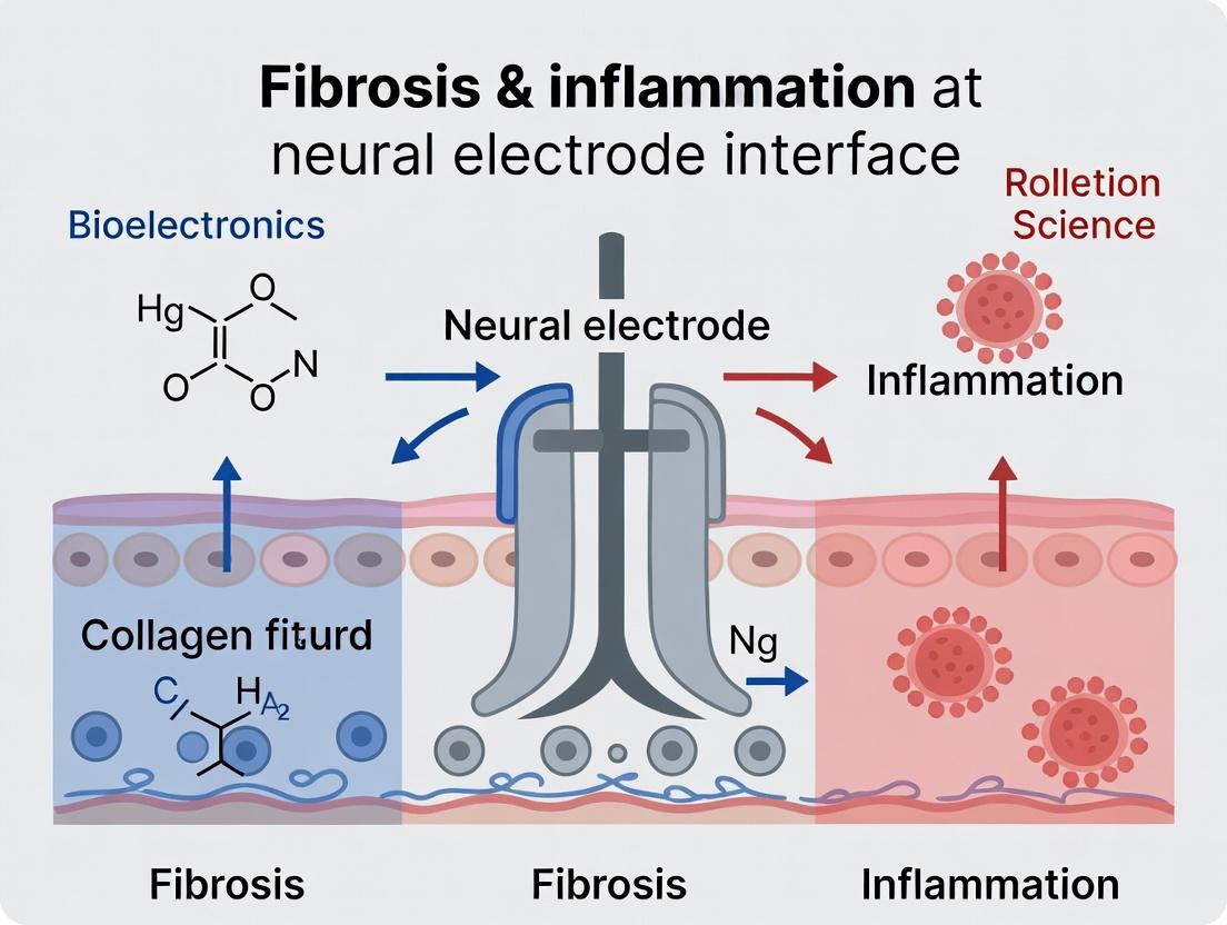
Breaking the Glial Scar: Advanced Strategies to Suppress Fibrosis and Neuroinflammation at Neural Electrode Interfaces
This article provides a comprehensive analysis of the persistent challenge of fibrotic encapsulation and chronic neuroinflammation at the neural electrode-tissue interface.
Breaking the Glial Scar: Advanced Strategies to Suppress Fibrosis and Neuroinflammation at Neural Electrode Interfaces
Abstract
This article provides a comprehensive analysis of the persistent challenge of fibrotic encapsulation and chronic neuroinflammation at the neural electrode-tissue interface. We detail the dual biological response—the foreign body reaction and reactive gliosis—that degrades recording fidelity and stimulation efficacy over time. For researchers and drug development professionals, we explore foundational mechanisms, current methodological approaches including pharmacological coatings, material innovations, and device design. The article critically examines troubleshooting strategies for existing implants, presents comparative data on validation techniques in preclinical models, and synthesizes future directions for creating stable, high-performance bioelectronic neural interfaces.
The Biological Battlefield: Understanding Fibrosis and Inflammation at the Neural Interface
Technical Support Center
Troubleshooting Guide: Common In-Vivo Experimental Issues
Q1: My chronic neural recordings show a steady decline in single-unit yield and signal amplitude over 4-6 weeks. What is the most likely cause and how can I confirm it?
A: This is a classic symptom of the Foreign Body Response (FBR). The decline is primarily due to insulating glial scar formation and neuronal loss/dampening around the electrode. To confirm:
- Perform post-mortem immunohistochemistry. Target: GFAP (astrocytes), Iba1 (microglia), NeuN (neurons).
- Quantify impedance spectroscopy. A persistent rise in low-frequency impedance (1 kHz) indicates tissue encapsulation, while changes at 1 kHz can reflect cellular fouling.
- Analyse signal metrics. Correlate the drop in spike amplitude and count with your histological and impedance data.
Q2: My immunofluorescence shows intense microglial activation at week 1, but by week 4, I see a dense GFAP+ scar with few neurons nearby. Is this expected progression?
A: Yes, this is the standard temporal progression of the FBR.
- Acute Phase (Days 1-7): Blood-brain barrier disruption, serum protein absorption, robust activation of microglia (Iba1+, CD68+) and astrocytes.
- Chronic Phase (Weeks 2+): Formation of a stabilized scar. Astrocytes (GFAP+, CSPG+) form a dense mesh. Microglia may remain active at the interface or become less prominent. A neuropil-deficient zone establishes around the implant.
Q3: What are the key quantitative benchmarks for a "severe" glial scar versus a "mild" one?
A: While benchmarks vary by model and implant, the following table provides typical ranges:
Table 1: Quantitative Histological Benchmarks for Gliosis Severity
| Metric | Mild Response | Severe Response | Measurement Method |
|---|---|---|---|
| Astrocyte Density (GFAP+) | < 25% increase from baseline | > 100% increase from baseline | Fluorescence intensity in peri-implant zone (50µm radius). |
| Microglial Activation (Iba1+) | Primarily ramified morphology | > 60% display amoeboid/phagocytic morphology | Cell count & morphology index in peri-implant zone. |
| Neuronal Density (NeuN+) | > 80% of baseline density | < 50% of baseline density | Neuron count in 50µm radius versus distal tissue. |
| Scar Thickness | < 50 µm | > 100 µm | Distance from implant surface to normalized GFAP signal. |
Q4: Which signaling pathways are most critical in driving this detrimental response?
A: Multiple pathways converge. Key players include:
- Pro-inflammatory Cytokine Signaling: IL-1β, TNF-α, and IL-6 release via microglial NLRP3 inflammasome activation.
- MAPK/NF-κB Pathway: A central hub activated by implant-induced injury, leading to pro-inflammatory gene transcription.
- TGF-β Signaling: Promotes the transition to chronic astrogliosis and extracellular matrix (ECM) deposition.
Diagram Title: Core Signaling Pathways in FBR-Driven Electrode Failure
Frequently Asked Questions (FAQs)
Q: What is the most reliable protocol for quantifying glial scarring around my electrode? A: Use a standardized immunohistochemistry and image analysis pipeline. Protocol: Peri-Implant Gliosis Quantification
- Perfusion & Sectioning: At endpoint, transcardially perfuse with 4% PFA. Extract and post-fix brain. Section tissue containing the implant tract at 30µm thickness using a cryostat.
- Immunostaining: Use free-floating sections. Block in 10% normal serum + 0.3% Triton X-100. Incubate in primary antibodies (chicken anti-GFAP, rabbit anti-Iba1, mouse anti-NeuN) for 48h at 4°C. Use appropriate fluorescent secondary antibodies.
- Imaging: Acquire z-stack images (20x objective) concentric to the implant tract. Use consistent laser power/gain settings across all samples.
- Analysis: Using ImageJ/FIJI:
- Create a 50µm radial bin mask from the tract edge.
- Measure mean fluorescence intensity for GFAP and Iba1 in each bin.
- Count NeuN+ nuclei in the same bins.
- Normalize intensity values to a distal, unaffected brain region in the same section.
Q: Are there any in-vitro models to pre-test my novel anti-fibrotic electrode coating? A: Yes, primary glial culture assays are valuable for screening. Protocol: Primary Microglia/Astrocyte Co-culture Response Test
- Culture: Isolate primary mixed glia from P0-P2 rat cortices. Seed your test electrode material or coated substrates in culture wells.
- Challenge: Add 100 ng/mL LPS to stimulate an immune response. Include uncoated implant material as a positive control and tissue culture plastic as a negative control.
- Readout (24-72h):
- ELISA: Measure TNF-α, IL-6 in supernatant.
- Immunocytochemistry: Fix and stain for Iba1 (microglia) and GFAP (astrocytes). Quantify cell morphology (ramified vs. amoeboid) and proliferation indices.
- qPCR: Analyze gene expression of Il1b, Tnf, Gfap, Tnfaip6.
Q: What key reagents are essential for studying the FBR?
Table 2: Research Reagent Toolkit for FBR & Gliosis Research
| Reagent / Material | Primary Function | Example Application |
|---|---|---|
| Anti-GFAP Antibody | Labels reactive astrocytes. | Quantifying astrogliosis thickness and intensity via IHC. |
| Anti-Iba1 Antibody | Labels all microglia/macrophages; morphology indicates activation state. | Assessing microglial recruitment and phagocytic activity. |
| Anti-NeuN Antibody | Labels neuronal nuclei. | Quantifying neuronal density and loss around implant. |
| Chondroitin Sulfate Proteoglycan (CSPG) Antibody | Labels inhibitory ECM components of the glial scar. | Assessing mature scar maturity and inhibitory character. |
| LPS (Lipopolysaccharide) | Toll-like receptor agonist; potent inflammatory stimulus. | In-vitro or in-vivo challenge to model/amplify neuroinflammation. |
| Minocycline | Microglial activation inhibitor. | In-vivo intervention study to dissect microglial role in FBR. |
| Dexamethasone | Broad-spectrum anti-inflammatory glucocorticoid. | Positive control for suppressing acute inflammatory phase of FBR. |
| Fluorescent-tagged Dextrans | Tracers for vascular permeability. | Assessing blood-brain barrier integrity post-implantation. |
| Polyimide / Silicon Neural Probes | Standard experimental implant substrates. | Negative controls vs. novel coated/treated probes in in-vivo studies. |
Q: How do I differentiate between the effects of inflammation (microglia) and scarring (astrocytes) on my signal? A: Use targeted pharmacological interventions and timeline studies.
- To test microglial role: Administer minocycline (50 mg/kg/day, i.p.) during the first week post-implantation. Compare signal degradation and histology to vehicle controls.
- To test astrocytic scarring: Analyze your signal metrics at specific time points (e.g., 1wk vs. 4wk vs. 12wk) and correlate with the ratio of Iba1 intensity to GFAP intensity. A early signal drop with high Iba1 suggests inflammation is key. A later, continuous drop with rising GFAP suggests scarring is dominant.
Diagram Title: Integrated Workflow for Evaluating Electrode Degradation
Troubleshooting Guide & FAQs for Neural Electrode Interface Research
FAQ 1: How can I differentiate between persistent pro-inflammatory microglia (M1) and resolving/anti-inflammatory microglia (M2) at the electrode interface, and what are the key quantitative markers?
Answer: Reliable differentiation is critical for assessing the inflammatory state. The table below summarizes current key markers and their expression profiles.
Table 1: Key Markers for Microglial Phenotype Identification
| Phenotype | Key Surface Markers (Flow Cytometry) | Key Secreted Cytokines (Luminex/ELISA) | Key Transcription Factors (IHC/WB) |
|---|---|---|---|
| Pro-inflammatory (M1-like) | CD86, CD32, MHC-II | TNF-α, IL-1β, IL-6, CCL2 | NF-κB p65, STAT1, AP-1 |
| Anti-inflammatory (M2-like) | CD206, Arg1, YM1/2 | IL-10, TGF-β, IGF-1 | STAT3, STAT6, PPARγ |
Troubleshooting Note: Pure in vivo phenotypes are rare. Always use a panel of at least 3 markers from different categories (surface, secreted, intracellular) for confirmation. High background in immunofluorescence? Perform careful isotype controls and include a nuclear stain (DAPI) to identify cell bodies accurately.
FAQ 2: What is a robust protocol to quantify reactive astrogliosis (e.g., GFAP area, hypertrophy) around implanted electrodes in tissue sections?
Answer: Use a standardized immunohistochemistry (IHC) and image analysis workflow.
Experimental Protocol: Quantification of Reactive Astrogliosis
- Tissue Preparation: Perfuse-fix animals with 4% PFA. Extract and post-fix brain tissue containing the implant site for 24h. Section coronally (30-40 µm) using a vibratome.
- Immunohistochemistry: Perform free-floating IHC. Block in 5% normal goat serum + 0.3% Triton X-100 for 1h. Incubate in primary antibody (Chicken anti-GFAP, 1:1000) for 48h at 4°C. Incubate in Alexa Fluor 488-conjugated secondary antibody (1:500) for 2h at RT. Mount with DAPI-containing medium.
- Image Acquisition: Using a confocal microscope, acquire z-stacks (e.g., 2 µm steps) at 20x magnification in consistent regions (e.g., 0-100 µm from the implant border). Maintain identical laser power, gain, and offset across all samples.
- Image Analysis (FIJI/ImageJ):
- Create a maximum intensity projection.
- Subtract background.
- Apply a consistent threshold to create a binary mask of GFAP+ signal.
- Metrics: Calculate (a) % GFAP+ Area: (GFAP+ pixels / total image pixels) * 100. (b) Hypertrophy Index: Measure the average process thickness from skeletonized images.
Troubleshooting Note: If signal is weak, consider antigen retrieval (citrate buffer, 95°C, 20 min) before blocking. For high background, reduce Triton concentration or increase blocking time.
FAQ 3: Which techniques effectively identify the origin (CNS vs. peripheral) and activation state of fibroblasts contributing to fibrotic capsule formation?
Answer: Distinguishing pericytes, meningeal fibroblasts, and bone-marrow derived fibroblasts is challenging but possible with combinatorial labeling.
Table 2: Marker Panel for Fibroblast Origin and Activation
| Cell Origin / State | Proposed Markers | Notes & Caveats |
|---|---|---|
| Perivascular Fibroblasts | PDGFRβ+, CD13+, Collagen1+ | Often confused with pericytes. Look for location adjacent to, but not wrapping, capillaries. |
| Meningeal Fibroblasts | S100A4+, SLP1+, CD44+ | Can invade parenchyma post-injury. |
| Bone-Marrow Derived | CD45+, Col1a1-GFP+ (in fate-mapping models) | Requires genetic lineage tracing for definitive proof. |
| Activated Fibroblasts | α-SMA, Fibronectin-EDA, POSTN | α-SMA indicates myofibroblast differentiation and contractile activity. |
Experimental Protocol: Combinatorial Immunofluorescence
- Co-stain tissue sections with PDGFRβ (fibroblast marker), CD31 (endothelial, to exclude vessels), and α-SMA (activation).
- Use spectral imaging or sequential staining with careful antibody stripping to avoid cross-reactivity.
- Quantification: Report the density of PDGFRβ+ cells within a defined radius (e.g., 150 µm) from the implant, and the proportion co-expressing α-SMA.
FAQ 4: How do I assess changes in the perineuronal net (PNN) component of the ECM following electrode insertion and chronic implantation?
Answer: PNN integrity, often visualised via Wisteria Floribunda Lectin (WFA) or specific antibodies (e.g., against Aggrecan), is a key metric of ECM stability.
Experimental Protocol: PNN Integrity Analysis
- Labeling: Stain free-floating sections with WFA-Alexa Fluor 647 (1:200) and a neuronal marker (NeuN, 1:500) for 24h at 4°C.
- Imaging: Capture confocal z-stacks of the implant periphery and contralateral control regions at 40x.
- Analysis: Identify NeuN+ neurons. For each neuron, measure the mean intensity of WFA staining in a 2 µm shell surrounding the soma. Normalize to the mean intensity of control region neurons. A significant decrease indicates PNN degradation.
The Scientist's Toolkit: Research Reagent Solutions
Table 3: Essential Reagents for Neural Interface Cellular Research
| Reagent | Function/Application | Example Product/Catalog # |
|---|---|---|
| CD68 Antibody (IHC) | Labels phagocytic microglia/macrophages. Critical for assessing foreign body response. | Abcam, ab955 |
| GFAP Antibody | Standard marker for astrocyte cell bodies and processes. Use chicken or rabbit polyclonal for robust labeling. | Synaptic Systems, 173 002 |
| PDGFRβ Antibody | Primary marker for identifying fibroblasts and pericyte populations in CNS tissue. | R&D Systems, AF385 |
| WFA Lectin, Biotinylated | Binds to N-acetylgalactosamine in chondroitin sulfate proteoglycans of PNNs. | Vector Labs, B-1355 |
| Laminin Antibody | Labels basement membrane; useful for visualizing vascular structures and ECM deposition. | Sigma-Aldrich, L9393 |
| Cytokine Multiplex Assay | Quantify 20+ analytes (TNF-α, IL-1β, IL-4, IL-10, etc.) from small tissue homogenate samples. | Milliplex, MCYTOMAG-70K |
| Cell Fate Mapping Mouse Model | Pdgfrβ-CreER; Rosa26-tdTomato: Inducible lineage tracing of fibroblasts/pericytes. | Jackson Laboratory, Stock #018280 + 007909 |
| Hydrogel-Coated Electrodes | Test material to mitigate fibrosis. e.g., PEDOT, silk, or hyaluronic acid-based coatings. | Custom synthesis or commercial probes (e.g., NeuroNexus). |
Diagrams
Title: Cellular Cascade at Neural Electrode Interface
Title: Workflow for Histological Analysis Post-Implant
Troubleshooting Guide & FAQ
Q1: In our rodent model, we observe a rapid loss of neural signal amplitude within the first week post-implantation. What is the primary cause, and how can we mitigate it? A: This is characteristic of the acute inflammatory phase (Days 1-7). The primary culprits are activated microglia and infiltrating macrophages releasing reactive oxygen species (ROS) and pro-inflammatory cytokines (e.g., IL-1β, TNF-α), which create a toxic microenvironment for neurons and directly damage electrode surfaces. Mitigation Strategy: Pre-coat electrodes with anti-inflammatory agents (e.g., dexamethasone) or use engineered surfaces that resist protein fouling (e.g., PEGylated coatings). Ensure surgical sterility and minimize mechanical trauma during implantation.
Q2: Our histology shows thick, collagen-dense capsules (>50 µm) around chronic implants, blocking electrical communication. What experimental approaches can disrupt this fibrotic encapsulation? A: Chronic fibrosis (Weeks 4+) is driven by TGF-β1 signaling, activating fibroblasts into collagen-secreting myofibroblasts. Approaches include:
- Local Drug Delivery: Incorporate TGF-β receptor inhibitors (e.g., SB-431542) or angiotensin II receptor blockers (e.g., Losartan) into slow-release polymer coatings.
- Surface Modification: Use nanotopographies that minimize fibroblast adhesion and promote a more favorable glial phenotype.
- Conditional Knockdown: In transgenic models, use targeted knockdown of key fibrotic genes (e.g., CTGF, α-SMA) in the implant vicinity.
Q3: How do we quantitatively distinguish between the beneficial glial scar (which seals the blood-brain barrier) and the detrimental fibrotic scar? A: Use multiplex immunofluorescence and quantitative morphology analysis. Key markers are summarized below:
| Scar Component | Primary Cellular Actors | Key Marker Proteins | Typical Timeframe | Beneficial vs. Detrimental Role |
|---|---|---|---|---|
| Acute Inflammation | Microglia, Macrophages | IBA1, CD68, iNOS | Days 1-7 | Detrimental: Cytokine/ROS storm. |
| Glial Scar | Astrocytes | GFAP, CSPG4 | Days 5 - Weeks 2 | Mixed: Seals BBB but creates physical/chemical barrier. |
| Fibrotic Scar | Fibroblasts, Myofibroblasts | Collagen I/III, Fibronectin, α-SMA | Weeks 2+ | Detrimental: Primary cause of signal attenuation via insulation. |
Q4: Our in vitro model of macrophage activation on electrode materials shows high variance. What is a standardized protocol for this assay? A: Protocol: Macrophage Cytokine Profiling on Novel Substrates.
- Cell Source: Use immortalized murine macrophage cell line (RAW 264.7) or primary bone-marrow-derived macrophages (BMDMs).
- Material Preparation: Sterilize electrode material coupons (e.g., 1x1 cm). Place in 24-well plate. Add poly-D-lysine coated wells as a biological control.
- Seeding & Polarization: Seed cells at 50,000 cells/cm². After 24h, polarize towards pro-inflammatory (M1) state using 100 ng/mL LPS + 20 ng/mL IFN-γ for 48h. Include unstimulated (M0) control.
- Analysis:
- Secreted Cytokines: Collect supernatant. Analyze via ELISA for TNF-α, IL-1β, IL-6 (M1), and IL-10, TGF-β1 (M2, for alternate activation).
- Gene Expression: Lyse cells for qPCR of same targets.
- Morphology: Image using phase-contrast; M1 typically exhibits a flattened, spread morphology.
Q5: Which signaling pathways are most critical for driving the transition from inflammation to fibrosis, and are they druggable? A: The TGF-β/Smad pathway is the master regulator. Concurrently, the PDGF and Wnt pathways are critical co-activators of fibroblast proliferation and activation.
Title: Core Pathways from Inflammation to Fibrosis at Neural Interface
Q6: What are the best in vivo models for screening anti-fibrotic coatings on neural electrodes? A: The choice depends on throughput vs. fidelity.
- High-Throughput Screening: Use mouse dorsal root ganglion (DRG) implantation or subcutaneous wire implant model. Readouts: histology for collagen density (Masson's Trichrome, picrosirius red) at 2-4 weeks.
- Functional Validation: Use rat cortical microarray (Utah array) or single-wire implant in motor/sensory cortex. Readouts: chronic electrophysiological recording (single/multi-unit yield, impedance) over 8-12 weeks correlated with terminal histology.
Q7: Our impedance spectroscopy data shows a steady rise in low-frequency impedance but a drop in high-frequency impedance over time. How do we interpret this? A: This pattern is a classic signature of fibrotic encapsulation.
- Rise in Low-Frequency (e.g., 1 Hz - 1 kHz) Impedance: Reflects increased resistance to charge transfer due to the formation of insulating, collagen-rich tissue around the electrode.
- Drop in High-Frequency (e.g., 10 kHz - 1 MHz) Impedance: Indicates an increase in capacitive coupling due to the growth of a conductive but cell-dense tissue layer (glia, fibroblasts) close to the electrode surface, effectively increasing the electrode's surface area.
Research Reagent Solutions Toolkit
| Item | Function & Application |
|---|---|
| Dexamethasone | Synthetic glucocorticoid. Used in eluting coatings to suppress acute pro-inflammatory cytokine release from macrophages/microglia. |
| SB-431542 | Small-molecule inhibitor of TGF-β receptor I (ALK5). Used in vitro and in local delivery to block Smad2/3 signaling and myofibroblast differentiation. |
| Losartan | Angiotensin II receptor blocker (ARB). Shown to reduce collagen I synthesis and fibrotic capsule thickness in chronic implant models. |
| Puerarin | A natural isoflavone. Recent studies show it modulates microglial polarization towards anti-inflammatory M2 state, reducing acute toxicity. |
| PEG-Silane (e.g., mPEG-Si) | Used for creating anti-fouling, hydrophilic self-assembled monolayers on silicon/glass electrodes to reduce initial protein adsorption. |
| CNTF (Ciliary Neurotrophic Factor) | Neurotrophic factor. Co-delivery can promote neuronal survival and process outgrowth in the inflammatory milieu. |
| Cellulose Nanocrystal (CNC) Coatings | Emerging nanomaterial coating that reduces glial activation and improves chronic recording performance in vivo. |
| Iba1 & CD68 Antibodies | Standard markers for identifying and quantifying activated microglia and macrophages in tissue sections. |
| α-SMA (Alpha Smooth Muscle Actin) Antibody | Gold-standard marker for identifying activated myofibroblasts in the fibrotic capsule. |
| Picrosirius Red Stain | Histological stain for collagen. When viewed under polarized light, quantifies collagen density and maturation (red/orange vs. green birefringence). |
Title: Timeline of Neural Interface Failure Post-Implantation
Troubleshooting & FAQs for Neural Electrode Interface Research
Q1: In our in vitro glial scar model, TNF-α/IL-1β stimulation isn't yielding consistent NF-κB activation. What are common pitfalls?
A: Inconsistent NF-κB nuclear translocation is frequently due to reagent degradation or improper timing. TNF-α is highly labile; avoid freeze-thaw cycles. Use a fresh aliquot and confirm activity with a positive control (e.g., HeLa cells). For timing, perform a time-course experiment (15, 30, 60, 90 min). Measure phospho-IκBα or p65 nuclear localization via immunofluorescence. A common oversight is not pre-treating cells with a proteasome inhibitor (e.g., MG-132, 10 µM for 30 min prior) before harvesting for western blot to prevent IκBα degradation.
Q2: When assessing TGF-β-induced Smad2/3 phosphorylation in neural cell cultures, background is high. How can we improve specificity?
A: High background often stems from endogenous TGF-β in serum. Use serum-free media for at least 4 hours pre-stimulation and during TGF-β1 (recommended 2-10 ng/mL) treatment. For immunoblotting, use Tris-Glycine gels (not Bis-Tris) for optimal separation of phosphorylated Smad2/3 from total protein. Include a negative control with a TGF-β receptor I kinase inhibitor (SB-431542, 10 µM). Ensure fixation for ICC is performed with ice-cold methanol, not PFA, for better phospho-epitope preservation.
Q3: Our PDGF-BB chemotaxis assays with microglia/macrophages show poor migration. What parameters should we verify?
A: PDGF-BB-mediated chemotaxis requires precise gradient establishment.
- Concentration: Use a gradient of 10-50 ng/mL PDGF-BB in the lower chamber. The upper chamber should have serum-free media.
- Membrane: Use translucent polycarbonate membranes (8 µm pores for macrophages), not collagen-coated, which can trap cells.
- Cell Preparation: Cells must be in single-cell suspension. Let cells recover in suspension for 30 min post-trypsinization before loading.
- Incubation: Ensure a humidified, 5% CO2 environment for 4-6 hours. Include a positive control (e.g., 10% FBS) and a negative control (0.1% BSA in media).
Q4: We are co-stimulating with cytokines and see conflicting fibrotic responses. How do we dissect the crosstalk between TNF-α and TGF-β1 pathways?
A: The crosstalk is complex. TNF-α can inhibit TGF-β/Smad signaling via NF-κB-induced Smad7 expression. To dissect:
- Sequential vs. Concurrent Stimulation: Pre-treat with TNF-α (20 ng/mL, 2h) before adding TGF-β1, or add them simultaneously.
- Key Readout: Measure Smad7 mRNA (qPCR) at 2-4h post-TNF-α addition. If Smad7 is upregulated, it will inhibit subsequent p-Smad2/3.
- Rescue Experiment: Use siRNA against Smad7 or a dominant-negative IκB to block NF-κB. Then, observe if TGF-β1-induced α-SMA or collagen I expression is restored.
- Secreted Factors: Always check conditioned media for active TGF-β1 via ELISA, as TNF-α can activate latent TGF-β.
Experimental Protocols
Protocol 1: Assessing NF-κB Activation via Immunofluorescence in Primary Astrocytes
- Plate primary rat/mouse astrocytes on PDL-coated glass coverslips in 24-well plates.
- At 80% confluency, serum-starve (0.5% FBS) for 12 hours.
- Stimulate with recombinant TNF-α (10-20 ng/mL) or IL-1β (5-10 ng/mL) for 30 minutes.
- Aspirate media, wash 1x with PBS, and fix with 4% PFA for 15 min at RT.
- Permeabilize with 0.2% Triton X-100 in PBS for 10 min. Block with 5% normal goat serum for 1h.
- Incubate with primary antibody against p65/RelA (1:500) overnight at 4°C.
- Incubate with Alexa Fluor-conjugated secondary antibody (1:1000) and DAPI (1:5000) for 1h at RT.
- Mount and image. Quantify the nuclear-to-cytoplasmic fluorescence intensity ratio of p65 for ≥50 cells per condition.
Protocol 2: Measuring Active TGF-β1 from Neural Cell Conditioned Media via ELISA Note: Most ELISA kits detect total TGF-β1. To measure the active form, an acidification step is OMITTED.
- Culture relevant neural cells (astrocytes, microglia). At desired confluence, replace media with serum-free media.
- Apply your experimental conditions (e.g., electrode material conditioned media, pro-inflammatory stimuli) for 24-48h.
- Collect conditioned media and centrifuge at 2000xg for 10 min to remove debris.
- Immediately assay the supernatant using a TGF-β1 ELISA kit designed to detect active TGF-β (e.g., DuoSet ELISA, R&D Systems). Do not acid-activate the samples.
- Run standards and samples in duplicate. Express as pg of active TGF-β1 per mg of total cellular protein.
Protocol 3: Combined Inflammatory & Fibrotic Gene Expression Panel (qRT-PCR)
- Treat neural co-culture or primary glial cells with relevant stimuli.
- Isolate total RNA using a column-based kit with on-column DNase I treatment.
- Synthesize cDNA using a high-capacity reverse transcription kit.
- Prepare qPCR reactions with SYBR Green master mix and gene-specific primers (see table for key targets).
- Run in triplicate on a real-time PCR system. Use ∆∆Ct method for analysis with at least two stable reference genes (e.g., Gapdh, Hprt1).
Data Tables
Table 1: Common Cytokine & Growth Factor Concentrations for In Vitro Neural Models
| Reagent | Typical Working Concentration | Key Receptor | Primary Downstream Readout | Common Inhibitor (Concentration) |
|---|---|---|---|---|
| TNF-α | 10-50 ng/mL | TNFR1 | p-IκBα, Nuclear p65, IL-6 secretion | R-7050 (TNFRI inhibitor, 1-10 µM) |
| IL-1β | 5-20 ng/mL | IL-1R1 | p-p38 MAPK, NF-κB, COX-2 | IL-1RA (Anakinra, 100-500 ng/mL) |
| TGF-β1 | 2-10 ng/mL | TβRII/TβRI | p-Smad2/3, α-SMA, Fibronectin | SB-431542 (TβRI inhibitor, 10 µM) |
| PDGF-BB | 10-50 ng/mL | PDGFR-β | p-Akt, p-ERK, Cell Migration | Imatinib (PDGFR inhibitor, 1-5 µM) |
Table 2: Key Gene Targets for qPCR Analysis in Neural Fibrosis/Inflammation
| Pathway | Gene Symbol | Full Name | Function |
|---|---|---|---|
| Inflammation | Il6 | Interleukin 6 | Pro-inflammatory cytokine |
| Inflammation | Ccl2 | C-C Motif Chemokine Ligand 2 | Monocyte/microglia recruitment |
| Inflammation | Nos2 | Nitric Oxide Synthase 2 | M1 microglia marker, oxidative stress |
| Fibrosis | Acta2 | Actin Alpha 2, Smooth Muscle | α-SMA, myofibroblast activation |
| Fibrosis | Col1a1 | Collagen Type I Alpha 1 Chain | Extracellular matrix deposition |
| Fibrosis | Fn1 | Fibronectin 1 | Adhesive glycoprotein, ECM |
| Crosstalk | Smad7 | SMAD Family Member 7 | Inhibitory Smad, NF-κB target |
Pathway & Workflow Diagrams
Title: Inflammatory & Fibrogenic Pathway Crosstalk at Neural Interface
Title: Experimental Workflow for Neural Electrode Interface Fibrosis Research
The Scientist's Toolkit: Research Reagent Solutions
| Item | Supplier Examples | Function in Context |
|---|---|---|
| Recombinant Human/Mouse TNF-α, IL-1β, TGF-β1, PDGF-BB | PeproTech, R&D Systems | High-purity, carrier-free cytokines for precise in vitro stimulation of pathways. |
| Phospho-Specific Antibodies (p-IκBα Ser32, p-Smad2 Ser465/467, p-p65 Ser536) | Cell Signaling Technology | Critical for detecting pathway activation via western blot (WB) or immunofluorescence (IF). |
| α-Smooth Muscle Actin (α-SMA) Antibody | Abcam, Sigma-Aldrich | Gold-standard marker for identifying activated myofibroblasts/astrocytes in fibrosis. |
| TGF-β1 ELISA Kit (Active & Latent forms) | DuoSet (R&D Systems), Quantikine | Measures active vs. total TGF-β1 secreted in response to inflammatory stimuli or biomaterials. |
| SB-431542 (TGF-β Receptor I Inhibitor) | Tocris, Selleckchem | Selective ALK5 inhibitor used to confirm TGF-β-specific effects in crosstalk experiments. |
| R-7050 (TNF Receptor I Inhibitor) | Tocris, MedChemExpress | Selective inhibitor of TNFRI-mediated signaling, used to dissect TNF-α's role. |
| Transwell Permeable Supports (8 µm pore) | Corning Costar | For PDGF-BB chemotaxis/migration assays of microglia and macrophages. |
| Collagen I, Rat Tail (High Concentration) | Corning, Millipore | For generating 3D contraction assay gels to assess myofibroblast activity. |
| RNA Isolation Kit with DNase Step | RNeasy (Qiagen), NucleoSpin | High-quality RNA extraction essential for sensitive qPCR of inflammatory/fibrotic genes. |
| SYBR Green qPCR Master Mix | PowerUp (Applied Biosystems), iTaq (Bio-Rad) | For robust, sensitive detection of mRNA expression changes in pathway target genes. |
Technical Support Center
Troubleshooting Guide: Common In-Vivo Experimentation Issues
Issue 1: Sudden, Sustained Increase in Electrode Impedance
- Symptoms: A sharp rise (e.g., >100 kΩ at 1 kHz) in recorded impedance that does not recover, accompanied by a drastic drop in neural signal amplitude.
- Likely Cause: Formation of a dense, fibrotic capsule (gliosis and fibrosis) around the electrode. This insulating barrier increases the effective distance between the electrode and viable neurons.
- Diagnostic Steps:
- Measure electrochemical impedance spectroscopy (EIS) to distinguish between a damaged electrode (uniform impedance increase across frequencies) and a biological response (frequency-dependent changes).
- Perform post-mortem immunohistochemistry (IHC) for GFAP (astrocytes), Iba1 (microglia), and NeuN (neurons) at the implant site.
- Immediate Mitigation: None for the current experiment. For future implants, consider prophylactic drug-eluting coatings (e.g., anti-inflammatory dexamethasone).
Issue 2: Progressive Deterioration of Signal-to-Noise Ratio (SNR) Over Weeks
- Symptoms: Unit yield and SNR gradually decline over 4-8 weeks post-implantation. Signals become noisier and harder to isolate.
- Likely Cause: Chronic neuroinflammation leading to progressive neuronal loss and displacement from the electrode interface.
- Diagnostic Steps:
- Track single-unit yield and spike amplitude over time for each channel.
- Correlate with post-mortem histology quantifying neuronal density in concentric circles from the implant site.
- Immediate Mitigation: Ensure stable, biocompatible grounding. Review sterilization and surgical aseptic techniques to minimize initial infection/inflammation.
Issue 3: Complete Signal Loss on Specific Channels
- Symptoms: One or more channels show no neural activity, only noise, while others function.
- Likely Cause: Localized, severe fibrosis or a micro-hemorrhage isolating that specific contact. Alternatively, mechanical failure.
- Diagnostic Steps:
- Check impedance: an open circuit (>>1 MΩ) suggests wire/connector break. A very high but stable impedance suggests biofouling.
- Perform a cyclic voltammetry (CV) scan in a saline solution (post-explant) to check for electroactive surface area loss.
- Immediate Mitigation: If a hardware issue is ruled out, flag the channel as non-functional in your analysis pipeline.
Frequently Asked Questions (FAQs)
Q1: What is the typical timeline for these failure modes post-implantation? A1: The process is phase-dependent. Acute inflammation peaks within the first week. Chronic gliosis and fibrosis establish between 2-4 weeks, coinciding with impedance peaks and neuronal displacement. Long-term (>6 months) neuronal loss can be significant.
Q2: How can I experimentally distinguish between signal attenuation from fibrosis vs. neuronal loss? A2: This requires multimodal correlation. Combine longitudinal in vivo electrical recordings (impedance, SNR, unit count) with terminal histological analysis (see Table 1). A rise in impedance with stable unit count suggests fibrotic insulation. A drop in unit count with stable impedance suggests neuronal loss.
Q3: Are there quantitative benchmarks for "normal" vs. "problematic" impedance values? A3: Benchmarks vary by electrode geometry and material. The change is more critical than the absolute value. Monitor baseline impedance in PBS pre-implant. A post-implant increase of 1-2 orders of magnitude is common; increases >3 orders often correlate with severe encapsulation. See Table 2 for aggregated data.
Q4: What are the key signaling pathways involved in this inflammatory host response? A4: The primary pathways involve the activation of microglia and astrocytes via DAMPs (Damage-Associated Molecular Patterns), leading to pro-inflammatory cytokine release (TNF-α, IL-1β, IL-6) via NF-κB and STAT3 signaling, ultimately driving fibroblast activation and collagen deposition.
Q5: What are the best-practice experimental protocols for validating anti-fibrotic coatings? A5: A robust protocol requires in vitro, in vivo, and ex vivo analyses. Key steps include: (1) In vitro cytokine challenge on relevant cell lines (e.g., astrocytes), (2) In vivo implantation in rodent model (e.g., rat motor cortex) with longitudinal electrical monitoring, (3) Terminal perfusion-fixation and serial sectioning for IHC, (4) Quantitative image analysis of glial scarring and neuronal density.
Data Presentation
Table 1: Correlation of Electrical Metrics with Histological Outcomes
| Electrical Metric (Change at 4 weeks) | Associated Histological Finding (IHC) | Typical Consequence |
|---|---|---|
| Impedance at 1 kHz ↑ > 200% | GFAP+ & Iba1+ cell density ↑ > 300% from baseline | Severe Signal Attenuation |
| Single-Unit Yield ↓ > 70% | NeuN+ cell density ↓ > 50% within 100 µm radius | Neuronal Loss |
| Noise Floor (RMS) ↑ > 50% | CD68+ (activated microglia) intensity ↑ | Poor Signal-to-Noise Ratio |
| Charge Storage Capacity (CSC) ↓ > 60% | Collagen IV (fibrosis) deposition ↑ | Reduced Stimulation Efficacy |
Table 2: Aggregated Longitudinal Impedance Data from Rodent Studies (1 kHz)
| Time Post-Implant | Typical Impedance Range (kΩ) | Suggested Interpretation & Action |
|---|---|---|
| Day 0 (in PBS) | 50 - 150 | Establish experiment-specific baseline. |
| Week 1 | 200 - 500 | Acute inflammation phase. Monitor. |
| Week 2 - 4 | 500 - 2000+ | Peak fibrotic response. Expected signal degradation. |
| Week 6+ | Stabilizes between 300 - 1500 | Chronic encapsulation. Yield may continue to decline. |
Experimental Protocols
Protocol 1: Longitudinal In-Vivo Electrochemical Impedance Spectroscopy (EIS)
- Objective: Monitor the biofouling and tissue integration of implanted electrodes.
- Materials: Multichannel recording system with EIS capability (e.g., Intan RHS, Tucker-Davis), implanted neural array, anesthetized or behaving animal setup.
- Steps:
- Connect the headstage to the implanted pedestal under isoflurane anesthesia or in a freely moving setup.
- Set EIS parameters: Frequency range: 10 Hz - 100 kHz (log-spaced, 5-10 points per decade), Amplitude: 10-50 mV RMS (to avoid Faradaic processes).
- Record spectrum on each electrode channel, referencing a common ground (e.g., skull screw).
- Fit data to a simplified Randles equivalent circuit model to extract solution resistance (Rs) and charge transfer resistance (Rct).
- Repeat weekly. The increase in Rct is a key indicator of insulating tissue growth.
Protocol 2: Perfusion-Fixation & Histological Processing for Neural Implants
- Objective: Preserve brain tissue for analysis of gliosis, fibrosis, and neuronal density around the implant.
- Materials: Peristaltic pump, 0.1M Phosphate Buffer (PB), 4% Paraformaldehyde (PFA) in PB, sucrose gradients (10%, 20%, 30% in PB), cryostat, gelatin-coated slides.
- Steps:
- Deeply anesthetize the animal and transcardially perfuse with 100-200 mL ice-cold PBS, followed by 300-400 mL of ice-cold 4% PFA.
- Carefully extract the brain, keeping the implant in situ if possible for precise localization. Post-fix in 4% PFA for 24h at 4°C.
- Cryoprotect by sinking the brain in successive sucrose solutions (10% for 24h, 20% for 24h, 30% until it sinks) at 4°C.
- Snap-freeze the brain in OCT compound. Using a cryostat, section the tissue coronally (30-40 µm thickness) through the implant track.
- Mount sections on slides for staining (e.g., H&E, GFAP/Iba1/NeuN multiplex immunofluorescence).
Visualization: Signaling Pathways and Workflows
Diagram 1: Key Signaling in Neural Electrode Fibrosis
Diagram 2: Experimental Workflow for Interface Evaluation
The Scientist's Toolkit: Research Reagent Solutions
| Item | Function & Relevance |
|---|---|
| Anti-inflammatory Coatings (e.g., Dexamethasone, α-MSH) | Drug-eluting layers to suppress the acute inflammatory response, mitigating initial glial activation. |
| Conductive Polymers (e.g., PEDOT:PSS) | Coatings that lower interfacial impedance and increase charge injection capacity, improving signal fidelity. |
| Hydrogel Coatings (e.g., Hyaluronic Acid, PEG) | Soft, hydrating interfaces that mechanically mimic neural tissue, reducing shear stress and micro-motion. |
| Anti-fibotic Agents (e.g., Mitomycin C, 5-Fluorouracil) | Compounds incorporated to inhibit fibroblast proliferation and collagen synthesis directly. |
| IHC Antibodies (GFAP, Iba1, CD68, NeuN, Collagen IV) | Essential for labeling astrocytes, microglia, activated phagocytes, neurons, and fibrotic tissue post-mortem. |
| Fluorophore-conjugated Lectins (e.g., Isolectin B4) | Labels vasculature and microglia, useful for assessing vascular integration and immune response. |
| Live/Dead Cell Assay Kits (for in vitro) | Quantify cytotoxicity of electrode materials or eluted drugs on co-cultured neural cells. |
| Cytokine ELISA/Multiplex Array Kits | Measure concentrations of key inflammatory markers (TNF-α, IL-1β, IL-6) in tissue homogenates near the implant. |
Engineering the Solution: Material, Pharmacological, and Device-Based Anti-Fibrotic Strategies
Technical Support & Troubleshooting Hub
This support center provides guidance for common experimental challenges in fabricating and characterizing engineered neural interfaces, framed within a thesis focused on mitigating fibrosis and inflammation.
FAQ & Troubleshooting Guides
Q1: My PEDOT:PSS film electrodeposited on a neural electrode is non-uniform, showing patchy conductivity. What went wrong? A: This is often due to inconsistent electrochemical deposition parameters or contaminated substrate surfaces.
- Troubleshooting Steps:
- Clean Substrate: Ensure the metal electrode (e.g., gold, Pt/Ir) is meticulously cleaned. Perform sequential sonication in acetone, isopropanol, and deionized water (15 min each). Treat with oxygen plasma (5 min, 100W) to ensure a clean, hydrophilic surface.
- Solution Preparation: Filter the PEDOT:PSS monomer/oxidant solution (e.g., EDOT in PSS solution with sodium p-toluenesulfonate) through a 0.45 µm syringe filter before use to remove aggregates.
- Deposition Parameters: Use chronopotentiometry (constant current) instead of cyclic voltammetry for more uniform nucleation. Optimize current density and total charge passed. A common starting point is 0.1-0.5 mA/cm² for 10-50 mC.
- Prevention: Always perform a cleaning protocol (Steps 1 & 2) and characterize deposition parameters on a test substrate before using valuable electrode arrays.
Q2: My soft PDMS substrate with topographic features (e.g., pillars, grooves) delaminates from the conductive layer. How can I improve adhesion? A: Poor adhesion results from incompatible surface energies and lack of effective interfacial bonding.
- Troubleshooting Steps:
- Surface Activation: Treat cured PDMS with oxygen plasma (30 sec - 2 min) to create a reactive silica-like (Si-OH) surface layer.
- Primer Layer: Immediately after plasma treatment, apply an adhesion promoter. For metal deposition, use a molecular primer like (3-Aminopropyl)triethoxysilane (APTES). For conductive polymers, a thin layer of poly-L-lysine or a crosslinker like poly(dopamine) can be coated.
- Mechanical Interlocking: Design topography with undercuts or high aspect ratios (where possible) to provide physical anchoring points for subsequent layers.
- Prevention: Integrate the adhesion promoter step as a standard part of your fabrication workflow. Test adhesion via tape test (ASTM D3359) on sample batches.
Q3: In vitro cell culture on my conductive polymer surface shows low cell viability or poor neural cell attachment compared to controls. What factors should I investigate? A: This indicates potential cytotoxicity, often from leaching components or excessive surface roughness/scaffold stiffness.
- Troubleshooting Checklist:
- Leaching: Ensure thorough washing/electrochemical cycling of the polymer film in sterile PBS or cell culture medium to remove unreacted monomers, oxidants, and oligomers. Soak for 24-48 hours with multiple medium changes.
- Surface Charge: Verify if your conductive polymer's inherent surface potential (zeta potential) is strongly anionic or cationic, which can repel cell membranes. Consider blending with neutral, cell-adhesive polymers like laminin or collagen.
- Stiffness Mismatch: The composite stiffness may still be too high. Measure the effective Young's modulus via AFM nanoindentation. For neural tissue mimicry, aim for a modulus in the low kPa range (< 50 kPa). Incorporate more soft hydrogel components (e.g., gelatin, agarose) into your polymer blend.
- Topography Scale: If topographic features are larger than single cells (>> 20 µm), they may not effectively guide cell adhesion. Consider subcellular feature sizes (1-10 µm).
Q4: My engineered neural interface performs well in vitro, but I observe heightened inflammatory markers (e.g., GFAP, Iba1) and early fibrotic encapsulation in vivo. What surface properties should I re-evaluate? A: This is a core thesis challenge. The in vivo environment is more aggressive, and the foreign body response (FBR) must be actively managed.
- Key Investigation Areas:
- Dynamic Softness: The effective softness in vivo may change due to swelling or protein adsorption. Characterize the hydrated, protein-coated modulus.
- Topography at the Cellular Level: Inflammatory response is mediated by immune cells (microglia, macrophages). Submicron to low-micron topography (0.5 - 5 µm) can influence macrophage polarization from pro-inflammatory (M1) to anti-inflammatory (M2) phenotypes. Ensure your topography is present at this scale.
- Biofunctionalization: Are you releasing an anti-inflammatory agent (e.g., dexamethasone)? Check loading efficiency and release kinetics. A burst release may deplete the drug too early. Aim for sustained release over weeks.
- Chronic Electrical Stimulation: If applicable, test if your stimulation parameters (charge density, pulse shape) are causing faradaic reactions that damage tissue. Use charge-balanced, capacitive stimulation phases. Monitor impedance and voltage transients.
Data Presentation: Key Performance Metrics for Engineered Surfaces
Table 1: Impact of Surface Modulus on Glial Scarring Metrics in Rodent Models (2-month Implant)
| Surface Effective Modulus (kPa) | Astrocyte Activation (GFAP+ intensity) | Fibrotic Capsule Thickness (µm) | Neuronal Density near Interface (% of Sham) |
|---|---|---|---|
| Rigid Control (> 1 GPa) | 100% | 45 ± 12 | 55 ± 8 |
| 1000 | 85% | 38 ± 10 | 65 ± 9 |
| 100 | 60% | 25 ± 7 | 80 ± 10 |
| 10 | 40% | 15 ± 5 | 92 ± 6 |
Table 2: Conducting Polymer Coatings: Electrical & Biological Trade-offs
| Polymer Blend | Charge Capacity (mC/cm²) | Impedance at 1 kHz (kΩ) | Neurite Outgrowth (vs. Control) | Stability (Cycles to 80% Cap.) |
|---|---|---|---|---|
| PEDOT:PSS | 120 ± 20 | 2.5 ± 0.5 | 1.2x ± 0.1 | > 1 million |
| PEDOT:PSS + Laminin Peptide | 95 ± 15 | 3.0 ± 0.8 | 1.8x ± 0.2 | 500,000 |
| PPy/DBSA | 200 ± 30 | 1.8 ± 0.3 | 0.9x ± 0.1 | 200,000 |
| PPy + Dexamethasone | 180 ± 25 | 2.0 ± 0.4 | 1.5x ± 0.2 | 150,000 |
Experimental Protocols
Protocol 1: Electrodeposition of PEDOT:PSS on Microelectrode Arrays for Enhanced Charge Injection Objective: To create a uniform, low-impedance, and biocompatible conductive polymer coating. Materials: See "Scientist's Toolkit" below. Steps:
- Electrode Cleaning: As per FAQ Q1, Step 1.
- Electrochemical Setup: Use a standard 3-electrode setup in an electrochemical cell. The microelectrode is the working electrode. Use a Pt mesh counter electrode and an Ag/AgCl reference electrode.
- Solution Preparation: Prepare 0.01M EDOT and 0.1M PSS (in DI water). Stir for 1 hour. Filter with a 0.45 µm filter.
- Deposition: Use galvanostatic (constant current) deposition. Apply a current density of 0.25 mA/cm² until a total charge of 20 mC is passed. Gently stir the solution.
- Post-Processing: Rinse thoroughly with DI water. Cycle the coated electrode in 0.1M PBS (pH 7.4) between -0.6V and +0.8V (vs. Ag/AgCl) for 20 cycles at 100 mV/s to stabilize the film.
- Sterilization: For cell culture, soak in 70% ethanol for 20 minutes, then rinse 3x with sterile PBS.
Protocol 2: Fabrication of Micropillared PDMS Substrates via Soft Lithography Objective: To create soft (Young's modulus ~10 kPa) substrates with defined micron-scale topography. Steps:
- Master Fabrication: Create a silicon master with the negative of your desired pillars (e.g., 5 µm diameter, 10 µm height, 10 µm spacing) using standard photolithography and deep reactive ion etching (DRIE).
- Mold Silanization: Vapor-deposit trichloro(1H,1H,2H,2H-perfluorooctyl)silane on the silicon master for 1 hour to create an anti-adhesion layer.
- PDMS Mixing & Degassing: Mix base and curing agent (e.g., Sylgard 184) at a 30:1 or 40:1 w/w ratio to achieve softer elastomer. Degas under vacuum until all bubbles are removed.
- Replica Molding: Pour the mixture over the silicon master. Cure at 60°C for at least 12 hours.
- Demolding: Carefully peel the cured PDMS from the master. Cut to desired size.
- Surface Activation & Bonding: Activate the PDMS and your device substrate/glass coverslip with oxygen plasma (30 sec). Bring surfaces into contact immediately to form an irreversible bond.
Diagrams
Title: Foreign Body Response Pathway & Mitigation Strategies
Title: Integrated Workflow for Neural Interface Testing
The Scientist's Toolkit: Key Research Reagent Solutions
Table 3: Essential Materials for Surface Engineering Experiments
| Item | Function & Application | Example/Supplier |
|---|---|---|
| Sylgard 184 (PDMS) | Silicone elastomer for creating soft, flexible substrates with tunable modulus via base:curing agent ratio. | Dow Corning |
| (3-Glycidyloxypropyl)trimethoxysilane (GOPS) | Crosslinker for PEDOT:PSS, improving its mechanical stability and adhesion in aqueous environments. | Sigma-Aldrich |
| Poly-D-Lysine / Laminin | Standard cell adhesion promoters coated on substrates to improve attachment of neural cells in vitro. | Thermo Fisher Scientific |
| EDOT Monomer (3,4-Ethylenedioxythiophene) | The monomer used for electropolymerization to create PEDOT conductive polymer films. | Heraeus Clevios |
| Polystyrene Sulfonate (PSS) | Polymeric counter-ion and dopant for PEDOT, providing solubility and ionic conductivity. | Sigma-Aldrich |
| Dexamethasone Sodium Phosphate | A potent synthetic glucocorticoid, often loaded into coatings for localized anti-inflammatory release. | Tocris Bioscience |
| FluoroSilane (FOTS) | Used to silanize silicon masters to ensure clean release of molded PDMS. | Gelest, Inc. |
| Live/Dead Viability/Cytotoxicity Kit | Standard assay (Calcein AM/EthD-1) to quantitatively assess cell health on novel surfaces. | Thermo Fisher Scientific |
| Anti-GFAP & Anti-Iba1 Antibodies | Primary antibodies for immunofluorescent labeling of reactive astrocytes and microglia, respectively. | Abcam |
Technical Support Center: Troubleshooting & FAQs
FAQs & Troubleshooting Guides
Q1: My poly(lactic-co-glycolic acid) (PLGA) coating is releasing Dexamethasone too quickly (burst release) in vitro. How can I achieve a more sustained, linear release profile?
A: A high initial burst release is a common challenge with PLGA matrices.
- Primary Cause: Drug molecules located on or near the surface of the coating dissolve immediately upon immersion in aqueous media.
- Solutions:
- Optimize Coating Morphology: Increase coating thickness or implement a multi-layer design. A dense, impermeable top layer (e.g., pure PLGA or Parylene-C) over a drug-loaded layer significantly reduces initial burst.
- Modify Polymer Properties: Use PLGA with a higher lactic acid to glycolic acid ratio (e.g., 85:15 vs. 50:50). The more hydrophobic LA degrades slower. Also, a higher molecular weight PLGA will have slower degradation and diffusion rates.
- Adjust Loading Method: Use a double emulsion (W/O/W) method for hydrophilic drugs to encapsulate them more deeply within polymer microspheres before coating, rather than simple blend-and-spray.
Experimental Protocol: Coating Fabrication & In Vitro Release Test
- Objective: Fabricate a Dexamethasone-loaded PLGA coating on a neural electrode substrate and characterize its release kinetics.
- Materials: PLGA (e.g., 85:15, MW ~100kDa), Dexamethasone, dichloromethane (DCM), phosphate-buffered saline (PBS, pH 7.4), Tween-80, ultrasonic bath, spin coater/dip coater, UV-Vis spectrometer/HPLC.
- Method:
- Prepare a 5% (w/v) PLGA solution in DCM.
- Dissolve Dexamethasone in the solution at a 10:90 drug:polymer ratio.
- Sonicate the mixture for 5 minutes to ensure homogeneity.
- For spin coating: Deposit solution on electrode substrate and spin at 2000 rpm for 30 seconds. For dip coating: Immerse substrate and withdraw at a controlled speed (e.g., 100 mm/min).
- Dry coatings under vacuum for 48h to remove residual solvent.
- For release testing: Immerse coated substrate in 1 mL PBS + 0.1% Tween-80 (sink condition) at 37°C under gentle agitation.
- At predetermined time points (1h, 4h, 1d, 3d, 7d, 14d, 30d), remove and replace the entire release medium.
- Quantify Dexamethasone concentration using HPLC (λ=242 nm) or a validated UV-Vis assay.
Q2: I am co-loading Dexamethasone and an anti-fibrotic (e.g., 4-(Phenylamino)-pyrrolopyrimidine, RSD-1001). How do I prevent drug-drug interaction and ensure independent release kinetics?
A: Physical or chemical interactions between drugs in a single matrix can alter release and stability.
- Primary Cause: Drugs may crystallize together, form salts, or one drug may alter the local microenvironment (pH) affecting the other's solubility.
- Solutions:
- Spatial Separation: Use a multi-reservoir or layered coating architecture. Apply one drug in a base PLGA layer, seal with a thin barrier layer, then apply the second drug in a top PLGA layer. This allows temporal sequencing of release.
- Carrier-Based Separation: Pre-encapsulate each drug in separate nanoparticle systems (e.g., PLGA nanoparticles for Dexamethasone, liposomes for the anti-fibrotic). Then blend these distinct carriers into a single biocompatible hydrogel coating (e.g., hyaluronic acid).
- Combinatorial Particle Fabrication: Use coaxial electrospray to create core-shell particles where one drug is in the core and the other in the shell, providing distinct release phases.
Q3: My drug-coated neural electrode exhibits increased electrochemical impedance post-coating. Is this expected, and how can I mitigate it?
A: Yes, an increase is typical as the coating adds material between the electrode and tissue. The goal is to minimize and stabilize it.
- Primary Cause: The coating acts as an insulating layer. Burst release or coating degradation can cause fluctuating impedance.
- Solutions:
- Optimize Coating Thickness & Porosity: Aim for the thinnest, most uniform coating that provides the desired drug load. Incorporate pore-forming agents (e.g., polyethylene glycol (PEG)) that leach out, creating channels for ion conduction. Ensure coating does not completely cover the entire electrode surface; use precise microdeposition.
- Conductive Composite Coatings: Incorporate conductive materials like graphene oxide, carbon nanotubes, or PEDOT:PSS into the drug-polymer matrix. This creates a percolation network for electron/ion transport.
- Characterize Electrochemically: Always perform cyclic voltammetry and electrochemical impedance spectroscopy (EIS) in PBS pre- and post-coating, and track over time during drug release to correlate impedance with coating state.
Q4: How do I confirm the stability and bioactivity of the released anti-fibrotic agent from my coating?
A: Degradation during fabrication (e.g., solvent exposure, heat) or release (hydrolysis) can inactivate drugs.
- Primary Cause: Harsh organic solvents or high processing temperatures. Instability of the drug in aqueous, physiological-temperature environments over long periods.
- Solutions:
- Post-Release Analysis: Collect release medium and analyze using HPLC-MS not just for concentration, but for chemical integrity. Compare chromatograms and mass spectra with a pristine drug standard.
- In Vitro Bioactivity Assay: Treat relevant cells (e.g., primary astrocytes or fibroblasts) with your collected release medium. Perform a functional assay (e.g., TGF-β1-induced fibroblast proliferation assay for anti-fibrotics, or LPS-induced TNF-α reduction in macrophages for dexamethasone). Compare activity to fresh drug solutions of the same concentration.
- Use Stabilizing Excipients: Incorporate antioxidants (e.g., ascorbic acid) or stabilizers into the polymer matrix to protect the drug.
Table 1: Comparison of Common Polymer Carriers for Neural Drug Delivery
| Polymer | Degradation Time (Approx.) | Key Release Mechanism | Advantages for Neural Interface | Challenges |
|---|---|---|---|---|
| PLGA | 2 weeks - 6 months | Hydrolysis & Diffusion | Tunable, FDA-approved, sustained release | Acidic degradation products, burst release |
| Poly(ethylene glycol) (PEG) | Non-degradable or hydrolysable | Diffusion (hydrogel) | High hydrophilicity, reduces protein adsorption | Limited drug load, may swell |
| Poly(3,4-ethylenedioxythiophene) (PEDOT) | Non-degradable | Electrically-controlled | Conductive, excellent charge capacity, low impedance | Difficult to achieve long-term sustained release |
| Chitosan | Enzymatic degradation | Swelling & Diffusion | Bioadhesive, antimicrobial, gentle processing | Fast release, mechanical stability in vivo |
| Hyaluronic Acid (HA) | Enzymatic degradation | Swelling & Diffusion | Native ECM component, biocompatible | Very rapid dissolution without crosslinking |
Table 2: Example In Vivo Performance Metrics of Coated Electrodes
| Study (Key Drugs) | Coating System | Release Duration (Key Findings) | Neural Interface Outcome (vs. Uncoated) |
|---|---|---|---|
| Dexamethasone (Dex) | PLGA microspheres in PEG hydrogel | ~28 days (sustained) | Reduced glial fibrillary acidic protein (GFAP+) reactivity by ~70% at 2 weeks. |
| Dex + RSD-1001 | Layered PLGA/Parylene | Dex: ~14 days; RSD: ~30 days (sequential) | Combined 60% reduction in fibrotic capsule thickness and 50% increase in neuronal density at 8 weeks. |
| Alpha-MSH | Conducting polymer (PEDOT) | Electrically-triggered, on-demand | On-demand release reduced activated microglia (Iba1+) by 55% within 24h of triggering. |
Signaling Pathways & Experimental Workflows
Title: Dual-Drug Action on Glial Scar Pathways
Title: Coating Development Workflow
The Scientist's Toolkit: Research Reagent Solutions
Table 3: Essential Materials for Coating Development
| Item / Reagent | Function / Purpose | Example & Notes |
|---|---|---|
| PLGA (50:50, 75:25, 85:15) | Biodegradable polymer matrix for sustained release. | Lactel Absorbable Polymers. Higher LA:GA ratio = slower degradation. |
| Dexamethasone | Potent synthetic glucocorticoid; model anti-inflammatory. | Sigma-Aldrich D4902. Use in microparticle form for better encapsulation. |
| Small Molecule Anti-fibrotics | Inhibit fibroblast proliferation & collagen production. | e.g., RSD-1001 (Tocris), Pirfenidone (Sigma). Verify solubility in polymer solvent. |
| Poly(ethylene glycol) (PEG) | Pore-former, hydrogel component, surface modifier. | Use as PEG (MW 1k-10k) to create release channels or as diacrylate for UV-crosslinked hydrogels. |
| Parylene-C | Ultra-thin, biocompatible vapor barrier layer. | Specialty coating service (e.g., SCS). Critical for creating multi-layer, sequential release systems. |
| Conductive Polymer (PEDOT:PSS) | Enables electroactive coatings for combined drug delivery & recording. | Heraeus Clevios PH1000. Can be doped with drugs during electrochemical deposition. |
| Phosphate Buffered Saline (PBS) w/ Tween-80 | Standard in vitro release medium (sink condition). | 0.1% Tween-80 prevents drug adsorption to vial walls and maintains sink conditions. |
| Dichloromethane (DCM) / Chloroform | Common solvent for dissolving PLGA. | HPLC grade. Use in fume hood. DCM evaporates faster, affecting coating morphology. |
| HPLC System with C18 Column | Gold-standard for quantifying drug concentration and purity in release studies. | Method: Mobile phase = Acetonitrile/Water, detect Dex at λ=242 nm. |
| Electrochemical Workstation | For characterizing coated electrode performance (CV, EIS). | e.g., GAMRY, Biologic. Essential for correlating release with interface impedance. |
Technical Support Center: Troubleshooting & FAQs
This support center provides guidance for common experimental challenges in developing bioactive neural electrode coatings within fibrosis and inflammation research.
Troubleshooting Guides
Issue 1: Low Peptide Density on Coated Electrode
- Problem: Immobilized RGD or IKVAV peptides show poor cell adhesion in validation assays.
- Checkpoints:
- Verify silanization (for metal surfaces) or polymer activation protocol.
- Measure sulfhydryl groups if using maleimide-thiol chemistry (Ellman's assay).
- Confirm peptide solubility and stability in coupling buffer.
- Solution: Optimize linker concentration and incubation time. Use quartz crystal microbalance (QCM) for real-time density monitoring.
Issue 2: Cytokine (IL-1Ra, IL-10) Bioactivity Loss
- Problem: Released cytokine fails to reduce TNF-α secretion in LPS-stimulated microglia.
- Checkpoints:
- Check hydrogel crosslinking method; harsh UV or radical polymerization can denature proteins.
- Analyze release kinetics; burst release may deplete cytokine.
- Validate cytokine stability post-encapsulation via ELISA or activity assay.
- Solution: Switch to enzymatic or ionic crosslinking. Use heparin-based delivery for sustained release.
Issue 3: Hydrogel Swelling/Mechanical Instability
- Problem: ECM-mimicking hydrogel (e.g., gelatin-methacrylate) delaminates from electrode or changes impedance.
- Checkpoints:
- Measure equilibrium swelling ratio; >30 may indicate weak crosslinking.
- Check substrate pre-treatment for covalent bonding.
- Test compressive modulus via AFM; target neural tissue modulus (~0.1-1 kPa).
- Solution: Adjust photoinitiator concentration and UV dose. Incorporate adhesive peptides (e.g., DOPA) at the substrate interface.
Frequently Asked Questions (FAQs)
Q1: What is the recommended method for quantifying immobilized peptide density on a platinum-iridium electrode? A1: Use X-ray Photoelectron Spectroscopy (XPS) for elemental analysis of peptide-specific tags (e.g., nitrogen, sulfur). Alternatively, fluorescently-tagged peptides allow for quantification via fluorescence microscopy or a plate reader after cleavage from the surface. Typical successful densities range from 50 to 200 pmol/cm² for RGD peptides.
Q2: How can I control the release profile of an anti-inflammatory cytokine from a hyaluronic acid hydrogel? A2: The release profile is modulated by hydrogel crosslinking density and cytokine affinity. For sustained release over 14-21 days, incorporate cytokine-binding motifs (e.g., heparin) into the hydrogel network. For rapid release, use physically entrapped cytokines in a loosely crosslinked matrix. Characterize using an in vitro ELISA-based release assay in PBS at 37°C.
Q3: My ECM hydrogel is inhibiting electrical impedance of the microelectrode. How can I improve conductivity? A3: Incorporate conductive polymers (e.g., PEDOT:PSS) or carbon nanomaterials (e.g., graphene oxide) into the hydrogel formulation. Ensure homogeneous dispersion to prevent insulating aggregates. Always measure electrochemical impedance spectroscopy (EIS) post-coating; target a post-coating impedance increase of less than one order of magnitude at 1 kHz.
Q4: What is the best in vitro assay to predict anti-fibrotic coating performance? A4: A co-culture of murine microglia (BV-2) and astrocytes (C8-D1A) stimulated with LPS or TGF-β1. Measure key markers via qPCR or immunoassay after 72 hours. A successful coating should show >40% reduction in pro-fibrotic markers (e.g., collagen I, fibronectin) and pro-inflammatory cytokines (e.g., IL-6, TNF-α) compared to a bare electrode control.
Experimental Data & Protocols
Table 1: Characteristic Performance Data of Bioactive Coating Components
| Coating Component | Target Function | Typical Loading/Concentration | Key Outcome Metric | Optimal Value Range |
|---|---|---|---|---|
| RGD Peptide | Enhance neuronal adhesion | 100-200 pmol/cm² | Neurite extension length (PC-12 cells) | ≥ 50 µm after 48h |
| IL-1Ra Cytokine | Inhibit IL-1-mediated inflammation | 10-50 ng/mg hydrogel | Reduction in IL-6 from activated microglia | ≥ 60% reduction |
| GelMA Hydrogel | Mimic soft neural ECM | 5-10% (w/v) polymer | Compressive Modulus | 0.5 - 2.0 kPa |
| PEDOT Conductive Polymer | Maintain charge injection | 0.5-1.0% (w/v) in hydrogel | Electrochemical Impedance (1 kHz) | < 50 kΩ |
Detailed Protocol: Coating Neural Electrodes with Peptide-Functionalized Hydrogel
Objective: Apply an ECM-mimicking, cytokine-loaded hydrogel coating to a neural microelectrode to mitigate gliosis.
Materials: Neural microelectrode array, Gelatin-methacryloyl (GelMA), Lithium phenyl-2,4,6-trimethylbenzoylphosphinate (LAP) photoinitiator, recombinant IL-10, cysteine-terminated RGD peptide, UV light (365 nm, 5 mW/cm²).
Method:
- Surface Activation: Clean electrodes with O₂ plasma for 5 min. Incubate in 2% (v/v) (3-Aminopropyl)triethoxysilane (APTES) in ethanol for 1 hour. Rinse.
- Peptide Coupling: React surface amines with 2 mM Sulfo-SMCC crosslinker in PBS for 30 min. Wash. Incubate with 0.5 mM cysteine-RGD peptide in degassed PBS for 2 hours at RT.
- Hydrogel Precursor Solution: Dissolve GelMA (8% w/v) and LAP (0.25% w/v) in PBS at 40°C. Add recombinant IL-10 to a final concentration of 25 ng/µl. Mix gently.
- Coating & Crosslinking: Pipette the precursor solution onto the electrode site. Immediately expose to 365 nm UV light for 60 seconds to form a crosslinked hydrogel layer.
- Validation: Sterilize in 70% ethanol for 20 min. Rinse with sterile PBS. Characterize cytokine release via ELISA and impedance via EIS.
Visualizations
Diagram 1: Bioactive Coating Workflow
Diagram 2: Signaling Pathways at the Neural Interface
The Scientist's Toolkit: Research Reagent Solutions
| Item | Function & Relevance to Neural Coatings |
|---|---|
| Gelatin-Methacryloyl (GelMA) | Photocrosslinkable hydrogel that mimics the RGD-rich neural ECM. Tunable mechanical properties. |
| Sulfo-SMCC Crosslinker | Water-soluble, heterobifunctional reagent for covalently immobilizing thiol-containing peptides to amine-functionalized electrode surfaces. |
| Recombinant IL-1Ra (Anakinra) | Anti-inflammatory cytokine analog used to competitively inhibit the IL-1 receptor, mitigating acute neuroinflammation. |
| CGRGDS or CIKVAV Peptide | Cysteine-terminated peptides for controlled surface immobilization. Promote specific neuronal adhesion over glial adhesion. |
| Lithium Phenyl-2,4,6-Trimethylbenzoylphosphinate (LAP) | Efficient, cytocompatible photoinitiator for visible light crosslinking of hydrogels, preserving protein bioactivity. |
| PEDOT:PSS Dispersion | Conductive polymer used to dope hydrogel coatings, maintaining low electrochemical impedance for signal fidelity. |
Technical Support Center
Troubleshooting Guide & FAQ
Q1: During in vivo implantation of a bioresorbable electrode, I observe premature dissolution before the planned study endpoint. What could be the cause and how can I mitigate this?
A: Premature dissolution is often linked to local inflammatory response or incorrect polymer crystallinity.
- Cause Analysis: An acute inflammatory response can create a locally acidic microenvironment (pH drop), accelerating hydrolysis of poly(lactic-co-glycolic acid) (PLGA) or poly(lactic acid) (PLA) based substrates.
- Mitigation Protocol:
- Pre-implantation Sterilization: Use cold ethylene oxide gas over gamma irradiation, which can alter polymer chain structure and degradation kinetics.
- Surface Coating: Apply a thin, anti-inflammatory drug-eluting layer (e.g., dexamethasone-loaded parylene-C) to suppress initial immune response.
- Material Characterization: Verify the glass transition temperature (Tg) and crystallinity of your polymer batch via Differential Scanning Calorimetry (DSC). Higher crystallinity typically slows degradation. Annealing the device can increase crystallinity.
Q2: My ultraflexible electrode delaminates at the conductor-polymer substrate interface during cyclic bending tests. How can I improve adhesion?
A: Delamination indicates weak interfacial bonding.
- Solution 1 (Surface Modification): Treat the polyimide or parylene substrate with an O₂ plasma (50W, 30 sec) immediately before metal deposition (e.g., Au, Pt) to increase surface energy and promote mechanical interlocking.
- Solution 2 (Adhesion Layer): Always use a chromium (Cr, 5-10 nm) or titanium (Ti) adhesion layer between the substrate and the primary conductive metal. For bioresorbable electrodes, a thin magnesium oxide (MgO) or silicon dioxide (SiO₂) interlayer can improve adhesion to PLGA.
- Testing Protocol: Perform a standardized tape test (ASTM D3359) on a flat control sample before proceeding to dynamic bending.
Q3: Signal-to-noise ratio (SNR) deteriorates significantly after 2 weeks of chronic neural recording with a miniaturized electrode. Is this fibrosis-related?
A: Likely yes. Increased electrochemical impedance at the electrode-tissue interface due to fibrotic encapsulation is a primary cause.
- Diagnostic Steps:
- Measure Impedance: Perform electrochemical impedance spectroscopy (EIS) at 1 kHz in vivo or post-explant. A consistent rise > 50% from baseline suggests fibrous tissue growth.
- Histological Correlation: Fix the brain tissue, section, and stain for astrocytes (GFAP) and microglia (Iba1) to confirm glial scarring, and for collagen IV to confirm fibrotic capsule.
- Preventive Approach: Implement a surface coating with antifibrotic agents (e.g., curcumin-eluting hydrogels, or immobilize CX3CL1 fractalkine to modulate microglial response).
Q4: What is the recommended sterilization method for ultraflexible hydrogel-based electrodes without compromising mechanical or electrical properties?
A: Standard autoclaving can destroy hydrogel networks. Use one of these validated methods:
- Aseptic Processing: Fabricate in a Class II biosafety cabinet using sterile-filtered hydrogel precursors and ethanol-sterilized electronics.
- Low-Temperature Hydrogen Peroxide Plasma (e.g., Sterrad): Effective for devices sensitive to heat and moisture.
- Ethylene Oxide (EtO): Ensure complete degassing (≥48 hrs) to prevent residual toxicity.
Experimental Protocols for Key Investigations
Protocol 1: Assessing Fibrotic Encapsulation In Vivo
- Implantation: Sterilize electrode and implant into target rat/mouse brain region.
- Chronic Monitoring: Record neural signals and impedance weekly for 4-12 weeks.
- Perfusion & Fixation: At endpoint, transcardially perfuse with 4% paraformaldehyde (PFA).
- Sectioning: Cryosection tissue into 20 µm slices around the implant track.
- Immunohistochemistry: Stain with: Primary antibodies: Anti-GFAP (astrocytes), Anti-Iba1 (microglia), Anti-Colagen IV (fibrosis). Secondary antibodies: Use appropriate fluorophore-conjugated antibodies.
- Imaging & Quantification: Confocal microscopy. Quantify fluorescence intensity in concentric zones (0-50µm, 50-100µm, 100-150µm) from the implant interface using ImageJ.
Protocol 2: Electrochemical Characterization of Miniaturized Electrodes
- Setup: Use a standard 3-electrode cell (working electrode = your device, reference = Ag/AgCl, counter = Pt mesh) in 1X PBS.
- Cyclic Voltammetry (CV): Scan from -0.6V to 0.8V at 50 mV/s. Calculate the cathodic charge storage capacity (CSCc).
- Electrochemical Impedance Spectroscopy (EIS): Apply 10 mV RMS sinusoidal signal from 10 Hz to 100 kHz. Record impedance magnitude and phase at 1 kHz.
- Accelerated Aging for Bioresorbables: Immerse in PBS at 70°C. Perform EIS daily. Use data to model degradation kinetics at 37°C.
Table 1: Comparative Performance of Electrode Architectures in Chronic Studies
| Electrode Type | Material System | Feature Size (µm) | Initial Impedance @1kHz (kΩ) | Impedance Increase at 4 Weeks (%) | Recorded Single-Unit Yield at 4 Weeks | Reference (Year) |
|---|---|---|---|---|---|---|
| Conventional Microwire | Stainless Steel / IrOx | 75 | ~150 | > 300% | < 20% | (Historical Control) |
| Ultraflexible Mesh | PI/Au Nanoribbons | 5 x 20 | ~800 | ~50% | > 70% | Liu et al. (2023) |
| Miniaturized Probe | SIROF on Silicon | 15 | ~50 | ~200% | ~40% | Campbell et al. (2024) |
| Bioresorbable Array | Mg/PLGA/MgO | 50 | ~200 | N/A (dissolves) | Stable for 8 weeks | Zhang et al. (2023) |
Table 2: Anti-Fibrotic Coating Efficacy
| Coating Strategy | Coating Thickness (nm) | Gliotic Scar Thickness at 2 Weeks (µm) | Neuron Density within 100 µm (% of naive) | Drug Release Duration |
|---|---|---|---|---|
| Uncoated (Control) | N/A | 85.2 ± 12.1 | 45.3 ± 5.6 | N/A |
| PEDOT:PSS | 300 | 65.7 ± 9.8 | 58.1 ± 6.2 | N/A |
| Dexamethasone-eluting PLGA | 1000 | 42.5 ± 7.3* | 72.4 ± 8.1* | 14 days |
| Laminin Peptide Functionalized | 10 (monolayer) | 55.1 ± 8.4 | 80.5 ± 7.8* | Permanent |
*Statistically significant (p < 0.05) vs. control.
The Scientist's Toolkit: Research Reagent Solutions
| Item | Function/Application |
|---|---|
| Parylene-C dimer | Conformal, biocompatible coating for insulation and moisture barrier. |
| SU-8 2000 series photoresist | High-aspect-ratio mold for creating microelectrode patterns. |
| Hydrogel precursor (PEGDA, GelMA) | Forms soft, ionic conductive or drug-eluting coating to mitigate FBR. |
| Iridium oxide (IrOx) sputtering target | Forms high-charge-capacity coating for recording/stimulation sites. |
| Poly(L-lactide-co-glycolide) (PLGA) | Tunable, bioresorbable substrate material. 85:15 ratio degrades slower than 50:50. |
| Dexamethasone sodium phosphate | Potent anti-inflammatory drug for local elution to suppress early inflammation. |
| Anti-GFAP antibody (Chicken, polyclonal) | Primary antibody for labeling astrocytic glial scar. |
| NeuroTrace (Nissl Stain) | Fluorescent stain for identifying neuronal cell bodies near implant. |
| Tetramethylrhodamine (TAMRA) conjugate | Fluorophore for tracking degradation of bioresorbable polymers in vitro. |
| Phosphate Buffered Saline (PBS), 10X, Sterile | Standard electrolyte for in vitro electrochemical testing and in vivo rinsing. |
Visualizations
Diagram 1: Fibrosis Cascade & Intervention Points
Diagram 2: Bioresorbable Electrode Design Workflow
Technical Support Center: Troubleshooting & FAQs
This support center provides guidance for implementing combination therapies in experimental models of neural electrode interface fibrosis. Issues are framed within the thesis context of mitigating chronic inflammation and fibrotic encapsulation to improve long-term electrode functionality.
Frequently Asked Questions (FAQs)
Q1: In our rodent cortical electrode model, our dual-drug eluting coating (dexamethasone + pirfenidone) shows promising early anti-inflammatory effects but fails to reduce collagen deposition at 4 weeks. What could be the issue?
A: This is a common pharmacokinetic mismatch. Dexamethasone (immunosuppressant) acts rapidly, while pirfenidone (anti-fibrotic) requires sustained presence during the proliferative phase (days 7-28). Check your release kinetics profile.
- Troubleshooting Steps:
- Characterize Release: Use HPLC to assay the in vitro release profile of each drug from your coating over 30 days. Pirfenidone should show sustained release.
- Adjust Coating Architecture: Consider a multi-layer coating with a fast-release dexamethasone outer layer and a slow-release pirfenidone core.
- Validate In Vivo: Use serial microdialysis or explain devices at key timepoints (3, 7, 14, 28 days) to measure local drug concentration and correlate with histology (Iba1 for inflammation, Picrosirius Red for collagen).
Q2: When administering a systemic TLR4 inhibitor (TAK-242) alongside a locally delivered TGF-β siRNA, we observe off-target hepatic effects in our subjects. How can we improve specificity for the neural interface?
A: Systemic immunomodulation often carries off-target risks. The strategy should shift to maximizing local, combinatorial action.
- Troubleshooting Steps:
- Localize Delivery: Reformulate TAK-242 for local release from the electrode coating or a concomitant slow-release hydrogel deposited at the implant site.
- Sequential Dosing: Consider a protocol where a single, low systemic dose of TAK-242 is given at implantation to blunt the acute insult, followed by sustained local siRNA to block the chronic fibrotic pathway.
- Monitor Systemically: Continue hepatic enzyme assays (ALT/AST) to confirm reduced off-target impact with the new delivery method.
Q3: Our in vitro macrophage-fibroblast co-culture model shows synergy between IL-4 receptor antagonism and FAK inhibition, but this does not translate to our in vivo microelectrode array model. Why might this happen?
A: In vitro models often lack the complexity of the in vivo milieu, including redundant signaling pathways and the full cellular repertoire.
- Troubleshooting Steps:
- Audit the Cellular Landscape: Perform flow cytometry on explained tissue to identify all major immune cell types (microglia, macrophages, T-cells) present at the in vivo interface. Your in vitro model may lack key players.
- Check Pathway Redundancy: In vivo, other pathways (e.g., PDGF, IL-13) may compensate. Consider adding a broad-spectrum tyrosine kinase inhibitor (e.g., imatinib) to your combo in a pilot study.
- Re-evaluate Timing: The therapeutic window for hitting specific cell-state transitions in vivo is narrow. Perform a time-course study to administer drugs at different post-implant days (e.g., 0, 3, 7) to find the optimal schedule.
Experimental Protocol: Evaluating a Triple-Combination TherapyIn Vivo
Title: Protocol for Assessing Electrode Performance and Fibrosis After Combinatorial Drug Delivery.
Objective: To quantitatively evaluate the efficacy of a local combination therapy (Agent A: Anti-inflammatory, Agent B: Anti-fibrotic, Agent C: Pro-neuronal survival) on chronic neural electrode interface stability.
Materials: Sterile neural microelectrode arrays (e.g., Utah or Michigan style), adult Sprague-Dawley rats, drug-eluting polymer coating solution (PLGA/PEG), stereotaxic surgical setup, behavioral recording system, impedance spectrometer, perfusion setup, histology equipment.
Method:
- Coating Fabrication: Prepare a layered polymer coating. Load Agent A in the fast-degrading outer layer, Agent B in the mid-layer, and Agent C in the slow-degrading inner layer. Use ultrasonic dispersion for even distribution.
- Surgical Implantation: Aseptically implant coated arrays into the target cortex (e.g., motor cortex, M1) of anesthetized rats. Include control groups: uncoated, single-drug coated, and sham surgery.
- Longitudinal Monitoring:
- Weekly: Record neural signal quality (signal-to-noise ratio, single-unit yield) and measure electrode impedance at 1 kHz.
- Behavioral: If applicable, assess motor or cognitive task performance linked to the implanted region.
- Endpoint Analysis (at 12 weeks):
- Perfusion & Explanation: Transcardially perfuse with PBS followed by 4% PFA. Carefully explant the brain with the electrode array in situ.
- Histology: Section tissue. Perform immunohistochemistry for: CD68/Iba1 (macrophages/microglia), GFAP (astrocytes), Neurofilament (neurons), and Picrosirius Red (collagen I/III). Use confocal microscopy.
- Quantification: Calculate the glial scar thickness (μm), neuronal density within 150 μm of interface, and collagen density.
Data Presentation: Key Metrics from Recent Combination Therapy Studies
Table 1: Efficacy Metrics of Combinatorial Approaches in Rodent Neural Electrode Models
| Therapy Combination (Delivery Method) | Reduction in Glial Scar Thickness vs. Control | Improvement in Single-Unit Yield at 8 Weeks | Reduction in 1 kHz Impedance | Key Reference (Year) |
|---|---|---|---|---|
| Dexamethasone + PEDOT/PSS (Coating) | 45% | 120% | 60% | Salam et al. (2022) |
| Minocycline (Systemic) + IL-1Ra (Local Hydrogel) | 52% | 90% | 55% | He et al. (2023) |
| Anti-TNF-α Ab + Pirfenidone (Dual-Release Coating) | 61% | 180% | 70% | Chen & Zhong (2024) |
| TGF-β siRNA + Lovastatin (Nanoparticle Coating) | 58% | 150% | 65% | Park et al. (2023) |
Table 2: Troubleshooting Common Combination Therapy Challenges
| Observed Problem | Potential Root Cause | Suggested Solution | Validation Assay |
|---|---|---|---|
| Loss of neural signal amplitude by Week 2 | Excessive early immunosuppression allowing microglial apoptosis, disrupting homeostasis. | Titrate down the dose/concentration of the acute anti-inflammatory agent. | IHC for Caspase-3 in Iba1+ cells at Day 14. |
| Increased fibrous encapsulation despite therapy | Upregulation of alternative fibrotic pathways (e.g., PDGF, CTGF). | Add a broad-spectrum anti-fibrotic (e.g., nintedanib) or use pathway analysis to identify and target the alternative. | RNA-seq on explanted tissue vs. control. |
| Drug coating delamination | Poor adhesion between polymer layers or between coating and electrode substrate. | Optimize surface plasma treatment before coating and use a tie-layer polymer. | SEM imaging of coating cross-section and adhesion peel test. |
The Scientist's Toolkit: Research Reagent Solutions
Table 3: Essential Reagents for Combination Therapy Research
| Reagent/Material | Function/Application | Example Product/Catalog |
|---|---|---|
| Poly(lactic-co-glycolic acid) (PLGA) | Biodegradable polymer for controlled, multi-kinetic drug release from electrode coatings. | Sigma-Aldrich, 719900 |
| Pirfenidone (PF-064) | Small molecule anti-fibrotic agent; inhibits TGF-β1 synthesis and collagen deposition. | MedChemExpress, HY-B0676 |
| Recombinant IL-1 Receptor Antagonist (IL-1Ra) | Protein-based immunomodulator; competitively inhibits pro-inflammatory IL-1. | R&D Systems, 480-RA |
| TGF-β1 siRNAs | Silences expression of a master regulator of the fibrotic cascade. | Dharmacon, SMARTpool TGFB1 |
| Picrosirius Red Stain Kit | Histological stain for specific detection and quantification of collagen types I and III. | Abcam, ab150681 |
| Iba1 (Anti-AIF1) Antibody | Immunohistochemistry marker for identifying macrophages and microglia at the interface. | Fujifilm Wako, 019-19741 |
| Poly(3,4-ethylenedioxythiophene)-Poly(styrene sulfonate) (PEDOT:PSS) | Conductive polymer coating that can be doped with drugs, improving charge transfer and local delivery. | Heraeus, Clevios PH1000 |
| Matrigel Basement Membrane Matrix | Used for in vitro 3D co-culture models of the peri-electrode cellular environment. | Corning, 354234 |
Diagrams
Diagram 1: Core Signaling Pathways Targeted in Combination Therapy
Diagram 2: Experimental Workflow for In Vivo Validation
Diagram 3: Logic of a Sample Triple-Combination Therapy
Mitigating Failure: Practical Approaches for Enhancing Existing Electrode Longevity and Performance
This support center provides troubleshooting and FAQs for researchers measuring electrochemical, histological, and functional metrics of failure at bioelectronic interfaces, within the context of addressing fibrosis and inflammation.
Troubleshooting Guides & FAQs
Q1: During in vivo electrochemical impedance spectroscopy (EIS), my measurements show high variability and inconsistent Nyquist plots. What could be the cause? A: This is often due to unstable electrode-tissue contact or fluid leakage into the connector. First, ensure all skull screws for grounding/reference are securely placed and the headcap is completely sealed with multiple layers of dental acrylic. For chronic arrays, verify the integrity of the percutaneous connector or wireless module seal. Before recording, gently irrigate the craniotomy with warm saline to stabilize the local environment. Run EIS at a consistent time of day relative to anesthesia/awakening to control for circadian inflammatory cycles.
Q2: My immunohistochemical staining for reactive astrocytes (GFAP) and microglia (Iba1) shows high background "speckling" around the implant site in brain tissue sections. How can I improve specificity? A: Speckling is frequently caused by antibody aggregation or non-specific binding to damaged tissue. Increase the blocking time to 2 hours at room temperature using a solution of 5% normal serum (from the secondary antibody host species) + 3% BSA + 0.1% Triton X-100 in PBS. Prior to blocking, treat sections with a Sudan Black B solution (0.1% in 70% ethanol) for 5 minutes to reduce lipofuscin autofluorescence. Centrifuge primary and secondary antibody aliquots at 14,000g for 10 minutes before each use to remove aggregates.
Q3: The signal-to-noise ratio (SNR) of my neural recordings from a chronically implanted microelectrode array degrades rapidly after week 2, despite stable impedance. What are the potential functional failure mechanisms? A: Stable low-frequency impedance but declining SNR suggests neuronal loss or silencing near the electrode, rather than total encapsulation. This is a key functional metric of failure. Potential causes include:
- Progressive focal inflammation: Even with stable fibrosis, persistent cytokine release (e.g., TNF-α) can silence nearby neurons.
- Neuronal apoptosis: Check for increased caspase-3 expression near the interface via IHC.
- Synaptic dysfunction: The electrode may be intact, but local synaptic connectivity is lost. Troubleshoot by correlating with histology for neuronal nuclei (NeuN) and presynaptic markers (synaptophysin) at the explant site.
Q4: When performing cyclic voltammetry (CV) to assess coating stability on my electrodes, I notice an irreversible oxidation peak. Does this indicate coating failure? A: Not necessarily. An irreversible peak indicates a permanent chemical change. First, run CV in PBS alone (without neurotransmitters like dopamine) to determine if the peak is from the coating itself. For conductive polymer coatings like PEDOT, an irreversible positive shift in the oxidation peak often indicates over-oxidation and loss of conductivity—a sign of failure. Ensure your potential window is within the aqueous limit (-0.6V to 0.8V vs. Ag/AgCl) and scan rate is moderate (50 mV/s) to prevent artificial stress.
Q5: How do I distinguish between adaptive immune response (lymphocytes) and innate immune response (microglia/macrophages) in tissue sections around my implant? A: Use a multiplexed panel with canonical markers:
- Innate: Iba1 (all myeloid cells), CD68 (phagocytic macrophages), TMEM119 (resident microglia).
- Adaptive: CD3 (T-cells), CD20 (B-cells), FoxP3 (regulatory T-cells). A sustained presence of CD3+ T-cells beyond 4 weeks suggests a chronic adaptive response, which is linked to severe, progressive fibrosis. Optimize antigen retrieval for each antibody; for CD3, a high-pH Tris-EDTA buffer with 20-minute microwave retrieval is often required.
Table 1: Timeline of Key Interface Failure Metrics in Rodent Models
| Week Post-Implant | Average Impedance at 1 kHz (% Change from Baseline) | Glial Scar Thickness (µm, GFAP+) | Fibrous Capsule Thickness (µm, Collagen IV+) | Mean Single-Unit Yield (% of Day 7) | Peak SNR (µV) |
|---|---|---|---|---|---|
| 1 | +150% | 45 ± 12 | 8 ± 3 | 100% | 55 ± 8 |
| 4 | +320% | 110 ± 25 | 25 ± 7 | 45% | 28 ± 6 |
| 8 | +500% (often stabilizes) | 135 ± 30 | 40 ± 10 | 20% | 15 ± 5 |
| 12 | +480% | 140 ± 35 | 45 ± 12 | <10% | 10 ± 4 |
Table 2: Common Anti-Fibrotic Drug Candidates & Their Electrochemical Impact
| Drug/Delivery Method | Target Mechanism | Effect on 1-kHz Impedance (vs. Control at 4 wks) | Effect on Capsule Thickness | Notable Artifact/Issue |
|---|---|---|---|---|
| Dexamethasone (Eluting Coating) | Broad anti-inflammatory | -40% | -35% | Can increase electrode impedance over time due to coating insulation. |
| Minocycline (Systemic) | Microglial inhibition | -15% | -20% | Systemic side effects; modest functional improvement. |
| αCD11d mAb (Local Release) | Leukocyte adhesion blockade | -50% | -55% | Requires stable hydrogel carrier; effective but costly. |
| PLGA Nanoparticles (VEGF) | Promote angiogenesis | +10% (due to increased vasculature) | -25% | Risk of unintended angiogenesis away from site. |
Experimental Protocols
Protocol 1: Combined In Vivo EIS and Functional Recording Session
- Preparation: Anesthetize animal (e.g., isoflurane 1.5-2% in O2). Secure in stereotaxic frame if acute. Confirm anesthesia depth via pedal reflex.
- Connection: Attach pre-amplifier/headstage to implanted connector. Apply conductive gel (e.g., SignaGel) to skull screw contacts if necessary.
- EIS Measurement: Using a potentiostat (e.g., Intan RHD2000 with EIS capability), apply a 10 mV RMS sinusoidal signal across a frequency range of 10 Hz to 50 kHz. Use a two-electrode configuration: working = electrode of interest, reference/counter = skull screw/remote wire. Save magnitude and phase data.
- Functional Recording: Immediately switch to recording mode. Acquire neural data (wideband 0.1 Hz to 7.5 kHz) for at least 300 seconds during a consistent state (e.g., stable anesthesia). Apply online common-average referencing if using multi-electrode arrays.
- Post-processing: Fit EIS data to a modified Randles circuit model to extract parameters (solution resistance Rs, tissue resistance Rt, double-layer capacitance Cdl). For functional data, compute SNR as (peak-to-peak spike amplitude) / (2 * std of background noise).
Protocol 2: Multimodal Histological Analysis of Explanted Neural Interface
- Perfusion & Extraction: Deeply anesthetize animal. Transcardially perfuse with 200 mL cold PBS (pH 7.4) followed by 300 mL of 4% paraformaldehyde (PFA) in PBS. Carefully explant the intact brain with the implant in situ.
- Sectioning: Place brain in 4% PFA for 24h at 4°C, then transfer to 30% sucrose in PBS until sunk. Embed in OCT. Using a cryostat, section coronally (30 µm thickness) through the entire implant track. Collect serial sections in 6 series.
- Immunofluorescence Staining: For one series, perform antigen retrieval in citrate buffer (95°C, 20 min). Block (see Q2). Incubate in primary antibody cocktail (e.g., Chicken anti-GFAP, Rabbit anti-Iba1, Mouse anti-NeuN) for 48h at 4°C. Incubate in appropriate fluorophore-conjugated secondary antibodies for 2h at RT. Include DAPI.
- Imaging & Quantification: Image using a confocal microscope with consistent settings. For glial scar, measure GFAP+ intensity as a function of distance from the implant edge (e.g., 0-100 µm, 100-200 µm). For neuronal density, count NeuN+ nuclei in concentric bins.
Diagrams
Title: Signaling Pathway from Implant to Interface Failure
Title: Experimental Workflow for Assessing Interface Health
The Scientist's Toolkit: Research Reagent Solutions
Table 3: Essential Materials for Interface Health Assessment Experiments
| Item | Function/Application in Research | Example Product/Catalog |
|---|---|---|
| Poly(3,4-ethylenedioxythiophene) Polystyrene sulfonate (PEDOT:PSS) | Conductive polymer coating to lower electrode impedance and improve charge injection limits. | Heraeus Clevios PH1000 |
| Isolectin B4 (Griffonia simplicifolia), Alexa Fluor conjugates | Labels microglia and vasculature in rodent tissue sections. Useful for quantifying vascular integration near implants. | Thermo Fisher Scientific I21412 |
| Dextran-Biotin, Fluorescent (e.g., Texas Red) | Intravenous injection prior to perfusion traces functional vasculature and assesses blood-brain barrier leakage around implants. | Vector Laboratories B-1170 |
| Fast Green FCF | Visual dye added to drug solutions for confirming local injection placement into the peri-implant space. | Sigma-Aldrich F7252 |
| Phosphate Buffered Saline (PBS), Electrochemical Grade | For in vitro and in vivo electrochemical testing. Low ionic contaminant levels prevent electrode corrosion and artifact. | Sigma-Aldrich 79383 |
| Osmium Tetroxide (OsO4) | Post-fixative for TEM analysis of ultrastructural interface (e.g., neuron-electrode cleft, myelin damage). Extreme caution required. | Electron Microscopy Sciences 19150 |
| Poly(lactic-co-glycolic acid) (PLGA) 50:50, ester-terminated | Biodegradable polymer for controlled local delivery of anti-inflammatory drugs (e.g., dexamethasone) from electrode surfaces. | Lactel Absorbable Polymers B6012-1 |
| Recombinant Human TGF-β1 Protein | Positive control for inducing fibroblast activation and collagen production in in vitro fibrosis models (e.g., with neural progenitor cells). | PeproTech 100-21 |
Troubleshooting Guide & FAQ
Q1: After systemic dexamethasone administration, our neural signal quality improves initially but degrades rapidly after cessation. Why does this happen, and what are the alternatives? A: Systemic corticosteroid delivery suppresses the global inflammatory response but does not alter the chronic foreign body reaction locally at the electrode-tissue interface. Upon withdrawal, inflammation and fibrosis often rebound. Consider local, sustained-release strategies. Key quantitative data from recent studies is summarized below:
Table 1: Efficacy of Systemic vs. Local Dexamethasone Delivery on Neural Electrode Performance
| Intervention Method | Study Duration | Signal Amplitude Change | Electrode Impedance Change | Histological Outcome (Fibrosis Thickness) | Key Limitation |
|---|---|---|---|---|---|
| Systemic IP Injection (Bolus) | 2 weeks | +40% at Week 1; -20% at Week 2 | -30% at Week 1; +50% at Week 2 | 85 µm | Rebound inflammation post-cessation |
| Polymeric Coating (Local, Sustained) | 4 weeks | +25% sustained for 3 weeks | -20% sustained for 3 weeks | 45 µm | Initial burst release may deplete drug |
| Microfluidic Platform (Local, On-Demand) | 6 weeks | +35% upon activation | -40% upon activation | 55 µm | Requires complex implantation hardware |
Experimental Protocol: Evaluating Local Drug Eluting Coatings
- Coating Fabrication: Prepare a solution of poly(lactic-co-glycolic acid) (PLGA, 50:50, 10% w/v) and dexamethasone (10% w/w of polymer) in dichloromethane.
- Electrode Functionalization: Dip-coat sterile neural electrodes (e.g., Michigan array) into the solution. Use a programmable puller for consistent withdrawal speed (e.g., 100 µm/s).
- Characterization: Use in vitro elution testing in phosphate-buffered saline (PBS) at 37°C. Sample eluent at predetermined time points (1, 3, 7, 14 days) and quantify dexamethasone via HPLC.
- In Vivo Implantation: Sterilize coated electrodes via ethylene oxide. Implant into target brain region of anesthetized rat/mouse using standard stereotactic surgery.
- Assessment: Monitor electrophysiological signals (spike amplitude, SNR, impedance) weekly. Terminate cohorts at 2, 4, and 8 weeks for histology (H&E, Iba1 for microglia, GFAP for astrocytes, Masson's Trichrome for collagen).
Q2: Our local hydrogel-based drug delivery system is causing increased glial scarring, contrary to its intended purpose. What could be the cause? A: The hydrogel material itself may be eliciting an immune response. Common issues include incomplete crosslinking leading to cytotoxic monomer leaching, or a mechanical stiffness mismatch with brain tissue triggering a fibrotic response. Ensure biocompatibility testing precedes drug efficacy studies.
Q3: When using a convection-enhanced delivery (CED) catheter for local infusion near an implant, how do we differentiate the effects of the drug from the effects of fluid pressure/edema? A: Essential controls include:
- A vehicle-only CED group (infusing PBS or the drug's solvent).
- A sham catheter insertion group with no infusion.
- Monitoring infusion pressure in real-time to stay below 10 psi to minimize mechanical trauma.
- Using MRI or tissue dyes post-mortem to visualize infusion volume and distribution.
Table 2: Key Research Reagent Solutions
| Reagent / Material | Function & Application in Neural Interface Research |
|---|---|
| PLGA (50:50, 0.5-1.0 dL/g) | Biodegradable polymer for creating sustained-release drug-eluting coatings on electrodes. |
| Dexamethasone Sodium Phosphate | Potent synthetic glucocorticoid; primary drug candidate for suppressing acute inflammatory and chronic fibrotic responses. |
| Iba1 Antibody | Immunohistochemistry marker for identifying activated microglia/macrophages at the implant interface. |
| Masson's Trichrome Stain | Histological stain for visualizing collagen deposition (fibrosis/glial scar) around the implant site. |
| Parylene-C | Biostable, conformal insulating coating used as a base layer for neural electrodes; can be modified for drug loading. |
| Matrigel / Hyaluronic Acid Hydrogels | Used as soft, biocompatible matrices for local, sustained drug release or as protective barriers on electrodes. |
| Fluorogold / DiI | Neuronal tracers used to assess neuronal density and health at distances from the implant site post-treatment. |
Diagram 1: Signaling Pathways in Foreign Body Response
Diagram 2: Experimental Workflow for Testing Interventions
Thesis Context: This support content is framed within research aimed at mitigating fibrosis and chronic inflammation at the neural electrode-tissue interface through advanced material and drug delivery strategies.
Troubleshooting Guides
Issue: Premature Coating Delamination in In Vivo Models
- Problem: Coating fragments or fully detaches from electrode shank within days of implantation, compromising both mechanical stability and drug delivery.
- Potential Causes & Solutions:
- Cause: Inadequate substrate (electrode) surface preparation (cleaning, activation).
- Solution: Implement a rigorous pre-coating protocol: Sonicate in sequential baths of deionized water, isopropanol, and acetone (15 min each). Use oxygen plasma treatment (100W, 2 min) to increase surface energy and promote adhesion.
- Cause: Mismatch in mechanical modulus between stiff coating (e.g., pure Parylene C) and soft neural tissue.
- Solution: Develop a compliant interlayer or blend. Incorporate soft segments (e.g., Polyurethane) or use a gradient coating strategy, starting with a more adhesive, compliant layer.
- Cause: Excessive swelling of a polymeric coating due to aqueous absorption in vivo.
- Solution: Optimize cross-linking density (e.g., UV or thermal curing time/intensity) or incorporate hydrophobic nanoparticles (e.g., silica) to reduce water uptake.
- Cause: Inadequate substrate (electrode) surface preparation (cleaning, activation).
Issue: Burst Release Instead of Sustained, Linear Drug Elution
- Problem: >60% of loaded anti-inflammatory drug (e.g., Dexamethasone) releases within first 48 hours, leaving electrode unprotected during chronic fibrotic phase (weeks 2-4).
- Potential Causes & Solutions:
- Cause: Drug is primarily adsorbed onto the coating surface, not encapsulated within the matrix.
- Solution: Switch to a co-dissolution or emulsion-based fabrication method to ensure homogeneous distribution. For layer-by-layer coatings, increase the number of bilayers to trap drug more effectively.
- Cause: Coating porosity or degradation rate is too high.
- Solution: For degradable polymers (e.g., PLGA), use a higher molecular weight or L-lactide to glycolide ratio to slow hydrolysis. For hydrogels, increase polymer concentration or cross-linking.
- Cause: Drug particle size is too small.
- Solution: Use nano- or micro-precipitation to create uniform drug nanocrystals before incorporation, providing a more controlled dissolution front.
- Cause: Drug is primarily adsorbed onto the coating surface, not encapsulated within the matrix.
Issue: Loss of Bioactivity of Released Therapeutic Agent
- Problem: Released drug (e.g., anti-inflammatory cytokine) fails to suppress glial activation in post-explant cell culture assays.
- Potential Causes & Solutions:
- Cause: Degradation during coating fabrication (e.g., exposure to high temperature, organic solvents, or UV light).
- Solution: Use milder processing conditions. Consider supercritical CO2 for impregnation or aqueous-based electrospinning. Add stabilizing excipients (e.g., trehalose) to the drug-loading solution.
- Cause: Denaturation at the coating-tissue interface due to localized pH changes or enzymatic activity.
- Solution: Utilize a protective encapsulant for the drug, such as liposomes or cyclodextrin complexes, which are then embedded within the primary coating.
- Cause: Degradation during coating fabrication (e.g., exposure to high temperature, organic solvents, or UV light).
Frequently Asked Questions (FAQs)
Q1: What are the most critical in vitro tests to predict chronic in vivo coating performance? A: A tiered in vitro approach is essential:
- Accelerated Aging: Soak in PBS at 37°C with periodic agitation. Sample and assay release medium at defined intervals. Perform SEM/optical microscopy to track physical degradation.
- Adhesion Testing: Use a standardized tape test (ASTM D3359) or a more quantitative microscratch test to measure coating adhesion strength pre- and post-aging.
- Bioactivity Assay: Collect elution samples from the aging study and apply to a relevant cell culture model (e.g., LPS-stimulated microglia). Measure TNF-α or IL-1β reduction to confirm drug functionality over time.
Q2: How do I accurately model the desired in vivo drug release profile for a 6-month implant? A: Target a multi-phasic release profile:
- Phase 1 (Days 0-7): Moderate initial release (~20-30% of total load) to combat acute surgical trauma and initial immune cell recruitment.
- Phase 2 (Weeks 2-8): Sustained, linear release (~40-50% of load) to modulate the critical foreign body response and fibrosis formation phase.
- Phase 3 (Months 3-6): Low, maintenance release (~20-30% of load) to provide ongoing suppression of chronic inflammation. Utilize mathematical models (e.g., Korsmeyer-Peppas, Higuchi) fitted to in vitro data to guide coating design.
Q3: Which polymer combinations best balance durability with controlled release for neural interfaces? A: Hybrid or multilayer systems often outperform single polymers.
| Polymer System | Typical Durability | Release Kinetics Control | Key Consideration for Neural Implants |
|---|---|---|---|
| Parylene C (Base) | Excellent (Months-Years) | Poor (Unless used as barrier layer) | Biostable, USP Class VI, excellent barrier. Poor for direct drug loading. |
| PLGA | Fair to Good (Weeks-Months) | Good (Tunable via MW & ratio) | Degradation products can lower local pH, potentially causing irritation. |
| Chitosan/Hyaluronic Acid (LbL) | Good (Weeks) | Excellent (Tunable via bilayers) | Natural polymers, can be bioactive. Stability may require cross-linking. |
| Polyurethane/PEG Hybrid | Good to Excellent (Months) | Moderate to Good | Can be engineered for high toughness and compliance. PEG content reduces protein adsorption. |
Q4: How can I non-destructively monitor coating integrity and drug release in vivo? A: Direct non-destructive monitoring remains a challenge. Key strategies include:
- Fluorescent Tagging: Covalently tag a small fraction of the drug or coating polymer with a near-infrared (NIR) fluorophore. Use intravital imaging through cranial windows (limited by depth penetration).
- Implantable Microsensors: Co-fabricate miniaturized electrochemical sensors on the device to detect local biomarkers (e.g., ROS, pH) that correlate with inflammation and coating performance (still largely in research phase).
- Post-Explant Analysis: A gold standard. Plan explants at multiple time points for SEM, HPLC (for residual drug), and histological analysis of the tissue interface.
Experimental Protocol: AcceleratedIn VitroDurability and Release Kinetics Test
Objective: To simultaneously assess the physical durability and drug release profile of a coating under simulated physiological conditions.
Materials:
- Coated neural electrode samples.
- Phosphate Buffered Saline (PBS), pH 7.4, sterile.
- Incubator shaker set to 37°C and 60 rpm.
- HPLC vials and system (or plate reader for UV-Vis assay).
- Scanning Electron Microscope (SEM).
- Adhesion test kit (e.g., cross-cut tape).
Procedure:
- Baseline Characterization: Record initial sample mass (M0). Perform adhesion test (Rating 0-5) and SEM imaging on a designated baseline sample.
- Setup: Place each test sample in a separate vial with 10 mL of PBS. Seal vials.
- Incubation: Place all vials in the incubator shaker (37°C, 60 rpm).
- Sampling: At pre-defined intervals (e.g., 1, 3, 7, 14, 30 days), complete the following:
- a. Gently remove the sample from its vial with non-abrasive tweezers.
- b. Blot sample dry with lint-free wipes and record mass (Mt).
- c. Release Medium Analysis: Withdraw 1 mL of the PBS release medium, store at 4°C for analysis. Replace with 1 mL of fresh, pre-warmed PBS.
- d. Return sample to vial with fresh PBS.
- e. At selected intervals (e.g., days 7, 30), sacrifice n=2 samples for adhesion testing and SEM to visualize surface changes, cracking, or delamination.
- Analysis:
- Mass Loss: Calculate percentage mass loss: ((M0 - Mt) / M0) * 100%.
- Drug Release: Use HPLC/UV-Vis to quantify drug concentration in each sampled medium. Calculate cumulative release.
- Adhesion: Track adhesion rating change over time.
The Scientist's Toolkit: Research Reagent Solutions
| Reagent / Material | Function in Coating/Drug Release Research |
|---|---|
| Parylene C | A vapor-deposited, biocompatible polymer providing a uniform, pin-hole free barrier coating. Serves as a durable base layer or a diffusion-limiting top coat. |
| PLGA (75:25, high MW) | A biodegradable copolymer. The 75:25 Lactide:Glycolide ratio and high molecular weight provide a slower degradation rate, suitable for release over several weeks to months. |
| Dexamethasone Sodium Phosphate | A potent, water-soluble synthetic glucocorticoid. A model anti-inflammatory drug used to suppress glial scar formation around implants. |
| (3-Aminopropyl)triethoxysilane (APTES) | A silane coupling agent. Used to functionalize metal (Pt, IrOx) or silicon oxide surfaces to promote adhesion of subsequent polymer layers. |
| Poly(ethylene glycol) (PEG) Diacrylate | A cross-linkable hydrophilic polymer. Used to create hydrogel coatings or to incorporate into networks to reduce protein fouling and modulate swelling/release. |
| Chitosan (Low MW, >90% Deacetylated) | A cationic natural polymer. Used in layer-by-layer assemblies or hydrogel coatings; can provide intrinsic anti-microbial properties and mucoadhesion. |
| Fluorescein Isothiocyanate (FITC)-Dextran | A fluorescent tracer molecule of various molecular weights. Used as a model compound to study release kinetics and coating permeability without drug cost/complexity. |
Diagrams
Diagram 1: Key Pathways in FBR Targeted by Drug-Eluting Coatings
Diagram 2: Hybrid Coating Development Workflow
Troubleshooting Guides & FAQs
FAQ 1: Why is persistent inflammation observed around my chronically implanted neural electrode despite stable impedance initially? Answer: This is a classic sign of micromotion-induced inflammation. Even small, cyclic movements at the tissue-electrode interface—caused by breathing, vascular pulsation, or behavioral activity—shear adjacent cells. This sustained mechanical insult activates mechanosensitive ion channels (e.g., Piezo1) and disrupts the cytoskeleton in macrophages and glial cells, perpetuating a pro-inflammatory state (M1 macrophage phenotype, reactive astrogliosis) and preventing transition to a healing phase. Initial impedance may be stable post-surgical recovery, but chronic micromotion drives fibrosis.
FAQ 2: How can I experimentally distinguish inflammation caused by initial surgical trauma from inflammation driven by ongoing micromotion? Answer: Utilize a combination of longitudinal in vivo monitoring and endpoint histology with specific temporal markers.
- Longitudinal Monitoring: Track impedance spectroscopy and neurotransmitter sensing (e.g., dopamine) fidelity weekly. Surgical trauma recovery typically shows impedance stabilization by 2-3 weeks. A subsequent rise in low-frequency impedance or loss of neurochemical signal suggests renewed inflammation/glial encapsulation from micromotion.
- Endpoint Histology: Use timed administration of proliferation markers (e.g., EdU). Cells proliferating >4 weeks post-implant are likely responding to chronic stimuli like micromotion. Immunostain for specific markers: high iNOS/CD86 indicates M1 macrophages (acute/mechanical insult), while CD206/Arg1 indicates M2 (repair). Micromotion sites show persistent iNOS+ cells adjacent to the electrode track.
FAQ 3: What are the primary in vitro models for studying cellular response to micromotion, and what are their limitations? Answer: The main models are:
- Cyclic Stretch Chambers: Cells seeded on flexible membranes undergo defined uniaxial/biaxial stretch. Good for studying mechanotransduction in astrocytes or microglia.
- Moving Substrate Platforms: Cells grown on plates that translate laterally to simulate shear. Better for simulating gliding motion.
- Moving Probe/AFM Systems: A coated probe cycles against a cell monolayer, simulating a moving electrode surface.
Limitations: These models often oversimplify the complex 3D, multi-cellular environment of the brain. They may not replicate the precise strain magnitudes (typically 1-10%) and frequencies (1-100 Hz) relevant to in vivo micromotion. Co-culture systems are recommended to study cell-cell interactions.
FAQ 4: My anti-inflammatory drug coating improves acute outcomes but fails long-term. Why? Answer: Most drug-eluting coatings release their payload over days to weeks, addressing only the initial surgical inflammation phase. They are depleted before chronic micromotion becomes the dominant inflammatory driver. The mechanical mismatch is a continuous problem requiring either a permanent mechanical solution or a triggered release system that responds to micromotion itself (e.g., shear-sensitive nanoparticles, piezoelectric release).
Table 1: Common Micromotion Parameters & Observed Cellular Responses
| Parameter | Typical Range In Vivo | Relevant In Vitro Model | Key Cellular Outcome |
|---|---|---|---|
| Displacement Amplitude | 5 - 100 µm | Moving substrate (10-50 µm) | Astrocyte activation (>10 µm), Neurite process beading |
| Strain Magnitude | 1 - 5% | Cyclic stretch (1-10%) | M1 Macrophage Polarization (>2% strain) |
| Frequency | 0.1 - 100 Hz | Cyclic stretch (0.5 - 2 Hz) | Elevated IL-1β release from microglia (at 1 Hz) |
| Force | 0.1 - 10 mN | AFM/indentation | Piezo1 channel activation, Ca2+ influx |
Table 2: Efficacy of Mitigation Strategies in Preclinical Models
| Strategy | Example Implementation | Reduction in Gliotic Scar Thickness | Effect on Single-Unit Yield (Week 12) | Key Limitation |
|---|---|---|---|---|
| Soft Conductive Coatings | PEDOT:PSS on PtIr | ~35% reduction vs. bare | ~25% improvement | Long-term stability under cycling |
| Tetherless Microsystems | Free-floating µLED/electrode mesh | ~50% reduction | ~40% improvement | Complex implantation, power delivery |
| Anti-Inflammatory Elution | Dexamethasone-releasing PLGA | ~30% reduction (Week 4 only) | No significant long-term benefit | Transient effect, depleted by Week 6 |
| Tissue-Integrating Coatings | Porous Silk Fibroin | ~40% reduction | ~30% improvement | Variable biodegradation rate |
Experimental Protocols
Protocol 1: Quantifying Micromotion-Induced Strain in a Brain Tissue Simulant Objective: To map strain fields generated by a cycling microelectrode in a viscoelastic phantom. Materials: Polyacrylamide hydrogel (modulus ~1 kPa), 50 µm diameter tungsten wire, micro-actuator, fluorescent beads (0.5 µm), confocal microscope. Method:
- Embed fluorescent beads uniformly in hydrogel.
- Implant wire electrode to desired depth.
- Cycle electrode laterally (e.g., 20 µm amplitude, 1 Hz) using actuator.
- Acquire time-lapse 3D image stacks (z-stack every 0.1s over several cycles).
- Use Particle Image Velocimetry (PIV) software to calculate displacement vectors for bead clusters between frames.
- Derive Lagrangian strain tensor fields from displacement gradients to visualize localized strain hotspots.
Protocol 2: Assessing Macrophage Mechano-Activation in a Cyclic Stretch Co-culture Objective: To test if a soft electrode coating reduces pro-inflammatory gene expression in macrophages under simulated micromotion. Materials: RAW 264.7 macrophages, BV-2 microglia, flexible silicone culture plates, biaxial stretch system, qPCR reagents, soft conductive coating (e.g., graphene-PVA composite). Method:
- Coat flexible plates with either (a) rigid standard material (Ti control) or (b) soft conductive coating.
- Seed macrophages/microglia on coated plates and culture for 24h.
- Subject plates to 2% cyclic biaxial stretch at 1 Hz for 48h. Include static controls.
- Harvest cells for RNA extraction.
- Perform qPCR for M1 markers (TNF-α, IL-1β, iNOS) and M2 markers (Arg1, CD206).
- Normalize to housekeeping genes and compare fold-change expression between coated/stretched groups.
Signaling Pathways & Workflows
Diagram 1: Micromotion-Induced Pro-Inflammatory Signaling
Diagram 2: Strategy Testing Workflow for Materials
The Scientist's Toolkit: Research Reagent Solutions
| Item | Function & Relevance to Micromotion Research |
|---|---|
| Viscoelastic Hydrogels (e.g., PDMS, Polyacrylamide) | Tunable brain-mimetic substrates for in vitro micromotion simulation and strain measurement. |
| Piezo1 Agonist (Yoda1) & Antagonist (GsMTx4) | Pharmacological tools to probe the role of mechanosensitive ion channels in glial activation. |
| EdU (5-ethynyl-2'-deoxyuridine) | Thymidine analogue for labeling proliferating cells in vivo to identify chronic response zones. |
| Shear-Sensitive Nanoparticles | Drug carriers (e.g., containing Dexamethasone) that release payload in response to fluid shear stress at the interface. |
| Conductive Polymer Coatings (PEDOT:PSS, Graphene-PVA) | Softer, more compliant electrode coatings that reduce mechanical impedance mismatch. |
| Anti-inflammatory Cytokine Antibody Array | Multiplex tool to profile shifts from pro-inflammatory (IL-1β, TNF-α) to pro-healing (IL-4, IL-10) signals. |
| Micro-Electromechanical Systems (MEMS) Actuator | For precise, controlled application of micromotion-scale displacements in benchtop models. |
Preventing Infection and Secondary Inflammatory Cascades at the Implant Site
Technical Support Center: Troubleshooting FAQs
FAQ 1: I observe persistent fibrous encapsulation around my neural implant in rodent models. How can I distinguish between infection-driven vs. standard foreign body response inflammation?
- Answer: Persistent, severe neutrophilic infiltration beyond 1 week post-implantation, pus formation, and systemic signs (e.g., weight loss, lethargy) suggest infection. The standard foreign body response typically shifts to macrophages and fibroblasts by day 5-7. Perform:
- Microbiological Culture: Aseptically explant the device, sonicate in sterile PBS, and plate on agar.
- Histopathology with Special Stains: Use H&E to assess cell types and Gram stain to visualize bacteria.
- Systemic Marker Assay: Elevated serum IL-6 and CRP are stronger indicators of infection than localized inflammation.
FAQ 2: My anti-inflammatory drug eluting coating is failing to mitigate glial scarring in vivo. What are the key troubleshooting steps?
- Answer: Failure can occur at the coating, drug release, or target engagement level. Investigate systematically:
- Coating Integrity: Use SEM to check for cracks/delamination pre- and post-insertion.
- Drug Release Kinetics: Perform in vitro elution assay in PBS at 37°C using HPLC to confirm sustained release profile matches design.
- Bioactivity Verification: Test released drug in vitro on primary microglia to confirm it retains anti-inflammatory (e.g., TNF-α suppression) efficacy.
- In Vivo Timing: Ensure the drug release window aligns with the peak inflammatory phase (days 1-7 post-implant).
FAQ 3: How do I quantify the extent of the secondary inflammatory cascade (e.g., oxidative stress, cytokine storm) at the peri-electrode site?
- Answer: A multi-modal approach is required. Use this protocol for precise quantification:
FAQ 4: What are the best practices for pre-implant sterilization and handling to minimize infection risk?
- Answer: Establish and adhere to a strict aseptic protocol.
- Device Sterilization: Use ethylene oxide (EtO) gas or low-temperature hydrogen peroxide plasma for polymer-coated electrodes. Avoid autoclaving if coatings are heat-sensitive.
- Surgical Field: Use sterile drapes, instruments, and proper skin disinfection (e.g., alternating chlorhexidine and isopropyl alcohol).
- Prophylaxis: Consider a single pre-operative dose of a broad-spectrum antibiotic (e.g., Cefazolin, 5 mg/kg IP for rodents) following institutional animal protocol approvals.
- Negative Control: Include a "sham" surgery group to baseline surgical trauma-induced inflammation.
Table 1: Comparative Efficacy of Anti-Inflammatory Coatings for Neural Electrodes
| Coating/Drug | Release Duration (Days) | % Reduction in GFAP+ Scar Thickness (vs. Bare) | % Reduction in Iba1+ Density (vs. Bare) | Key Cytokine Downregulated | Common Pitfall |
|---|---|---|---|---|---|
| Dexamethasone/PLLA | 14-21 | ~40-50% | ~50-60% | TNF-α, IL-1β | Late-stage fibroblast proliferation unaffected |
| Minocycline/HA Hydrogel | 7-10 | ~30-40% | ~60-70% | IL-1β, MMP-9 | Short release window |
| IL-1Ra / Peptide Nanofilm | 5-7 | ~20-30% | ~40-50% | IL-1β | Rapid burst release |
| PEDOT/Curcumin | Continuous (conductive) | ~25-35% | ~35-45% | TNF-α, COX-2 | Complex electrochemical deposition |
Table 2: Inflammatory Cell Timeline at Neural Implant Site (Rodent Model)
| Post-Implant Phase | Dominant Cell Types | Key Soluble Mediators | Primary Function & Consequence |
|---|---|---|---|
| Acute (0-24 hrs) | Neutrophils, M1 Microglia | ROS, IL-1β, TNF-α | Initial defense; can cause bystander neuronal damage. |
| Subacute (3-7 days) | M1/M2 Microglia, Macrophages | IL-1β, TNF-α, IL-6, TGF-β | Phagocytosis, antigen presentation; drives chronic inflammation if unresolved. |
| Chronic (>1 week) | Reactive Astrocytes, Fibroblasts | TGF-β, CSPGs, Fibronectin | Glial/fibrotic scar formation; electrode insulation and signal degradation. |
Experimental Protocols
Protocol: In Vitro Assessment of Anti-fouling Coatings Aim: To test the ability of a modified coating to repel protein adsorption and microglial adhesion.
- Substrate Preparation: Coat glass coverslips or neural probe mimics with your experimental coating (e.g., PEG, zwitterionic polymer). Include an uncoated control.
- Protein Adsorption Assay: Immerse substrates in 1 mg/mL fluorescently tagged (e.g., FITC) bovine serum albumin (BSA) in PBS for 1 hour at 37°C.
- Quantification: Rinse gently, image with fluorescent microscopy, and quantify mean fluorescence intensity (MFI) per area. Lower MFI indicates anti-fouling property.
- Primary Microglia Adhesion Assay: Seed GFP-expressing primary microglia onto substrates at 50,000 cells/cm² in culture medium.
- Adhesion Quantification: After 24 hours, gently rinse off non-adherent cells. Count adherent cells (GFP+ clusters) in 5 random fields per sample.
Protocol: Evaluating Oxidative Stress In Vivo Aim: To measure reactive oxygen species (ROS) generation peri-implant.
- In Vivo DHE Injection: At time point (e.g., day 3 post-implant), inject dihydroethidium (DHE, 5 mg/kg, IP) 1 hour before perfusion. DHE is oxidized by superoxide to fluorescent ethidium.
- Tissue Processing: Perfuse-fix with 4% PFA, section brain, and mount.
- Imaging & Analysis: Image implant track using a Cy3 filter. Quantify fluorescence intensity in concentric zones (0-50 µm, 50-100 µm, 100-150 µm from track edge) using ImageJ. Normalize to sham surgery DHE signal.
Diagrams
Diagram 1: Key Inflammatory Pathways at Neural Interface
Diagram 2: Experimental Workflow for Implant Evaluation
The Scientist's Toolkit: Research Reagent Solutions
Table 3: Essential Reagents for Implant Inflammation Studies
| Item | Function/Application | Example Product/Catalog |
|---|---|---|
| Primary Antibodies (IHC/IF) | Label specific cell types and inflammatory markers. | Anti-Iba1 (Wako), Anti-GFAP (Abcam), Anti-CD68 (Bio-Rad), Anti-IL-1β (R&D Systems) |
| Cytokine Multiplex Assay | Quantify multiple inflammatory cytokines from small tissue lysates. | Bio-Plex Pro Mouse Cytokine 8-plex (Bio-Rad), MSD V-PLEX Proinflammatory Panel (Meso Scale) |
| Hydrogel for Local Delivery | Biocompatible vehicle for sustained drug release at implant site. | Hyaluronic Acid (HA) hydrogels, Poly(lactic-co-glycolic acid) (PLGA) microparticles |
| Conductive Polymer Coating | Improve electrode interface; can be doped with anti-inflammatories. | Poly(3,4-ethylenedioxythiophene) (PEDOT) dispersion. |
| Reactive Oxygen Species Probe | Detect oxidative stress in situ in tissue sections. | Dihydroethidium (DHE), MitoSOX Red (for mitochondria). |
| Microglial Cell Line | In vitro screening of coating biocompatibility and drug efficacy. | BV-2 cell line (murine), HMC3 cell line (human). |
| Matrigel or Collagen I | Model the brain extracellular matrix for in vitro 3D cell invasion assays. | Corning Matrigel Matrix, Rat Tail Collagen Type I. |
| Slow-Release Pellet | In vivo positive control for anti-inflammatory effect. | Dexamethasone sustained-release pellet (Innovative Research of America). |
Bench to Brain: Validating and Comparing Anti-Fibrotic Strategies in Preclinical and Clinical Models
Technical Support Center
Troubleshooting Guide: Glial Cell Culture and HTS Assays
Q1: My primary murine microglial cultures show excessive clumping and low viability after 24 hours. What could be the cause? A: This is often due to activation-induced aggregation. Key troubleshooting steps:
- Check enzymatic dissociation: Over-digestion with trypsin can increase activation. Use a gentle papain-based neural tissue dissociation system and limit mechanical trituration.
- Validate media components: Ensure serum is heat-inactivated. Consider using lower serum concentrations (e.g., 2-5%) or shifting to defined serum-free microglial media after initial plating. Contaminating LPS in media or supplements is a common culprit.
- Assess coating: For purity, avoid poly-L-lysine alone; use a combination (e.g., PDL + laminin). Ensure coating is thoroughly rinsed.
Q2: In my high-throughput screening of anti-fibrotic coatings, I'm getting high variability in the astrocyte proliferation (Ki67) assay. How can I improve consistency? A: High variability in HTS often stems from coating application or cell seeding.
- Coating Uniformity: Use automated non-contact dispensers for coating application to 384-well plates. Include a positive control well (e.g., poly-D-lysine) and a negative control well (BSA-only) on every plate.
- Cell Seeding: Use a multichannel pipette with reagent reservoirs calibrated for viscosity. Implement a "seeding and settling" protocol: seed, let plates sit undisturbed for 20 min at RT, then transfer to incubator.
- Data Normalization: Use a per-plate Z-score or B-score normalization to correct for systematic row/column effects common in HTS. Raw data should be transformed using:
Z' = (X - Median_{plate}) / MAD_{plate}where MAD is the Median Absolute Deviation.
Table 1: Common HTS Normalization Methods
| Method | Formula | Best For | Impact on Variability |
|---|---|---|---|
| Z-Score | (X - μ)/σ | Single plate, normal data | Reduces plate-wide SD to 1. |
| B-Score | Complex (row/col median polish) | Plates with spatial drift | Removes row/column artifacts. |
| % of Control | (X / Neg Control Mean)*100 | Easier biological interpretation | Can amplify edge effect errors. |
Q3: My ELISA for TGF-β from activated astrocytes is consistently below the detection limit, even with stimulation. A: This is typically a sample collection issue. TGF-β is often secreted in a latent complex.
- Sample Activation: You must activate latent TGF-β to measure the total amount. Transiently acidify your conditioned media (e.g., add 1N HCl to pH 3.0, incubate 10 min, then neutralize with 1N NaOH). This step is critical.
- Concentration: Concentrate conditioned media 5-10X using 10kDa MWCO centrifugal concentrators.
- Stimulation Positive Control: Always include a well stimulated with 10 ng/mL recombinant TGF-β1 as a protocol control.
Q4: When testing polymer coatings, my fluorescent live/dead assay shows high background fluorescence, interfering with quantification. A: This indicates dye adsorption or autofluorescence from the coating material.
- Optimize Rinsing: Increase PBS rinse steps post-staining from 2 to 4 times. Include a gentle agitation step.
- Include Coating-Only Controls: Process wells with coating but no cells through the entire staining protocol to measure background. Subtract this value from experimental wells.
- Switch Dyes: Consider switching from ethidium homodimer-1 to a far-red DNA stain like DRAQ7, which may have less interference with polymer coatings.
FAQs
Q: What is the optimal glial co-culture ratio for modeling the neuroinflammatory response to electrode materials? A: For a representative in vitro model of the glial scar, a ratio of 1 microglia : 3 astrocytes is commonly used. Seed astrocytes first, allow adherence for 4-6 hours, then add microglia. This ratio mimics the cellular composition at an early-stage injury response.
Q: Which high-throughput assay is most predictive of in vivo fibrotic encapsulation? A: No single assay is fully predictive. A tiered HTS approach is recommended (see workflow below). The most correlative single assay is the secreted collagen type I assay (e.g., C1CP ELISA from conditioned media), as it directly measures a key fibrotic matrix component.
Q: How do I validate that my coating reduces the pro-fibrotic M2 phenotype in microglia? A: Use a multi-parameter flow cytometry panel. Key markers include:
- Pro-inflammatory (M1): CD86, iNOS, MHC-II.
- Pro-fibrotic/Resolution (M2): CD206, ARG1, TGF-β (intracellular).
Table 2: Key Markers for Glial Phenotyping
Cell Type Phenotype Surface Marker Secreted/Functional Marker Microglia Pro-Inflammatory (M1) CD86, CD32 TNF-α, IL-1β, iNOS Microglia Pro-Fibrotic (M2) CD206, CD163 TGF-β1, ARG1, IGF-1 Astrocyte Reactive (A1) N/A C3, GBP2 Astrocyte Reactive (A2) N/A PTX3, SERPINA3N
Experimental Protocol: Tiered HTS of Anti-Fibrotic Coatings
Title: Sequential HTS Workflow for Neural Interface Coatings
1. Primary Murine Mixed Glial Culture Isolation (for HTS Feeders)
- Materials: P0-P2 C57BL/6 mouse pups, HBSS, Papain Dissociation System, DMEM/F-12 + 10% FBS, 1% Pen/Strep.
- Protocol: Dissociate cortices in papain (30 min, 37°C). Triturate gently. Pellet, resuspend in media. Seed in T75 flasks at 1.5 brains/flask. Change media at 3 days, then twice weekly. At confluency (10-14 days), shake flasks at 200 rpm for 2h to remove microglia. Adherent layer is enriched astrocytes. Re-shake overnight to harvest remaining microglia. Culture separately for defined co-cultures.
2. High-Throughput Coating Screening (384-well format)
- Day 1 - Coating: Apply 20 µL of coating solution/well via acoustic dispenser. Incubate 1h at 37°C. Aspirate, rinse 2x with sterile PBS.
- Day 1 - Seeding: Seed U-87 MG astrocytes or primary astrocytes at 5,000 cells/well in 40 µL. Centrifuge plates (300 x g, 1 min). Incubate 24h.
- Day 2 - Stimulation: Add 10 ng/mL TGF-β1 in fresh media. Incubate 48h.
- Day 4 - Endpoint Assay:
- Option A (Proliferation): Quantify dsDNA using PicoGreen reagent per manufacturer's protocol. Fluorescence read (ex/em ~480/520).
- Option B (Fibrosis): Collect conditioned media for Pro-Collagen I C1CP ELISA. Acid-activate samples before assay.
- Data Analysis: Normalize data using B-score correction. Calculate % inhibition relative to TGF-β-stimulated positive control (uncoated well).
Title: Key Signaling Pathways in Glial-Driven Fibrosis
The Scientist's Toolkit: Key Research Reagent Solutions
Table 3: Essential Materials for Glial Scar & Coating Research
| Reagent/Material | Function & Role in Thesis Research | Example Product/Catalog |
|---|---|---|
| Papain-Based Dissociation Kit | Gentle isolation of viable primary microglia and astrocytes from rodent neural tissue. | Worthington Papain Dissociation System |
| Poly-D-Lysine (PDL) & Laminin | Standard coating for neuronal/glial culture. Serves as a positive control for coating HTS. | Corning PDL & Mouse Laminin |
| Recombinant Human TGF-β1 | Gold-standard cytokine to induce pro-fibrotic activation in astrocytes in vitro. | PeproTech TGF-β1 |
| PicoGreen dsDNA Assay Kit | High-throughput, sensitive quantification of cell number/DNA content for proliferation/cytotoxicity on coatings. | Invitrogen Quant-iT PicoGreen |
| Procollagen I C1CP ELISA Kit | Direct measurement of the major fibrotic collagen matrix deposited by activated astrocytes. | Takara Bio C1CP ELISA Kit |
| CellEvent Caspase-3/7 Green | Live-cell, HTS-compatible reagent for quantifying apoptosis induced by coating materials. | Thermo Fisher Scientific |
| Iba1 & GFAP Antibodies | Standard markers for immunohistochemistry/high-content imaging to quantify microglial and astrocyte activation. | Fujifilm Wako Iba1; MilliporeSigma GFAP |
| 384-Well, Tissue Culture Treated, Black Plates | Essential vessel for HTS assays, enabling imaging and fluorescence reads. | Corning 384-Well Black Plates |
Technical Support Center: Troubleshooting & FAQs
FAQ 1: How do I decide between a rodent (rat/mouse) and a non-human primate (NHP) model for initial testing of a novel anti-fibrotic neural coating? Answer: The choice depends on your research phase and specific question. Use rodents for high-throughput screening of material biocompatibility and initial efficacy of anti-inflammatory drugs. Transition to NHP studies for final validation of functional electrode performance and chronic inflammatory response, as their neuroanatomy, immune system, and fibrous tissue response are more analogous to humans. See Table 1 for a direct comparison.
FAQ 2: In a rat cortical implant model, we observe high variability in glial fibrillary acidic protein (GFAP) signal around the electrode tract. What are potential causes and solutions? Answer: High GFAP variability can stem from surgical technique inconsistency, electrode micromotion, or individual animal immune response differences.
- Troubleshooting Steps:
- Surgical Protocol: Standardize craniotomy size, durotomy method, and insertion speed using a stereotaxic frame with a hydraulic microdrive.
- Histology: Ensure consistent perfusion fixation times and post-fixation periods. Use the same antibody lot and optimize immunohistochemistry (IHC) protocol with appropriate positive/negative controls.
- Analysis: Implement blinded, automated image analysis for consistent region-of-interest (ROI) definition around the implant site.
FAQ 3: Our anti-inflammatory drug reduces fibrosis in mouse models but shows no effect in NHP studies. Why might this translation fail? Answer: This common issue often relates to species-specific differences in drug pharmacokinetics, target expression, or disease pathophysiology.
- Investigation Guide:
- Pharmacokinetics: Verify drug reaches the target tissue in NHPs at effective concentrations. Consider different dosing regimens or delivery systems (e.g., controlled-release coatings).
- Target Relevance: Confirm the drug’s molecular target (e.g., a specific cytokine receptor) is expressed and plays a similar role in NHP vs. human glial and fibroblast activation.
- Model Fidelity: Ensure your rodent fibrosis model is appropriately challenged. The NHP chronic response may involve more complex signaling.
FAQ 4: What are the key histological markers to quantify both inflammation and fibrosis in neural interface studies? Answer: A multi-target approach is essential. See Table 2 for standard markers.
FAQ 5: How can we minimize confounds from anesthesia and postoperative analgesia in electrophysiological recordings in both species? Answer: Anesthetics (e.g., isoflurane) and analgesics (e.g., buprenorphine) can modulate neural activity and inflammation.
- Protocol Recommendation: Standardize anesthetic type, concentration, and duration across all subjects. For chronic recordings, allow a sufficient washout period post-anesthesia before data acquisition. Document and account for all administered drugs in your analysis.
Data Presentation
Table 1: Comparative Analysis of Rodent vs. NHP Models for Neural Electrode Studies
| Parameter | Rodent (Rat/Mouse) Model | Non-Human Primate (Marmoset/Rhesus) Model |
|---|---|---|
| Translational Relevance | Moderate. Useful for mechanistic pathways. | High. Close neuroanatomical & immunological homology to humans. |
| Typical Cohort Size (n) | 10-20 (easier for statistical power) | 3-6 (due to cost & ethical constraints) |
| Study Duration (Chronic Implant) | 1-6 months common | 6 months - 2+ years feasible |
| Tissue Reaction Timecourse | Accelerated; fibrotic capsule may form in weeks. | More human-like; chronic inflammation unfolds over months. |
| Electrophysiological Yield | High single-unit yield, but less complex signals. | Lower yield per probe, but signals more akin to human. |
| Cost & Regulatory Burden | Relatively lower. IACUC approval. | Very high. Strict USDA/OLAW regulations, specialized facilities. |
| Primary Use Case | Screening material/ drug efficacy, understanding acute inflammation. | Validating long-term safety & function, translational biomarker discovery. |
Table 2: Key Histopathological Markers for Neural Interface Evaluation
| Process | Target Cell/Structure | Common Markers (Species: R=Rodent, N=NHP) | Quantitative Method |
|---|---|---|---|
| Acute Inflammation | Microglia | IBA1 (R, N), CD68 (R, N) | Cell count/density, morphometric analysis. |
| Astrogliosis | Astrocytes | GFAP (R, N), Vimentin (R, N) | Intensity measurement, scar thickness. |
| Fibrosis | Fibroblasts / ECM | Collagen I & IV (R, N), Fibronectin (R, N), α-SMA (R, N) | Capsule thickness, area fraction of staining. |
| Neuronal Health | Neurons | NeuN (R, N), MAP2 (R, N) | Neuronal density/distance from implant. |
Experimental Protocols
Protocol 1: Standardized Histological Processing & Analysis of Implanted Neural Electrode Site
- Perfusion & Fixation: Deeply anesthetize subject. Transcardially perfuse with 200-500 mL (rodent) or 2-4 L (NHP) of 0.1M PBS (pH 7.4), followed by equal volume of 4% paraformaldehyde (PFA) in PBS. Extract brain and post-fix in 4% PFA for 24-48h at 4°C.
- Sectioning: Cryoprotect brain in 30% sucrose solution. Embed in OCT compound. Coronally section (30-40 µm thick) through the implant region using a cryostat.
- Immunohistochemistry (IHC): Perform free-floating IHC. Block sections in 5% normal serum/0.3% Triton X-100 for 1h. Incubate in primary antibody (e.g., rabbit anti-IBA1, 1:1000) in blocking solution for 48h at 4°C. After PBS washes, incubate with species-appropriate fluorophore-conjugated secondary antibody (1:500) for 2h at RT. Include DAPI for nuclei.
- Imaging & Quantification: Image using a confocal or epifluorescence microscope with consistent settings. For glial scarring, capture z-stacks at the electrode interface. Use software (e.g., ImageJ, QuPath) to quantify cell density within a 100µm ROI from the implant track and measure fluorescence intensity normalized to background.
Protocol 2: Functional Electrophysiology Assessment in Chronic NHP Implants
- Implant & Recording: Utilize a commercial microelectrode array (e.g., Utah array, Neuropixels) implanted in primary motor cortex (M1). Allow 2-4 weeks for surgical recovery and initial stabilization.
- Task & Data Acquisition: Train NHP on a visuomotor reaching task. During sessions, record broadband neural data (≥30 kHz sampling rate) synchronously with kinematic task events (cue, movement onset, reward).
- Signal Stability Metric: Calculate the daily signal-to-noise ratio (SNR) and number of isolatable single units. Track the waveform shape and firing rate of chronically tracked units over weeks.
- Correlation with Histology: Upon study termination, perform perfusion and histology (Protocol 1). Correlate electrophysiological signal decay over time with the degree of astrogliosis and fibrosis at the explant site.
Mandatory Visualization
Title: Tissue Response Cascade to Neural Implant
Title: Translational Research Workflow for Neural Interfaces
The Scientist's Toolkit: Research Reagent Solutions
| Item | Function/Application in Fibrosis & Inflammation Research |
|---|---|
| Iba1/AIF1 Antibody | Labels microglia/macrophages for quantifying innate immune response at implant site. |
| GFAP Antibody | Standard marker for reactive astrocytes, used to measure astroglial scar formation. |
| Picrosirius Red Stain | Histochemical stain that specifically highlights collagen fibrils (fibrosis) under polarized light. |
| Cytokine Multiplex Assay (e.g., Luminex) | Quantifies a panel of pro/anti-inflammatory cytokines (IL-1β, IL-6, TNF-α, IL-10) from tissue homogenate. |
| Hydrogel-Based Drug Delivery Coatings (e.g., PEG) | Used as a vehicle for local, sustained release of anti-inflammatory (e.g., dexamethasone) agents from electrodes. |
| Neuropixels or Utah Array | High-density electrophysiology probes for functional validation of tissue health in chronic NHP studies. |
| 3D In Vitro Glial-Fibroblast Co-culture Model | Preclinical screening platform to test material/drug effects on cell-cell interactions driving fibrosis. |
Troubleshooting Guides & FAQs for Neural Electrode Interface Experiments
FAQ 1: How do I quantify fibrosis thickness around my implanted electrode in a rodent model? A: Standard quantification involves post-mortem histology. Perfuse-fix the brain, section it (e.g., 40 µm thick) around the implant tract, and stain for glial fibrillary acidic protein (GFAP) for astrocytes and Iba1 for microglia. Use confocal microscopy and image analysis software (e.g., ImageJ) to measure the intensity and radial distance of the stained sheath from the electrode surface. Common issues include non-uniform staining, which can be mitigated by optimizing antibody concentrations and including appropriate positive/negative controls in each batch.
FAQ 2: My drug-eluting coating degrades too quickly in vivo. How can I modify the release kinetics? A: Rapid degradation often points to the polymer's hydrolysis rate or swelling properties. Consider blending your primary polymer (e.g., PLGA) with a more hydrophobic polymer (e.g., PCL) to slow water ingress. Alternatively, increase the coating's thickness or create a multi-layer coating with a pure polymer barrier layer. Characterize the revised coating using in vitro elution testing in PBS at 37°C with agitation, measuring drug concentration via HPLC at regular intervals to generate a new release profile before proceeding to in vivo testing.
FAQ 3: My combinatorial approach shows high efficacy but also unexpected neural toxicity. How do I isolate the cause? A: Systematically deconstruct the combination. Perform in vitro assays with primary neuronal cultures:
- Expose cultures to the material substrate alone.
- Expose to the drug(s) at the concentration released from the coating.
- Expose to the combination. Assess viability (e.g., Calcein-AM/ethidium homodimer assay), neurite outgrowth, and pre-/post-synaptic marker expression (e.g., via immunocytochemistry). This will help identify if toxicity is due to a degradation product, a drug-material interaction, or the drug itself at the localized high concentration.
FAQ 4: Electrical impedance spectroscopy shows a rapid increase post-implantation. Does this definitively indicate fibrosis? A: Not definitively. A rapid rise (within 1-2 weeks) is more indicative of the acute inflammatory phase (protein adsorption, activated microglia/astrocytes) and edema. Chronic fibrosis (weeks 2-8+) typically contributes to a sustained, gradual increase in impedance. Use impedance as a functional correlate, but always pair it with terminal histology for the specific time point. Ensure your saline solution during in vivo measurements is consistent, as ionic concentration affects readings.
FAQ 5: How do I choose a control for my pharmacological release study? A: A rigorous control is crucial. Use a "blank" coating group where the electrode is coated with the identical polymer/delivery matrix but without the active therapeutic agent. This controls for any effects of the coating material itself and the implantation trauma. A sham surgery (craniotomy only) and an uncoated electrode group are additional benchmarks for understanding the baseline foreign body response.
Experimental Protocols
Protocol 1: In Vitro Characterization of Anti-Inflammatory Drug Release from Conductive Hydrogel. Objective: To quantify the release profile of Dexamethasone from a PEDOT:PSS/Hyaluronic acid hydrogel coating.
- Coating Preparation: Synthesize PEDOT:PSS/HA hydrogel via electrochemical deposition on gold electrode sites. Load by soaking in 1 mg/mL Dexamethasone-phosphate solution for 24h at 4°C.
- Elution Study: Immerse coated electrode in 1.5 mL of phosphate-buffered saline (PBS, pH 7.4) at 37°C with gentle shaking (50 rpm).
- Sampling: At predetermined times (1, 3, 6, 24, 48, 72, 168 hrs), remove and replace the entire elution buffer with fresh PBS.
- Quantification: Analyze collected samples using ELISA for Dexamethasone concentration. Plot cumulative release versus time.
Protocol 2: Histological Evaluation of Foreign Body Response in Rat Brain. Objective: To assess astrocytic and microglial activation around implanted neural probes at 4 weeks post-implantation.
- Perfusion & Fixation: Deeply anesthetize rat and transcardially perfuse with 0.9% saline followed by 4% paraformaldehyde (PFA) in PBS.
- Brain Extraction & Sectioning: Extract brain, post-fix in 4% PFA for 24h, then cryoprotect in 30% sucrose. Section coronally (40 µm thickness) through the implant region using a cryostat.
- Immunohistochemistry: Perform free-floating staining. Block sections in 5% normal goat serum, then incubate in primary antibodies: rabbit anti-GFAP (1:1000) and chicken anti-Iba1 (1:500) for 48h at 4°C. Incubate with appropriate fluorescent secondary antibodies (e.g., Alexa Fluor 488, 594).
- Imaging & Analysis: Acquire z-stack images using a confocal microscope. Using ImageJ, measure the GFAP+ and Iba1+ fluorescence intensity as a function of radial distance from the electrode track.
Data Presentation
Table 1: Summary of Strategy Efficacy on Key Metrics at 4 Weeks Post-Implantation
| Strategy | Impedance Increase (%) | Fibrosis Thickness (µm) | Neuronal Density (%) within 50 µm) | Key Advantage | Key Limitation |
|---|---|---|---|---|---|
| Material-Only (e.g., Soft Probe) | 150 ± 25 | 45 ± 8 | 65 ± 5 | Biocompatibility, Long-term stability | Limited bioactive intervention |
| Pharmacological (e.g., DEX-eluting) | 120 ± 30 | 30 ± 6 | 75 ± 7 | Potent acute anti-inflammatory effect | Transient effect, potential systemic side effects |
| Combinatorial (Soft + DEX) | 90 ± 20 | 20 ± 5 | 85 ± 4 | Synergistic effect, sustained improvement | Increased complexity of fabrication |
Table 2: Research Reagent Solutions Toolkit
| Reagent / Material | Function / Role in Research | Example Vendor |
|---|---|---|
| Poly(3,4-ethylenedioxythiophene):Polystyrene sulfonate (PEDOT:PSS) | Conductive polymer coating for electrodes; improves charge injection and can be functionalized. | Heraeus |
| Poly(D,L-lactide-co-glycolide) (PLGA) | Biodegradable polymer used for controlled drug-eluting coatings on implants. | Sigma-Aldrich |
| Dexamethasone Sodium Phosphate | Potent synthetic glucocorticoid; used to suppress acute inflammatory and fibrotic responses. | Cayman Chemical |
| Recombinant Human TGF-β1 | Cytokine used in vitro to activate fibroblasts and model pro-fibrotic signaling pathways. | PeproTech |
| Anti-GFAP Antibody, Chicken Polyclonal | Primary antibody for labeling and quantifying astrocyte activation in tissue sections. | Abcam |
| Iba-1 Antibody, Rabbit Polyclonal | Primary antibody for labeling and quantifying microglia/macrophages in tissue sections. | Fujifilm Wako |
| Fluorophore-conjugated Secondary Antibodies (e.g., Alexa Fluor series) | For detection of primary antibodies via fluorescence microscopy. | Thermo Fisher |
Visualizations
Title: Therapeutic Strategies Blocking Fibrosis Cascade
Title: Experimental Workflow for Strategy Evaluation
Advanced Histological and Imaging Techniques for Quantifying Fibrotic Capsule Thickness and Cellular Density
Technical Support Center: Troubleshooting & FAQs
FAQ: Sample Preparation & Sectioning
Q1: During cryosectioning of the brain tissue with the implanted electrode tract, my tissue consistently fractures or delaminates around the cavity. How can I improve integrity?
- A: This is common due to differing mechanical properties. Ensure optimal embedding: Perfuse and cryoprotect with 30% sucrose until the tissue sinks. Use a gradient OCT embedding method: Fill the mold partially with OCT, position the tissue with the tract parallel to the cutting plane, and allow it to freeze slightly before covering with more OCT. This reduces stress. For paraffin embedding, use a slow, graded ethanol dehydration series (70%, 80%, 90%, 95%, 100% x3) with 1-2 hours per step to prevent shrinkage artifacts around the implant site.
Q2: My immunofluorescence staining for immune markers (e.g., Iba1, GFAP, CD68) shows high background autofluorescence around the lesion site. How do I mitigate this?
- A: Tissue injury induces autofluorescence. Implement chemical reduction: Treat sections with a 0.1% Sudan Black B in 70% ethanol solution for 20 minutes post-staining but before mounting. Rinse thoroughly. Alternatively, use a true multispectral imaging system to unmix the fluorescence signal from the autofluorescence background computationally. Also, ensure optimal fixation; over-fixation in PFA increases autofluorescence.
FAQ: Imaging & Quantification
Q3: When using confocal microscopy to measure capsule thickness, I find significant variability depending on the plane of measurement. What is a standardized approach?
- A: Manual measurement from a single plane is highly variable. Adopt a systematic random sampling protocol:
- Acquire z-stacks of the entire capsule around the implant tract at 40x or 63x magnification.
- Use software (e.g., FIJI/ImageJ) to generate maximum intensity projections.
- Apply a radial analysis macro: Draw 8-12 equidistant radii from the centroid of the implant tract outward. Measure capsule thickness along each radius (defined by the transition from high GFAP/CD68 to normal tissue).
- Average the measurements from multiple, non-adjacent sections per sample (e.g., every 10th section over a 1mm span).
Standardized Measurement Protocol Table:
Parameter Recommendation Rationale Objective 40x (NA 1.3) or 63x (NA 1.4) oil Balances resolution and field of view. Sampling Measure every 10th section, 5 sections per sample Avoids over-representing one region. Radial Lines 12 per section, 30-degree intervals Ensures circumferential coverage. Thresholding Consistent auto-threshold (e.g., Otsu) across all images Removes operator bias in defining capsule edge.
- A: Manual measurement from a single plane is highly variable. Adopt a systematic random sampling protocol:
Q4: My cell density counts (e.g., nuclei within the capsule) are inconsistent between observers. How can I automate this?
- A: Manual counting is prone to error. Implement an automated segmentation pipeline in FIJI:
- Pre-processing: Apply a Gaussian blur (σ=1) to reduce noise.
- Nuclei Segmentation: Use the "Weka Trainable Segmentation" plugin to train a classifier distinguishing nuclei from background, or use the "StarDist" plugin for deep learning-based segmentation of DAPI/Hoechst channels.
- Region of Interest (ROI) Definition: Manually or semi-automatically define the fibrotic capsule ROI based on the collagen (Sirius Red) or GFAP signal.
- Quantification: Use the "Analyze Particles" function on the segmented nuclei mask, constrained to the capsule ROI. Outputs include cell count and density (cells/area).
- A: Manual counting is prone to error. Implement an automated segmentation pipeline in FIJI:
Troubleshooting Guide: Common Artifacts & Solutions
| Problem | Possible Cause | Solution |
|---|---|---|
| Non-uniform staining across the capsule. | Inadequate antibody penetration due to dense collagen. | Use longer incubation times (overnight at 4°C), include 0.3% Triton X-100 in all antibody buffers, and consider antigen retrieval (citrate buffer, pH 6.0, 20 min heating) even for fluorescence. |
| Inability to co-localize specific cell types (e.g., macrophages) with fibrotic markers. | Antibody species/host compatibility issues or signal crosstalk. | Use sequential staining with polymer/tyramide amplification systems. Stain for collagen first, image, then stain for cellular markers. Use primary antibodies from different hosts (e.g., rabbit anti-Iba1, chicken anti-GFAP). |
| Poor signal-to-noise in deep tissue imaging of the capsule. | Light scattering in thick tissue sections. | Use confocal microscopy with spectral detection to eliminate bleed-through. For capsules >50µm, consider two-photon microscopy for deeper penetration and reduced photobleaching outside the focal plane. Mount with anti-fade medium containing DAPI. |
Research Reagent Solutions Toolkit
| Reagent / Material | Function in Fibrosis Analysis |
|---|---|
| Anti-GFAP Antibody (chicken or rabbit) | Astrocyte marker; delineates reactive glial scar boundaries. |
| Anti-Iba1 / Anti-CD68 Antibody | Labels activated microglia and infiltrating macrophages within the cellular layer of the capsule. |
| Sirius Red F3B Stain | Binds to fibrillar collagen (Types I and III); essential for visualizing and quantifying the acellular, collagenous component of the capsule under polarized or brightfield light. |
| Hoechst 33342 or DAPI | Nuclear counterstain for defining total cellularity and density within the capsule region. |
| Polymer-based IHC Detection Kit (e.g., HRP-polymer + Tyramide Signal Amplification) | Amplifies low-abundance antigen signals (e.g., cytokines, growth factors) within the dense, non-specific background of the fibrotic tissue. |
| Triton X-100 (0.3-1.0%) | Detergent used in staining buffers to improve antibody penetration through lipid membranes and dense extracellular matrix. |
| ProLong Diamond Antifade Mountant | Preserves fluorescence photostability, critical for long imaging sessions and z-stack acquisition. |
Experimental Protocol: Integrated Workflow for Capsule Analysis
Title: Quantification of Peri-Electrode Fibrotic Capsule Metrics. 1. Tissue Preparation: Perfuse-fix animal with 4% PFA. Extract brain with implant in situ. Post-fix 24h, then transfer to 30% sucrose for 48h. Embed in OCT. 2. Sectioning: Cryosection coronally at 20µm thickness. Collect serial sections on Superfrost Plus slides. Store at -80°C. 3. Staining (Multiplex Immunofluorescence & Histology): * Fix slides in cold acetone for 10 min. * Perform antigen retrieval (if needed). * Block in 10% normal serum + 0.3% Triton for 1h. * Incubate in primary antibody cocktail (e.g., GFAP, Iba1) overnight at 4°C. * Incubate in appropriate secondary antibodies (Alexa Fluor conjugates) for 2h at RT. * Counterstain with DAPI and apply anti-fade mountant. * On a serial section, perform Sirius Red/Fast Green stain for collagen quantification. 4. Imaging: Acquire images using a confocal microscope (for IF) and a brightfield/polarized microscope (for Sirius Red). Use consistent laser/gain settings across samples. 5. Analysis: * Capsule Thickness: Use radial measurement method on GFAP/Sirius Red channels (see FAQ A3). * Cellular Density: Use automated nuclei segmentation within the capsule ROI (see FAQ A4). * Collagen Density: Measure the percentage area of Sirius Red birefringence under polarized light within the capsule ROI using thresholding in FIJI.
Visualization Diagrams
Title: Fibrotic Capsule Formation Pathway
Title: Experimental Workflow for Capsule Analysis
Correlating Biological Response with Electrophysiological Performance Metrics Over Time
Technical Support & Troubleshooting Center
Frequently Asked Questions (FAQs)
Q1: My chronically implanted electrode shows a progressive decline in signal-to-noise ratio (SNR) over 4 weeks. Is this always indicative of fibrosis?
A: Not exclusively. A declining SNR can result from multiple factors. While fibrotic encapsulation (increased impedance) is a primary cause, other issues like electrode material degradation, unstable headstage connection, or amplifier drift must be ruled out. Follow the diagnostic workflow in Diagram 1.
Q2: How can I distinguish between inflammation-driven signal loss and fibrosis-driven signal loss in my in vivo recordings?
A: Temporal correlation with specific biomarkers is key. Acute inflammation (Days 1-7) often correlates with increased low-frequency noise and microglia activation markers (Iba1). Chronic fibrosis (Weeks 2+) correlates with stable, high impedance and collagen IV/GFAP upregulation. Implement concurrent weekly electrochemical impedance spectroscopy (EIS) and endpoint immunohistochemistry as per Protocol 1.
Q3: What is the most reliable electrophysiological metric to correlate with immunohistochemical data for fibrosis?
A: The phase angle at 1 kHz, derived from EIS, has shown high correlation with fibrous capsule thickness in recent studies (see Table 1). Single-unit yield and electrode sensitivity (η) are also strong correlates.
Q4: My control electrodes and drug-coated electrodes show similar impedance at 1 kHz, but different unit counts. What does this mean?
A: Impedance at 1 kHz is sensitive to the bulk conductive properties of the tissue interface but may not capture local neuron-electrode coupling. Different unit counts with similar impedance suggest differences in the local cellular microenvironment (e.g., surviving neurons vs. glial scar). Analyze higher frequency impedance (10 kHz) and check for peri-electrode neuronal nuclei (NeuN) density.
Troubleshooting Guides
Issue: Sudden Loss of All Electrophysiological Signals Mid-Experiment.
- Check 1: Verify headstage and commutator connections. Gently reseat all connections.
- Check 2: Test the recording system with a dummy electrode or signal generator to isolate the fault to the implant vs. the hardware.
- Check 3: If hardware is functional, perform a quick in vivo EIS sweep. A catastrophic increase in impedance (>5 MΩ at 1 kHz) suggests acute inflammatory response or electrode failure.
- Check 4: Consult the experimental workflow (Diagram 2) to see if this time point coincides with a known inflammatory peak.
Issue: High Variability in Neural Signal Amplitude Between Subjects with Identical Implants.
- Step 1: Ensure consistent surgical placement (stereotaxic coordinates, dura handling, insertion speed).
- Step 2: Post-sacrifice, verify implant location histologically. Variability often stems from placement relative to the target layer.
- Step 3: Normalize electrophysiological metrics (like SNR) to the Day 0 or Day 1 post-implant baseline for each subject, not to an absolute value.
- Step 4: Correlate the variability with subject-specific immunohistochemical markers (e.g., CD68 for macrophages). See Protocol 2.
Issue: Poor Correlation Between Weekly EIS Metrics and Terminal Histology.
- Step 1: Confirm that your EIS model (e.g., Randles circuit) is appropriate for your electrode geometry. Incorrect modeling gives inaccurate parameter extraction.
- Step 2: Ensure precise alignment of the final EIS measurement time point with sacrifice and perfusion. A delay can allow biological changes.
- Step 3: Verify that the histology sectioning plane precisely captures the electrode tract. Use guide holes or deposited dye.
- Step 4: Use multiple, spatially correlated histological metrics (capsule thickness, cell density within 100 µm) instead of a single measure.
Data Presentation
Table 1: Correlation Coefficients (Pearson's r) Between Electrophysiological Metrics and Histological Outcomes (Summarized from Recent Literature)
| Electrophysiological Metric | Fibrous Capsule Thickness | Neuronal Density (within 50µm) | Microglia/Macrophage Activation (%) | Astrocyte Reactivity (GFAP Intensity) | ||
|---|---|---|---|---|---|---|
| Impedance at 1 kHz ( | Z | ₁ₖₕ₂) | +0.78 to +0.92 | -0.65 to -0.80 | +0.60 to +0.75 (Acute) | +0.70 to +0.85 |
| Phase Angle at 1 kHz | -0.85 to -0.95 | +0.55 to +0.70 | -0.50 to -0.65 | -0.75 to -0.90 | ||
| Single-Unit Yield | -0.90 to -0.98 | +0.80 to +0.95 | -0.70 to -0.85 (Chronic) | -0.85 to -0.97 | ||
| Electrode Sensitivity (η) | -0.82 to -0.94 | +0.75 to +0.88 | -0.65 to -0.78 | -0.80 to -0.92 | ||
| Signal-to-Noise Ratio (SNR) | -0.75 to -0.90 | +0.70 to +0.85 | -0.60 to -0.75 | -0.72 to -0.88 |
Experimental Protocols
Protocol 1: Concurrent Weekly In Vivo EIS and Recording for Longitudinal Correlation
- Animal Preparation: Anesthetize animal and secure in stereotaxic frame/sedation chamber.
- Connection: Attach a custom EIS-recording headstage to the implanted pedestal.
- EIS Measurement: Using a potentiostat (e.g., Intan RHS, NeuroNexus), apply a 10 mV RMS sinusoidal sweep from 10 Hz to 100 kHz. Perform in triplicate.
- Data Fitting: Fit the averaged spectrum to an equivalent circuit model (e.g., modified Randles: [Rs(Cdl[RetZw])]) to extract parameters like Ret (charge transfer resistance) and Cdl (double-layer capacitance).
- Immediate Recording: Immediately following EIS, perform a 10-minute spontaneous neural recording session. Calculate mean SNR and single-unit yield.
- Data Logging: Log all metrics with a timestamp relative to implant date.
- Terminal Time Point: At the final time point (e.g., 8 weeks), proceed directly to perfusion fixation following the final EIS/recording session.
Protocol 2: Tissue Processing & Quantitative Histology for Electrode-Tissue Interface
- Perfusion & Extraction: Transcardially perfuse with 4% paraformaldehyde (PFA). Carefully extract the brain with the implant intact using a wide craniotomy.
- Electrode Removal: Gently retract the electrode array, leaving the tissue tract.
- Sectioning: Cryoprotect tissue, embed in OCT. Section coronally (40 µm thickness) through the entire implant tract.
- Immunostaining: Perform sequential immunofluorescence staining on every 3rd section.
- Primary Antibodies: Chicken Anti-GFAP (Astrocytes), Rabbit Anti-Iba1 (Microglia), Mouse Anti-NeuN (Neurons), Rat Anti-CD68 (Macrophages), Rabbit Anti-Collagen IV (Fibrosis).
- Secondary Antibodies: Use species-specific Alexa Fluor conjugates (488, 555, 647).
- Imaging: Acquire high-resolution z-stacks (confocal/2-photon) at the electrode tract and in contralateral control regions.
- Quantification:
- Capsule Thickness: Measure GFAP+/Collagen IV+ dense zone radially from the tract edge in 4 quadrants.
- Cell Densities: Count Iba1+, CD68+, and NeuN+ cells in concentric shells (0-50 µm, 50-100 µm, 100-150 µm) from the tract.
- Intensity: Measure mean fluorescence intensity for GFAP in a 150 µm perimeter.
Visualizations
Diagram 1: SNR Drop Diagnostic Workflow
Diagram 2: Timeline of Bio-& Electrophysiological Changes
The Scientist's Toolkit: Research Reagent Solutions
| Item | Function in Research | Example Vendor/Catalog |
|---|---|---|
| Conductive Polymer Coating (PEDOT:PSS) | Improves charge injection limit, reduces interfacial impedance, can be doped with anti-inflammatory drugs. | Heraeus Clevios, Sigma-Aldrich |
| Anti-inflammatory Drug (Dexamethasone) | Synthetic glucocorticoid loaded into coatings to suppress acute inflammatory response post-implant. | Sigma-Aldrich D4902 |
| Iba1 Antibody | Labels all microglia/macrophages via ionized calcium-binding adapter molecule 1 for immunohistochemistry. | Fujifilm Wako 019-19741 |
| GFAP Antibody | Labels reactive astrocytes (glial fibrillary acidic protein) to quantify astrogliosis and scar formation. | Agilent Dako Z0334 |
| Collagen IV Antibody | Specific marker for basement membrane and fibrotic capsule collagen deposition. | Abcam ab6586 |
| NeuN Antibody | Labels neuronal nuclei to quantify neuronal survival and density around the implant. | Millipore Sigma MAB377 |
| In Vivo Potentiostat | Enables electrochemical measurements (EIS, CV) in live, behaving animals. | Intan Technologies RHS stim/recording controller |
| Fluoropolymer-coated Arrays | Chronic implants with bioactive fluoropolymer coatings designed to mitigate glial scarring. | NeuroNexus, Blackrock Microsystems |
| Matrigel Basement Membrane | Used in pre-coating or as a carrier for therapeutic molecules to promote neural integration. | Corning 356231 |
| Biotinylated Hyaluronic Acid | Coating material to create a soft, biocompatible interface that mimics neural tissue modulus. | Creative PEGWorks |
Conclusion
Achieving stable, long-term integration of neural electrodes requires a multipronged strategy that proactively modulates the host immune and fibrotic response from implantation onward. Foundational understanding reveals that targeting both the acute inflammatory phase and the subsequent chronic fibrotic encapsulation is critical. Methodological advances in soft materials, controlled drug release, and bioactive surfaces show significant promise but must be optimized for durability and safety. Troubleshooting emphasizes the need for real-time monitoring of the interface state. Comparative validation in physiologically relevant models remains the gold standard for translation. The future lies in smart, adaptive interfaces that dynamically respond to the local biological environment, paving the way for lifelong, high-fidelity brain-computer interfaces and neuromodulation therapies. This convergence of neurobiology, materials science, and pharmaceutical engineering defines the next frontier in bioelectronic medicine.